Aortic Valve Leaflets Tee: An Evidence-Based Patient Guide

TEE uses sound waves, not radiation, to create detailed images of the aortic valve from the esophagus. Short-axis and long-axis views allow clinicians to evaluate leaflet number, movement, shape, and valve function.
Key Takeaways
- TEE uses sound waves, not radiation, to create detailed images of the aortic valve from the esophagus.
- Short-axis and long-axis views allow clinicians to evaluate leaflet number, movement, shape, and valve function.
- The test is commonly used when standard chest ultrasound images are unclear or more detailed valve assessment is needed.
- Sedation is usually used, and most people go home the same day with a responsible adult.
- TEE is generally safe, but patients should discuss swallowing problems, bleeding risks, allergies, and medicines with their care team.
Aortic valve leaflets TEE refers to viewing the three flaps of the aortic valve with transesophageal echocardiography (TEE), a specialized ultrasound performed through the esophagus. It helps clinicians assess how the valve opens and closes and identify problems such as aortic stenosis, aortic regurgitation, infection, or structural abnormalities.
Overview: What Does Aortic Valve Leaflets TEE Mean?
Aortic valve leaflets TEE describes the assessment of the aortic valve’s leaflets using transesophageal echocardiography, often called TEE. The aortic valve sits between the heart’s main pumping chamber, the left ventricle, and the aorta, which carries oxygen-rich blood to the body. In most people, it has three thin tissue flaps, called leaflets, that open to let blood leave the heart and close to prevent blood from flowing backward.
TEE is an ultrasound examination in which a flexible probe is passed gently through the mouth into the esophagus after the throat is numbed and sedation is given. Because the esophagus lies directly behind the heart, the probe can provide high-resolution images that may be harder to obtain with an ultrasound performed through the chest wall.
The test is not a treatment by itself. It is a diagnostic procedure that can clarify the cause and severity of valve concerns, guide decisions about monitoring or treatment, and help plan procedures when needed. It may be used alongside a standard transthoracic echocardiogram, electrocardiogram, cardiac CT, MRI, or cardiac catheterization, depending on the clinical question.
How TEE Shows the Aortic Valve Leaflets
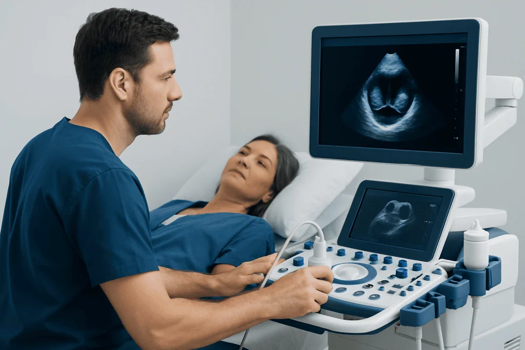
TEE creates images by sending harmless high-frequency sound waves from the probe and receiving the echoes that return from heart structures. A trained cardiologist interprets images from several probe positions and angles. Doppler ultrasound is also used to measure the direction and speed of blood flow across the valve.
An aortic valve leaflets short axis TEE view looks across the valve, often allowing the clinician to see the leaflets in a circular arrangement. This view can help identify whether there are three leaflets or an anatomical variation such as a bicuspid aortic valve, where two leaflets are present. It may also show leaflet thickening, calcification, restricted opening, abnormal tissue, or a gap that contributes to leakage.
An aortic valve leaflets TEE long axis view shows the valve from the side and helps assess how far the leaflets move, whether they meet properly when closed, and how blood travels through the left ventricular outflow tract and into the aorta. Looking at aortic leaflets on TEE in more than one plane is important because valve disease can be complex and no single image provides the full picture.
Why It May Be Recommended: Candidacy and Clinical Uses

A clinician may recommend TEE when a standard echocardiogram does not give sufficiently clear images or when closer inspection of the valve is likely to change care. It can be especially useful if body shape, lung disease, prior chest surgery, or other factors limit ultrasound images obtained through the chest wall.
Common reasons for evaluating aortic valve leaflets include suspected aortic stenosis, in which the valve becomes narrowed; aortic regurgitation, in which blood leaks backward through the valve; a bicuspid valve; or suspected infective endocarditis. In endocarditis, TEE can help look for infection-related growths, abscesses, or damage around a valve. These findings are interpreted alongside symptoms, blood tests, cultures, and other imaging.
TEE may also be performed before, during, or after heart valve surgery or a catheter-based valve procedure to help plan treatment and assess the result. If a structural valve problem is found, the care team may discuss appropriate next steps, including monitoring, medication for related symptoms or conditions, or aortic valve replacement when clinically indicated. The exact plan depends on the valve problem, its severity, overall health, and individual goals.
What Happens During the Procedure
Before the examination, the patient usually receives instructions not to eat or drink for several hours. The care team reviews medical history, current medicines, allergies, dental work, prior problems with sedation, and symptoms such as difficulty swallowing. Blood-thinning medicines should never be stopped without specific instructions from the clinician who prescribed them.
At the appointment, an intravenous line is often placed for sedating medicine. Monitoring equipment checks heart rhythm, blood pressure, and oxygen levels. The throat is sprayed or gargled with a local anesthetic, and a mouth guard protects the teeth and probe. Most patients receive moderate sedation, which promotes relaxation and drowsiness while allowing them to breathe independently.
Once the patient is comfortable, the clinician guides the lubricated probe into the mouth and esophagus. The patient is not expected to swallow the probe actively, and the examination team continuously monitors comfort and safety. Images are recorded from selected positions; the imaging part often takes less than an hour, although preparation and recovery add time to the visit.
Patients should arrange for an adult to take them home after sedated TEE. They should not drive, make important decisions, drink alcohol, or operate machinery until the effects of sedation have fully worn off, following the care team’s instructions.
Results, Benefits, and What They Can Show
The major benefit of TEE is detailed visualization of the valve and surrounding heart structures. It can help distinguish normal leaflet anatomy from thickening, calcification, limited movement, prolapse, perforation, or poor leaflet closure. Doppler findings may help estimate the degree of narrowing or backward leakage and show how valve disease affects blood flow.
A report may describe leaflet number and appearance, how well the leaflets open and close, the size of the valve opening, and whether there is regurgitation. It may also comment on the aortic root, ascending aorta, left ventricular function, other heart valves, and the presence of clots or infection-related abnormalities when relevant.
Results are considered in context. For example, symptoms, physical examination findings, routine echocardiography, CT or MRI results, and overall surgical risk can all affect treatment decisions. A result suggesting valve disease does not automatically mean a procedure is needed; many people benefit from scheduled follow-up and repeat imaging.
Related evaluations may be relevant for people with broader heart or blood-vessel concerns, such as assessment of aortic stenosis or aortic regurgitation. A cardiologist can explain what the findings mean for the individual patient.
Recovery Timeline and Possible Risks
After the probe is removed, the patient remains in a recovery area until alert enough to leave safely. A numb throat, mild sore throat, dry mouth, bloating, or temporary hoarseness can occur and usually settles within a day or two. Eating and drinking should wait until the throat numbness has worn off and swallowing feels normal, to reduce the risk of choking.
Most people return home the same day. Depending on sedation and the reason for the examination, they may be advised to rest for the remainder of the day and return to usual activities the following day. If TEE was performed during surgery or another procedure, recovery follows the plan for that broader treatment.
Serious complications are uncommon, but TEE can occasionally cause breathing problems related to sedation, a reaction to medication, irregular heartbeat, bleeding, dental injury, or injury to the throat or esophagus. Risk may be higher in people with significant esophageal disease, recent upper digestive tract surgery, known narrowing of the esophagus, or certain bleeding conditions. The team weighs these factors before recommending the test.
Contact the clinical team promptly after discharge for persistent or worsening throat or chest pain, trouble swallowing, fever, vomiting blood, black stools, breathing difficulty, or any symptom that feels severe or unusual. These symptoms are not expected after routine recovery and require timely assessment.
Preparing Well and When to Seek Medical Care
Patients can help make TEE safer and more useful by following fasting instructions carefully and bringing an accurate list of medicines, supplements, allergies, and medical conditions. It is particularly important to tell the team about reflux with swallowing difficulties, esophageal varices, previous radiation to the chest or neck, prior esophageal surgery, loose teeth, sleep apnea, pregnancy, and past reactions to anesthesia or sedation.
Medical care should be sought urgently for chest pain, fainting, severe shortness of breath, sudden weakness, or other symptoms that could suggest an emergency. For non-emergency symptoms that can occur with significant valve disease—such as increasing breathlessness during routine activity, reduced exercise tolerance, ankle swelling, palpitations, dizziness, or new fatigue—a timely appointment with a doctor is appropriate.
People already diagnosed with an aortic valve condition should attend recommended follow-up visits even if they feel well. Regular surveillance helps clinicians identify changes before symptoms become limiting and supports shared decision-making about timing of treatment.
Acibadem International’s multidisciplinary cardiology and cardiac surgery specialists at JCI-accredited hospitals assess valve conditions and provide diagnostic and treatment planning for international patients. Care plans should always be individualized after review by a qualified clinician.
Frequently asked questions
Is aortic valve leaflets TEE painful?
TEE is usually not painful, although the throat may feel briefly uncomfortable as the probe is placed. A numbing medicine and sedation are commonly used to improve comfort. Mild throat soreness afterward is possible and typically temporary.
Why is TEE used instead of a regular echocardiogram?
A regular echocardiogram is performed through the chest and is often the first test used to assess heart valves. TEE places the ultrasound probe in the esophagus, closer to the heart, which can provide clearer and more detailed images in selected situations. The two tests may provide complementary information.
What does a short-axis view of the aortic valve show?
The short-axis view looks across the aortic valve and can show the arrangement and number of leaflets. It is useful for assessing whether leaflet opening is restricted and for identifying structural features such as a bicuspid valve. Findings must be interpreted along with other TEE views and Doppler measurements.
How long does it take to recover after TEE?
Most people recover at home on the day of the test. Throat numbness and sedation may last for several hours, so eating should wait until swallowing is normal and driving should be avoided for the rest of the day. Many patients resume usual activities the next day if their clinician agrees.
Can TEE diagnose aortic valve stenosis or leakage?
TEE can provide detailed images that support the diagnosis and assessment of aortic stenosis or aortic regurgitation. Doppler ultrasound helps evaluate blood flow through and around the valve. Clinicians typically combine TEE findings with symptoms, a standard echocardiogram, and other tests when needed.
Who may not be suitable for TEE?
TEE may not be appropriate, or may need special precautions, for people with major esophageal narrowing, active bleeding, a recent tear or surgery involving the esophagus, or some swallowing disorders. The care team reviews these risks before the procedure. Alternative imaging may be considered when TEE is not suitable.
References
- American Heart Association
- American College of Cardiology
- American Society of Echocardiography
- European Society of Cardiology
- National Heart, Lung, and Blood Institute
This article is for general information only and is not a substitute for professional medical advice. Please consult a qualified doctor about your individual situation.
Explore treatments in Turkey — costs, top hospitals & a free quote
JCI-accredited · board-certified surgeons · reply within 24h
Add us as a Preferred Source to see more of our trusted health content across Google Search, AI Overviews and Discover.
Check your numbers in seconds
BMI, calories, due date, blood pressure and 30+ more clinical calculators — free, instant, doctor-reviewed ranges.