Barium Enema — Explained by Medical Evidence, Not Myths
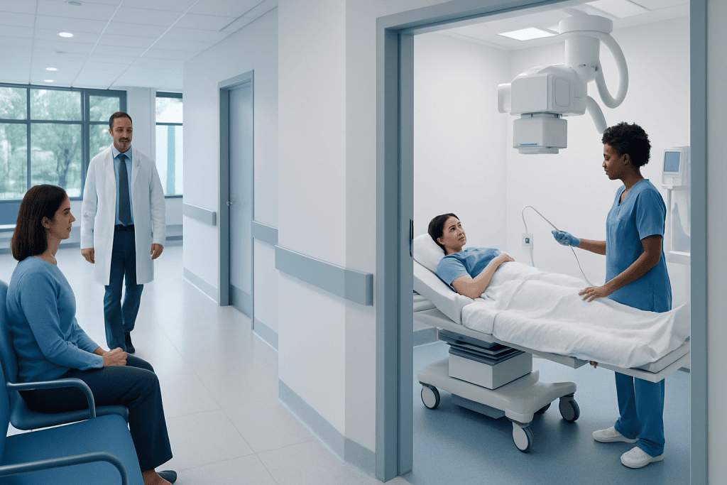
A barium enema is a lower gastrointestinal imaging test that uses barium contrast, and sometimes air, to show the colon on X-rays. It may be used to investigate symptoms such as changes in bowel habits, unexplained abdominal pain, bleeding, or suspected structural problems.
Key Takeaways
- A barium enema is a lower gastrointestinal imaging test that uses barium contrast, and sometimes air, to show the colon on X-rays.
- It may be used to investigate symptoms such as changes in bowel habits, unexplained abdominal pain, bleeding, or suspected structural problems.
- Preparation usually includes a bowel-cleansing plan so the colon is as empty as possible for clearer images.
- The test can cause temporary cramping or pressure, but it is usually completed without sedation.
- Results may suggest the need for follow-up tests, often including colonoscopy, if an abnormality is found.
A barium enema is an imaging test that outlines the colon and rectum with a contrast liquid so they can be seen on X-ray. Although it is used less often than in the past, it can still help doctors evaluate certain bowel symptoms and structural changes in the large intestine.
Overview: what a barium enema is and why it is done
A barium enema is a medical imaging test used to examine the large intestine, which includes the colon and rectum. During the test, a liquid called barium sulfate is introduced into the rectum through a small tube. Barium coats the lining of the bowel and makes it more visible on X-ray images, helping doctors look for changes in shape, narrowing, irritation, or other abnormalities.
This test is also called a lower gastrointestinal series. In some cases, air is gently added after the barium to expand the colon and improve detail on the images. This is sometimes referred to as a double-contrast barium enema. The goal is not treatment, but clearer visualization of the inside contour of the bowel.
Barium enema is not the first choice for every patient today, because other tests such as colonoscopy and advanced imaging are often available. Even so, it still has a role in selected situations, especially when a doctor wants to assess bowel structure or when another test is not suitable. The value of the test depends on the person’s symptoms, medical history, and the clinical question being asked.
When doctors may recommend a barium enema
A doctor may recommend a barium enema when symptoms suggest a problem in the large intestine and imaging of the colon is needed. It can help evaluate persistent constipation, changes in bowel habits, unexplained abdominal pain, rectal bleeding, or concern about a narrowing or blockage. It may also be considered after an incomplete colonoscopy, depending on the situation and local practice.
The test may show structural conditions such as diverticula, abnormal widening or narrowing of the bowel, some inflammatory changes, or masses that alter the shape of the colon. It can also provide useful information in certain congenital or postoperative bowel problems. If a person has symptoms linked to chronic bowel disease, the doctor may compare the best test options, including imaging and endoscopy.
Because a barium enema mainly shows the outline of the bowel rather than taking tissue samples, it does not replace colonoscopy when biopsy or direct treatment is needed. If an abnormality is seen, the next step is often direct visualization with colonoscopy. In some cases, cross-sectional imaging such as CT colonography may also be discussed as part of the diagnostic plan.
How to prepare for a barium enema

Preparation is an important part of a barium enema because stool left in the colon can make the images harder to interpret. The bowel usually needs to be emptied beforehand. A healthcare team will give specific instructions, which may include a low-fiber or clear-liquid diet for a short period, along with laxatives or other bowel-cleansing steps.
Patients should tell their doctor about all medicines, allergies, previous bowel surgery, and whether there is any chance of pregnancy. Certain medications may need to be adjusted before the test, but this should only be done on medical advice. If the person has severe constipation, active bowel symptoms, or recent rectal procedures, the imaging team may tailor the preparation.
It is also helpful to ask practical questions in advance, such as when to stop eating, whether regular medications can be taken with water, and what to expect afterward. Good preparation helps improve image quality and reduces the chance that the test will need to be repeated. Following the instructions closely is one of the simplest ways to make the examination more accurate.
What happens during the procedure
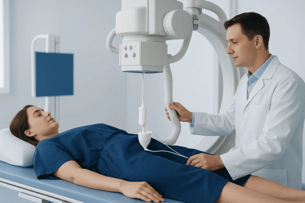
A barium enema is usually performed in a radiology department by trained imaging staff. The patient changes into a gown and lies on an X-ray table. Initial images may be taken first. A small lubricated tube is then gently placed into the rectum so the barium can flow into the colon. If a double-contrast study is planned, a small amount of air may be introduced after the barium.
During the test, the patient may be asked to turn from side to side or hold different positions so images can be taken from several angles. It is common to feel fullness, pressure, or cramping as the colon fills. These sensations can be uncomfortable but are usually brief. Staff members guide the patient through each step and explain when to stay still or hold the breath.
The test often takes less than an hour, though timing can vary. Sedation is usually not needed, so most people can go home the same day without a recovery period. After the images are complete, the tube is removed and the patient can use the bathroom to pass the barium. Additional images may sometimes be taken afterward to see how the bowel empties.
Compared with descriptions that focus only on discomfort, it is more helpful to understand that the procedure is structured and closely supervised. The main aim is to obtain clear information while keeping the patient as comfortable as possible. Communicating with the radiology team during the test can help them adjust positioning and provide reassurance.
Risks, side effects, and what the test can and cannot show
Barium enema is generally considered safe when used appropriately, but like any medical test, it has some limitations and risks. Temporary bloating, cramping, and mild constipation afterward are fairly common. Stools may appear pale or whitish for a short time as the barium leaves the body. Drinking fluids afterward is often advised unless a doctor has recommended fluid restriction for another reason.
Less common but more serious risks include bowel obstruction from retained barium, allergic-type reactions to materials used during the procedure, or injury to the bowel. A perforation of the colon is rare, but this is one reason the test is not suitable for everyone. It may be avoided in people with suspected bowel perforation, severe active inflammation, certain recent surgeries, or acute severe illness affecting the colon.
The test can show the shape and contour of the large bowel, but it cannot collect tissue samples or remove polyps. This is an important distinction. If the concern is a condition such as colon cancer or inflammatory disease, a barium enema may suggest an abnormality, but further evaluation is often necessary to confirm what it is. For inflammatory conditions such as ulcerative colitis, doctors often rely on endoscopy, biopsy, and clinical assessment in addition to imaging.
Understanding the results and possible next steps
A radiologist reviews the images and sends a report to the referring doctor. The results may be described as normal, or they may note findings such as narrowing, outpouchings, filling defects, irregularity of the bowel wall, or changes in bowel position. These findings do not always point to a single diagnosis, but they help guide what should happen next.
If the study is normal, the doctor will consider whether the symptoms might be due to causes not seen on this test. If the study is abnormal, the next step often depends on the pattern seen and the person’s symptoms. For example, a suspicious narrowing or mass-like finding may lead to further examination with colonoscopy or, in selected cases, additional imaging such as MRI if nearby structures also need evaluation.
Results are best understood in context rather than in isolation. A symptom such as rectal bleeding, for instance, may require a different follow-up plan than constipation alone. Doctors combine the imaging report with examination, blood tests, family history, and other factors before making a diagnosis or recommending treatment.
Aftercare, self-care, and when to seek medical care
After a barium enema, most people can return to their usual activities unless their doctor advises otherwise. Drinking extra fluids may help the body clear the barium and reduce constipation. Some people find that their abdomen feels slightly bloated for a short time, but this generally settles. Stool color may stay light for a day or two.
Patients should follow any instructions provided by the radiology team, especially if they were told to use a mild laxative afterward. If bowel movements do not return or constipation becomes more noticeable, it is sensible to contact a healthcare professional for advice. It is best not to ignore symptoms simply because the test itself is over.
Medical care should be sought promptly if there is severe abdominal pain, persistent vomiting, fever, inability to pass stool or gas, significant rectal bleeding, or marked abdominal swelling after the procedure. These symptoms are not expected and may need urgent assessment. People should also contact a doctor if their original bowel symptoms continue, worsen, or return.
For people who need coordinated assessment, Acibadem International’s multidisciplinary specialists and JCI-accredited hospitals diagnose and treat bowel conditions for international patients. If symptoms point toward a broader digestive problem, related evaluation may include specialist care in Crohn's disease or other colorectal conditions, depending on the findings.
Frequently asked questions
Is a barium enema painful?
Most people do not describe it as painful, but it can feel uncomfortable. Common sensations include pressure, fullness, and temporary cramping as the colon fills with barium and sometimes air. The discomfort usually improves once the test is finished.
How long does a barium enema take?
The examination is often completed within about 30 to 60 minutes, although timing can vary. Preparation before the appointment is usually more time-consuming than the test itself. The radiology team can explain the expected schedule for the individual patient.
Do patients need anesthesia or sedation for a barium enema?
Sedation is usually not required for this test. Because patients stay awake and can usually resume normal activities afterward, recovery is often simple. If a person has special needs or concerns, they should discuss them with the doctor in advance.
What is the difference between a barium enema and a colonoscopy?
A barium enema uses contrast and X-rays to outline the colon, while colonoscopy uses a camera to directly view the inside of the bowel. Colonoscopy can also take biopsies and remove some polyps, which a barium enema cannot do. That is why an abnormal barium enema often leads to colonoscopy for confirmation.
Can a barium enema detect colon cancer?
It can sometimes show changes that raise concern for colon cancer, such as a narrowing or a mass-like outline. However, it does not confirm cancer on its own because it cannot take a tissue sample. Suspicious findings usually need follow-up, often with colonoscopy and biopsy.
What should patients do after the test?
They should follow the instructions given by the imaging team, which often include drinking fluids to help pass the barium. Mild bloating or pale stools can happen for a short time. If severe pain, fever, vomiting, or inability to pass stool develops, medical advice should be sought promptly.
This article is for general information only and is not a substitute for professional medical advice. Please consult a qualified doctor about your individual situation.
Persistent digestive symptoms? Get evaluated in Turkey
JCI-accredited · board-certified surgeons · reply within 24h
Add us as a Preferred Source to see more of our trusted health content across Google Search, AI Overviews and Discover.

More from the Health Library
Diarrhea That’s Green — Explained by Medical Evidence, Not Myths

Acne Prone Skin: A Complete Medical Overview

Nervous Stomach: A Complete Medical Overview

Toenail: What Patients Need to Know

Body Acne: An Evidence-Based Guide for Patients