Brain Vessel Embolization: What Interventional Neurology Treats Step by Step

Brain vessel embolization is performed through a thin catheter inserted into a blood vessel, usually in the wrist or groin. It is commonly used for brain aneurysms, arteriovenous malformations, dural arteriovenous fistulas, and certain highly vascular tumors.
Key Takeaways
- Brain vessel embolization is performed through a thin catheter inserted into a blood vessel, usually in the wrist or groin.
- It is commonly used for brain aneurysms, arteriovenous malformations, dural arteriovenous fistulas, and certain highly vascular tumors.
- The procedure uses imaging guidance and materials such as coils, liquid embolic agents, particles, or stents depending on the condition.
- Recovery is often faster than with open surgery, but careful follow-up is still important.
- Risks and benefits vary by diagnosis, vessel anatomy, and overall health, so individualized planning is essential.
Brain vessel embolization is a minimally invasive, image-guided procedure used to block or reduce abnormal blood flow in the brain. It can help treat conditions such as aneurysms, arteriovenous malformations, dural fistulas, and some tumors while avoiding open surgery in many cases.
Overview
Brain vessel embolization is a minimally invasive procedure that treats abnormal or harmful blood vessels in and around the brain. It is performed by specialists in interventional neurology, interventional neuroradiology, or endovascular neurosurgery using real-time imaging to guide very small catheters through the blood vessels. The aim is to block, reduce, or redirect blood flow in a precise area while protecting normal brain circulation.
This approach is used for several conditions, including certain brain aneurysms, arteriovenous malformations, dural arteriovenous fistulas, and some tumors that have a rich blood supply. In some cases, embolization is the main treatment. In others, it is used before surgery or radiosurgery to make treatment safer or more effective.
Because the procedure is done from inside the blood vessels, it usually does not require a large incision in the skull. Many patients appreciate that this often means a shorter hospital stay and recovery period compared with traditional open surgery. Still, it remains a highly specialized treatment that requires careful planning, expert imaging, and close monitoring.
What Conditions Can Brain Vessel Embolization Treat?
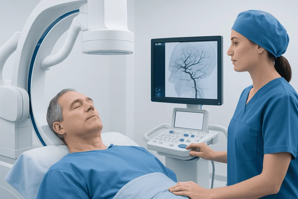
Doctors may recommend brain vessel embolization when a blood vessel abnormality creates a risk of bleeding, seizures, headaches, pressure on nearby structures, or other neurological symptoms. One of the best-known uses is for aneurysms, where the goal is to seal off the bulging part of the vessel so blood no longer enters it. Depending on the aneurysm’s shape and location, treatment may involve coils, stent-assisted techniques, or flow-diverting devices as part of aneurysm treatment.
Another important use is in arteriovenous malformations, or AVMs. These are abnormal tangles of arteries and veins that can disrupt normal blood flow and may bleed. Embolization can reduce the size of an arteriovenous malformation or block selected feeding vessels before surgery or focused radiation.
Dural arteriovenous fistulas are abnormal connections between arteries and veins in the covering of the brain. These can sometimes cause pulsating noise in the ear, headaches, eye symptoms, or bleeding risk depending on their drainage pattern. Embolization is often a key treatment for these lesions. The procedure may also be used before removal of certain vascular tumors to reduce blood loss during surgery.
How the Procedure Is Done Step by Step

Before the procedure, the care team reviews symptoms, brain scans, blood tests, medications, allergies, and overall medical history. Patients may need MRI, CT, or a detailed angiogram to map the blood vessels. The doctor also explains the reason for embolization, what material may be used, possible alternatives, and the expected benefits and risks.
On the day of treatment, the patient is positioned on an angiography table and connected to monitors. The procedure is usually done under sedation or general anesthesia, depending on the condition and complexity. After the skin is cleaned and numbed, the doctor inserts a small catheter into an artery, often at the wrist or groin, and carefully guides it toward the blood vessels in the neck and brain using X-ray imaging.
Once the target vessel is reached, contrast dye is injected so the team can clearly see the blood flow pattern. The embolization material is then delivered with great precision. This may include tiny platinum coils, liquid embolic agents that harden in place, small particles, glue-like substances, or a stent or flow diverter when needed. The exact method depends on the diagnosis, vessel anatomy, and treatment goal.
After the abnormal vessel or blood flow channel has been treated, the team performs more imaging to confirm the result. The catheter is removed, and pressure or a closure device is used at the access site. Patients then go to a recovery area or intensive monitoring unit so the team can watch blood pressure, neurological status, and the puncture site closely.
Preparation, Tests, and Diagnosis
Diagnosis and planning are a major part of safe embolization. Brain blood vessel problems can be evaluated with CT angiography, MR angiography, and especially catheter angiography, also called cerebral angiography. This test gives detailed images of the arteries and veins and often provides the most accurate information for treatment planning.
The doctor will also review medicines that affect bleeding or clotting, such as blood thinners or antiplatelet drugs. Some patients need special instructions about when to stop or continue these medicines. Kidney function, contrast dye reactions, and pregnancy status may also be discussed because they can affect procedural planning.
In many cases, treatment decisions are made by a multidisciplinary team. This may include interventional neurologists, neurosurgeons, neurologists, neuroradiologists, anesthesiologists, and intensive care specialists. For complex conditions, the team compares embolization with other options such as monitoring, open surgery, or stereotactic radiosurgery. When needed, detailed cerebral angiography helps define the safest route and technique.
Benefits, Risks, and Recovery
The main benefit of brain vessel embolization is that it can treat a problem from inside the blood vessels without a large surgical opening. For many patients, this can mean less tissue disruption, shorter hospitalization, and earlier return to daily activities. It may also make a later surgery safer by reducing blood flow to the abnormal area first.
At the same time, embolization is not risk-free. Possible complications can include bleeding, stroke, vessel injury, spasm of a blood vessel, reactions to contrast dye, infection, or incomplete closure of the abnormal vessel. The exact risk depends on the condition being treated, the location of the lesion, the materials used, and the patient’s general health.
Recovery varies from person to person. Some patients stay only a short time in the hospital, while others need longer observation, especially after treatment of a ruptured aneurysm or a complex vascular malformation. Mild bruising or soreness at the catheter entry site is common. The team usually gives guidance on activity, hydration, medications, and warning signs to watch for once the patient goes home.
Follow-up imaging is often necessary because some treated vessels can reopen or change over time. Repeat angiography, MRI, or CT scans may be scheduled to confirm that treatment remains effective. When embolization is part of a broader care plan, further AVM treatment or surgery may still be recommended.
Treatment Materials and Techniques
Different embolization tools are chosen for different problems. Coils are commonly used for aneurysms and work by filling the aneurysm sac so blood flow slows and clotting occurs inside it. In some situations, a stent is placed to support the vessel wall or help keep coils in position. Flow-diverting devices may be used for selected aneurysms to reroute blood away from the bulge over time.
Liquid embolic agents are often used for AVMs and dural fistulas. These materials travel into tiny abnormal channels and then solidify, helping close off the problem vessels. Particles may be used when the goal is to reduce blood supply to a tumor before surgery. The choice depends on how large the vessel is, how fast blood is moving, and how close the lesion is to healthy brain tissue.
Sometimes embolization is planned as one stage of treatment rather than the final step. For example, a complex AVM may be partially embolized first and then treated with microsurgery or radiosurgery. In other cases, a single session is enough. The most suitable strategy is individualized and based on safety, effectiveness, and the long-term treatment plan.
Follow-Up, Self-Care, and When to Seek Medical Help
After discharge, patients are usually advised to rest, avoid heavy lifting for a short period, and follow instructions about wound care and medicines. Drinking enough fluids may be recommended unless a doctor has said otherwise. If antiplatelet or blood-thinning medicines are prescribed, they should be taken exactly as directed and not stopped without medical advice.
Ongoing follow-up is important even when a patient feels well. Some vascular conditions need repeat scans months or years later to confirm they remain sealed or stable. People who have underlying risk factors such as high blood pressure, smoking, or vascular disease may also be encouraged to manage these closely to protect overall blood vessel health.
Urgent medical attention is needed if a person develops sudden weakness, difficulty speaking, severe new headache, loss of vision, confusion, seizure, chest pain, heavy bleeding at the catheter site, or worsening swelling and pain. These symptoms do not always mean a serious complication, but they should be checked promptly. A qualified doctor can advise whether urgent imaging or emergency care is needed.
For international patients seeking evaluation, Acibadem International’s multidisciplinary specialists and JCI-accredited hospitals diagnose and treat complex brain vascular conditions with advanced endovascular techniques, including endovascular treatment when appropriate.
Frequently asked questions
Is brain vessel embolization the same as brain surgery?
No. Brain vessel embolization is a minimally invasive endovascular procedure performed through a catheter placed in a blood vessel, usually in the wrist or groin. It may avoid open brain surgery in some cases, although some patients still need surgery or other treatments as part of a combined plan.
How long does brain vessel embolization take?
The procedure length depends on the condition being treated and how complex the blood vessel anatomy is. Some treatments are completed relatively quickly, while others take several hours. The care team can usually give a more specific estimate after reviewing the imaging.
Will the patient be awake during the procedure?
Some patients receive conscious sedation, while others have general anesthesia. The choice depends on the treatment type, the need to keep the patient still, and overall safety. The doctor and anesthesia team explain the plan before the procedure.
What are the main risks of brain vessel embolization?
Possible risks include stroke, bleeding, blood vessel injury, contrast dye reaction, and problems at the catheter entry site. However, the exact risk varies widely based on the diagnosis, the vessel location, and the patient’s health. Specialists weigh these risks carefully against the benefits of treatment.
How soon can normal activities be resumed?
Many people can return to light activities within a few days, but timing varies depending on the condition treated and the hospital stay. More complex cases may need a longer recovery period. The treating team provides individualized guidance about work, exercise, and travel.
Can an embolized brain vessel problem come back?
Sometimes it can. Certain aneurysms, AVMs, and fistulas may reopen, change, or need additional treatment over time, which is why follow-up imaging is important. Regular review helps doctors detect changes early and decide whether any further care is needed.
References
- World Health Organization
- American Stroke Association
- National Institute of Neurological Disorders and Stroke
- Society of NeuroInterventional Surgery
- Radiological Society of North America
This article is for general information only and is not a substitute for professional medical advice. Please consult a qualified doctor about your individual situation.
Interventional Neurology in Turkey — costs, top hospitals & a free quote
JCI-accredited · board-certified surgeons · reply within 24h
Add us as a Preferred Source to see more of our trusted health content across Google Search, AI Overviews and Discover.
Check your numbers in seconds
BMI, calories, due date, blood pressure and 30+ more clinical calculators — free, instant, doctor-reviewed ranges.

More from the Health Library



Related Specialists

Prof. Dr. Gültekin Karakuş
Cardiology
Prof. Dr. Vahit Emre Özden
Orthopedic Surgery & Traumatology
Dr. Hudheifa Yussuf Haji
Inpatient Clinic Physicians Clinical Service
Prof. Dr. Selami Çakmak
Orthopedic Surgery & Traumatology