Cerebral Angiography Before Neuroendovascular Treatment: What to Expect Step by Step
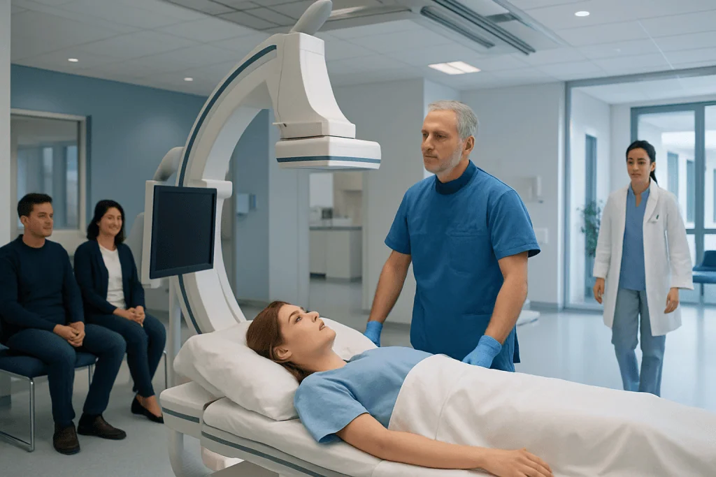
Cerebral angiography provides highly detailed images of brain blood vessels before treatment. The procedure is usually performed through a small catheter inserted in the wrist or groin.
Key Takeaways
- Cerebral angiography provides highly detailed images of brain blood vessels before treatment.
- The procedure is usually performed through a small catheter inserted in the wrist or groin.
- It helps plan neuroendovascular treatment for conditions such as aneurysms, AVMs, and vessel narrowing.
- Most patients go home the same day or after a short observation period, depending on the reason for the test.
- Following preparation and aftercare instructions can support a smoother recovery.
Cerebral angiography is a detailed imaging procedure used to examine the blood vessels of the brain and guide treatment planning. Before neuroendovascular treatment, it helps specialists understand the exact location, shape, and blood flow pattern of a vascular problem so care can be tailored safely and precisely.
Overview: Why cerebral angiography is done before treatment
Cerebral angiography, sometimes called a brain angiogram or catheter angiography, is a specialized imaging procedure that shows the blood vessels in and around the brain in very fine detail. It is often used before neuroendovascular treatment because it can reveal the exact anatomy of a blood vessel problem more clearly than many other imaging tests. This detail helps the care team decide whether treatment is needed and, if so, how to perform it as safely as possible.
During the procedure, a thin flexible tube called a catheter is guided through an artery, and contrast dye is injected while X-ray images are taken. The dye outlines the blood vessels so doctors can see their size, shape, direction, and blood flow. This can be especially helpful when evaluating conditions such as brain aneurysm, arteriovenous malformations, carotid or intracranial narrowing, fistulas, or other vascular changes.
Before a planned neuroendovascular procedure, cerebral angiography acts like a roadmap. It allows interventional specialists to understand where a blockage, bulge, leak, or abnormal connection is located and how nearby vessels branch. In some cases, the angiography is performed as a separate planning step; in others, it may be done immediately before treatment during the same session.
When doctors recommend it
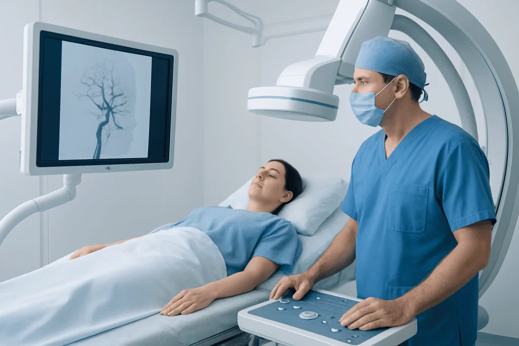
Doctors may recommend cerebral angiography when noninvasive imaging such as CT angiography or MR angiography does not provide enough detail, or when treatment is being considered. It is commonly used before procedures that treat aneurysms, vessel narrowing, stroke-related blockages, arteriovenous malformations, or dural arteriovenous fistulas. It helps answer practical questions about the blood vessels that matter during treatment.
For example, specialists may use cerebral angiography before aneurysm treatment to study the neck and shape of an aneurysm, or before AVM embolization to identify the arteries feeding an abnormal vessel tangle. It may also be part of planning for stroke thrombectomy or other catheter-based procedures involving the brain’s circulation.
Although cerebral angiography is highly informative, it is recommended selectively. The decision is based on the patient’s symptoms, earlier imaging results, medical history, and the type of treatment being considered. The goal is to gather the information needed to make treatment accurate and individualized.
How to prepare before the procedure

Preparation usually begins with a review of medical history, current symptoms, allergies, and medications. The care team will want to know about previous reactions to contrast dye, kidney problems, bleeding disorders, pregnancy, diabetes, or use of blood thinners. Blood tests may be requested to check kidney function and clotting, especially if treatment may follow soon after the angiogram.
Patients are often asked not to eat or drink for a period before the procedure, although the exact timing can vary. The doctor may advise continuing some medications and temporarily adjusting others, particularly anticoagulants, antiplatelet drugs, or diabetes medications. It is important to follow the individual instructions provided rather than relying on general advice alone.
Before the procedure starts, a hospital gown is usually provided and an intravenous line may be placed for fluids or medication. The team explains the steps, confirms consent, and checks basic vital signs. Many patients find it reassuring to ask in advance whether the catheter will likely be placed through the wrist or the groin, how long the visit may take, and whether someone should accompany them home.
Step by step: what happens during cerebral angiography
The procedure is usually performed in a specialized angiography suite by an interventional neuroradiologist, interventional neurologist, or neurosurgical endovascular specialist, together with nurses and technologists. The access site, most often the wrist or groin, is cleaned and numbed with local anesthetic. Mild sedation may be given to help the patient relax, but many people remain awake and able to respond during the test.
After the area is numbed, the doctor inserts a small sheath into the artery and carefully advances the catheter toward the arteries supplying the brain. X-ray guidance is used throughout, so the team can see the catheter’s position in real time. When the catheter is in place, contrast dye is injected and a series of images is captured from different angles. Patients may notice a brief warm sensation, a metallic taste, or a feeling of flushing when the dye is injected; these sensations are usually short-lived.
The specialist may study several arteries to build a complete picture of blood flow. The images can show aneurysms, narrowing, vessel blockages, abnormal connections, or the feeders and drainage pattern of an AVM. If a planned intervention is scheduled at the same time, the angiography findings guide the next stage, such as carotid artery stenting or another targeted endovascular procedure.
Once imaging is complete, the catheter is removed and pressure or a closure device is used to help seal the artery. The entire process may take around an hour or longer depending on complexity, whether multiple vessels must be studied, and whether treatment is performed in the same session.
Possible risks, side effects, and safety measures
Cerebral angiography is a commonly performed procedure in experienced centers, but like any invasive test it carries some risks. These can include bruising or bleeding at the access site, temporary discomfort, allergic reaction to contrast dye, and changes in kidney function in susceptible patients. There is also a small risk of blood vessel injury, blood clot formation, or stroke during the procedure.
To reduce risk, the team carefully reviews medical history and medications in advance, uses sterile technique, monitors the patient closely, and chooses equipment and contrast use thoughtfully. Imaging is performed in real time to guide catheter movement precisely. If the patient has a known contrast allergy or kidney concerns, additional precautions may be taken as appropriate.
Many people worry most about pain or serious complications. In practice, the local anesthetic usually limits discomfort at the access site, and the team continuously watches for any changes in blood pressure, heart rate, neurological status, or symptoms. Patients should report any pain, weakness, numbness, trouble speaking, chest discomfort, rash, or unusual feelings right away so the team can respond promptly.
Recovery and what to expect afterward
After cerebral angiography, the patient is moved to a recovery area for monitoring. The access site is checked for bleeding or swelling, and blood pressure, pulse, and general neurological status are observed. If the catheter was inserted through the groin, lying flat for a period may be necessary; wrist access often allows earlier sitting up, though activity restrictions still apply.
It is normal to have mild soreness, a small bruise, or some tiredness afterward. Drinking fluids may be encouraged to help clear the contrast dye, unless another medical condition limits fluid intake. Depending on the reason for the angiogram and whether treatment was done at the same time, the patient may go home the same day or stay longer for observation.
At home, strenuous activity, heavy lifting, and soaking the puncture site are often avoided for a short time. The care team provides instructions on medications, wound care, bathing, and when normal activities can resume. Near the end of the care pathway, patients seeking specialized treatment may appreciate knowing that Acibadem International’s multidisciplinary specialists in JCI-accredited hospitals diagnose and treat complex neurovascular conditions for international patients.
When to call a doctor and questions to ask
Patients should contact their doctor promptly if they develop increasing pain, swelling, active bleeding, fever, worsening bruising, numbness, weakness, speech difficulty, severe headache, chest symptoms, or signs of poor circulation in the limb used for access. These symptoms do not always mean a serious problem, but they should be assessed without delay. The hospital team will explain which symptoms require urgent attention and where to seek help after discharge.
It can also be helpful to ask a few practical questions before the procedure. Examples include whether the angiography is diagnostic only or may lead directly to treatment, how long recovery is expected to take, when regular medications should be restarted, and what restrictions apply afterward. Patients may also ask what the images are expected to show and how the results will influence the next treatment decision.
Clear communication can make the experience less stressful. Knowing the purpose of the test, how to prepare, and what symptoms to monitor afterward often helps patients feel more confident and involved in their care.
Frequently asked questions
Is cerebral angiography the same as a CT or MRI scan?
No. Cerebral angiography is a catheter-based procedure that provides very detailed, real-time images of the brain's blood vessels, while CT and MRI angiography are noninvasive imaging tests. Doctors may use cerebral angiography when they need more precise information for diagnosis or treatment planning.
Will cerebral angiography hurt?
Most patients feel a brief sting from the numbing medicine at the access site and then pressure rather than sharp pain. When contrast dye is injected, a warm or flushing sensation can occur for a few seconds. The care team works to keep the patient comfortable throughout the procedure.
How long does the procedure take?
The imaging part often takes about an hour, but total time at the hospital is usually longer because of preparation and recovery. If the anatomy is complex or if treatment is performed in the same session, the procedure may take more time.
Why is cerebral angiography needed before neuroendovascular treatment?
It gives specialists a detailed map of the blood vessels involved. This helps them choose the best treatment approach, select the right devices, and reduce unnecessary risk by understanding the exact anatomy before intervention.
How soon can normal activities be resumed?
Many people return to light daily activities within a day or two, but heavy lifting and strenuous exercise are often restricted for a short period. The timing depends on the access site used, overall health, and whether additional treatment was performed.
What should patients do if they take blood thinners?
They should tell their doctor well before the procedure and follow individualized instructions carefully. Blood thinners should never be stopped or changed without medical advice, because the safest plan depends on why the medication was prescribed and the procedure being performed.
References
- American Heart Association
- National Institute of Neurological Disorders and Stroke
- Radiological Society of North America
- Society of NeuroInterventional Surgery
- NHS
This article is for general information only and is not a substitute for professional medical advice. Please consult a qualified doctor about your individual situation.
Explore treatments in Turkey — costs, top hospitals & a free quote
JCI-accredited · board-certified surgeons · reply within 24h
Add us as a Preferred Source to see more of our trusted health content across Google Search, AI Overviews and Discover.