Dermatofibrosarcoma Protuberans: Diagnosis, Outlook, and Modern Treatment Approaches

Dermatofibrosarcoma protuberans is a rare skin and soft tissue sarcoma that tends to grow slowly. It often begins as a firm, painless patch or lump that enlarges over months or years.
Key Takeaways
- Dermatofibrosarcoma protuberans is a rare skin and soft tissue sarcoma that tends to grow slowly.
- It often begins as a firm, painless patch or lump that enlarges over months or years.
- Diagnosis usually requires a skin or soft tissue biopsy and imaging in selected cases.
- Surgery with clear margins is the main treatment and is important for reducing recurrence.
- Long-term follow-up matters because DFSP can come back locally even after treatment.
Dermatofibrosarcoma protuberans is a rare, usually slow-growing soft tissue cancer that starts in the skin and nearby tissues. It is often highly treatable, especially when diagnosed early and removed completely with carefully planned surgery.
Overview: what dermatofibrosarcoma protuberans is
Dermatofibrosarcoma protuberans is a rare type of cancer that develops in the deeper layers of the skin and the tissue just beneath it. Doctors often shorten the name to DFSP. It usually grows slowly and may be present for a long time before it is diagnosed, but it can extend into surrounding tissues if left untreated.
Although DFSP is a cancer, it behaves differently from many faster-growing skin cancers. It most often spreads locally rather than to distant organs, and complete treatment is commonly possible. The main challenge is that its edges can extend beyond what is visible on the skin, which is why accurate diagnosis and carefully planned treatment are so important.
DFSP can appear in adults of different ages and may occur on the trunk, shoulders, chest, abdomen, arms, legs, or less commonly the head and neck. Because it may resemble a scar, a cyst, or another harmless skin growth early on, specialist assessment is often needed to confirm the diagnosis.
Symptoms and how it may look on the skin
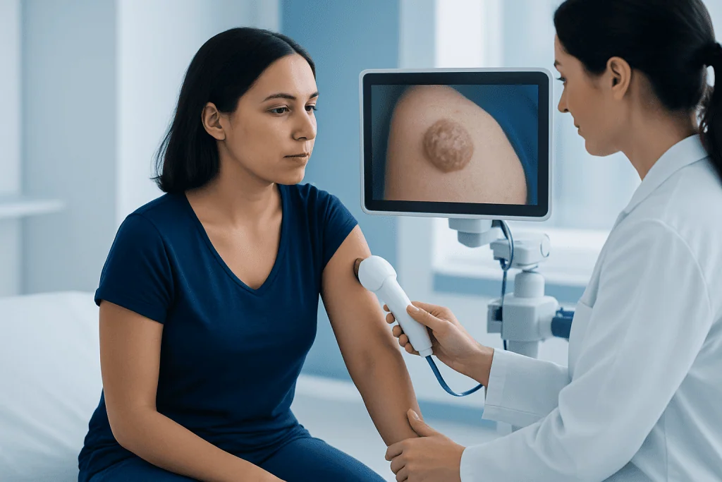
The most common sign of dermatofibrosarcoma protuberans is a firm area of skin that slowly enlarges. At first, it may look like a flat or slightly raised patch. Over time, it can become thicker, more nodular, or develop a protuberant, bumpy surface, which is where the name comes from.
Many people do not have pain, itching, or other symptoms in the early stages. The lesion may be skin-colored, reddish-brown, purple, or slightly darker than the surrounding skin. Because it tends to grow gradually, it may be mistaken for a benign lump or a scar, especially if it feels stable for a period before enlarging again.
Possible features include:
- A slowly enlarging plaque or lump
- Firm texture
- Painless growth
- Skin discoloration over the area
- Raised nodules developing within an older flat patch
In more advanced cases, the tumor may involve deeper tissues such as fat, fascia, or muscle. Any skin lesion that persists, changes, or continues to enlarge deserves medical evaluation, particularly if it has been present for months or years.
Causes, genetics, and risk factors

The exact cause of dermatofibrosarcoma protuberans is not fully understood. In many cases, the tumor is linked to a specific genetic change involving chromosomes 17 and 22. This change leads to abnormal signaling that encourages tumor cells to grow. It is usually not something a person inherits from their parents, but rather a change that develops in the tumor itself.
Most cases occur without a clear trigger. Occasionally, DFSP has been reported in areas of prior skin injury, surgical scars, burns, or trauma, but this does not mean that injury directly causes the cancer in most people. For the majority of patients, there is no obvious lifestyle-related cause.
Known or possible factors associated with DFSP include:
- Adult age, though it can occur at other ages
- A tumor-specific genetic rearrangement involving collagen and growth factor genes
- Rare occurrence in previous scarred or injured skin
It is important to know that DFSP is uncommon, and most lumps, bumps, or skin changes are not this condition. Still, a lesion that keeps growing should not be ignored, especially if it does not fit the pattern of a typical benign skin finding.
How diagnosis is confirmed
Diagnosis begins with a medical history and physical examination, often by a dermatologist, surgical specialist, or oncologist familiar with soft tissue tumors. Because DFSP can resemble other conditions, appearance alone is not enough to confirm it. A biopsy is needed to identify the tumor under the microscope.
The biopsy may be a punch biopsy, incisional biopsy, or excisional biopsy depending on the size and location of the lesion. Pathologists look for the typical spindle-shaped tumor cells and may use immunohistochemical markers, such as CD34, to support the diagnosis. In some cases, molecular testing is used to detect the characteristic gene fusion linked to DFSP.
Imaging is not always required for small, superficial tumors, but it can be helpful when the lesion is large, recurrent, near critical structures, or suspected to involve deeper tissues. Magnetic resonance imaging may be used to assess local extent before treatment. If there is uncertainty about the diagnosis, doctors may also consider other skin and soft tissue tumors, including skin cancer and certain benign fibrous growths.
Because complete removal is a key part of treatment, careful mapping of the tumor’s extent matters. Multidisciplinary review can be especially useful when the tumor is located on the face, scalp, hands, or other areas where tissue preservation and cancer control both need close attention.
Modern treatment approaches
The main treatment for dermatofibrosarcoma protuberans is surgery. The goal is to remove the entire tumor with a margin of healthy tissue around it, because DFSP can send out microscopic extensions beyond the visible lesion. Achieving clear margins lowers the chance that the tumor will come back in the same area.
Two common surgical strategies are wide local excision and margin-controlled surgery such as Mohs micrographic surgery in selected cases. Wide excision removes the tumor along with surrounding tissue, while Mohs-type approaches examine margins in stages to help preserve healthy tissue when appropriate. Depending on the location and size of the defect after removal, reconstruction may sometimes be needed using techniques related to plastic and reconstructive surgery.
If the tumor is difficult to remove completely, has recurred, or involves important structures, additional planning may be required. Some patients may benefit from care through a specialized oncology team. In selected advanced or unresectable cases, targeted therapy may be considered, especially when the tumor carries the characteristic genetic fusion. Radiotherapy may also be used in certain situations, such as positive margins when further surgery is not feasible.
When doctors suspect deeper tissue involvement, treatment planning may include advanced imaging and specialist surgical assessment similar to approaches used for sarcoma surgery. For international patients, Acibadem International’s multidisciplinary specialists at JCI-accredited hospitals diagnose and treat rare tumors such as DFSP with coordinated dermatology, pathology, surgical, and oncology care.
Outlook, recurrence, and follow-up
The outlook for dermatofibrosarcoma protuberans is often favorable, especially when the tumor is found early and removed completely. DFSP is known more for local recurrence than for spreading to distant parts of the body. This means careful treatment and regular follow-up are central to long-term management.
The risk of recurrence depends on factors such as tumor size, location, whether margins were clear, and whether the tumor has come back before. A less common, more aggressive change called fibrosarcomatous transformation can be associated with a higher risk of spread and may lead doctors to recommend closer monitoring and broader treatment planning.
Follow-up visits usually involve examination of the treated area and nearby skin and soft tissues. Patients are also encouraged to watch for any new lump, thickening, or scar-like change at or near the treatment site. Prompt reassessment helps detect recurrence at an earlier, more manageable stage.
Self-care after treatment and practical prevention steps
There is no proven way to prevent dermatofibrosarcoma protuberans from developing, because it is not strongly linked to common lifestyle factors. However, people can support early detection by paying attention to skin changes and seeking medical advice for any lesion that enlarges, hardens, or persists over time.
After treatment, self-care focuses on wound healing, scar care, follow-up attendance, and awareness of changes around the surgical site. Patients should follow their surgeon’s instructions closely and ask when it is safe to return to normal physical activity. If reconstruction was required, recovery guidance may be more individualized.
Helpful habits include:
- Checking the treated area regularly for new thickening or nodules
- Keeping scheduled follow-up appointments
- Reporting any change in the scar or nearby skin
- Protecting healing skin from irritation and unnecessary trauma
- Discussing any persistent concern with a qualified doctor rather than self-diagnosing
While general skin awareness is helpful, DFSP is not mainly caused by sun exposure in the way some other skin cancers are. Even so, routine skin protection and regular medical care remain good overall health practices.
When to seek medical care
Medical care should be sought for any skin patch, plaque, or lump that continues to grow, becomes firmer, or develops raised nodules. This is especially important if the area has been present for a long time and does not behave like a typical scar, cyst, or harmless skin bump.
People who have already been treated for DFSP should contact their doctor if they notice a new lump, thickening, discoloration, or discomfort near the surgical scar. Review is also important if pathology results show unclear margins or if there are questions about whether further treatment is needed.
Urgent evaluation is sensible if a lesion is rapidly changing, interfering with movement, bleeding repeatedly, or located in a cosmetically or functionally sensitive area such as the face or near a joint. A qualified specialist can confirm whether the lesion is DFSP or another condition and guide the next steps safely.
Frequently asked questions
Is dermatofibrosarcoma protuberans a serious cancer?
Dermatofibrosarcoma protuberans is a cancer, but it usually grows slowly and is often very treatable when diagnosed early. Its main concern is local recurrence, which is why complete removal and follow-up are important.
What does dermatofibrosarcoma protuberans look like?
It often begins as a firm, painless patch or thickened area of skin that slowly enlarges. Over time, it may become raised, nodular, or more noticeable in color and texture.
How is DFSP diagnosed?
DFSP is diagnosed with a biopsy, which allows a pathologist to examine the tissue under a microscope. Imaging such as MRI may be used if the tumor is large, recurrent, or suspected to involve deeper tissues.
What is the best treatment for dermatofibrosarcoma protuberans?
Surgery is the main treatment for most cases. The goal is to remove the tumor completely with clear margins, and in selected situations doctors may also consider targeted therapy or radiotherapy.
Can dermatofibrosarcoma protuberans come back after treatment?
Yes, DFSP can recur in the same area, especially if microscopic tumor cells remain after surgery. Regular follow-up helps detect recurrence early, when it is usually easier to treat.
Does DFSP spread to other parts of the body?
Distant spread is uncommon in classic DFSP. However, certain higher-risk forms, including tumors with fibrosarcomatous transformation, may behave more aggressively and need closer monitoring.
References
- National Cancer Institute
- American Cancer Society
- National Comprehensive Cancer Network
- DermNet
- World Health Organization
This article is for general information only and is not a substitute for professional medical advice. Please consult a qualified doctor about your individual situation.
Oncology care in Turkey — second opinion and treatment plan
JCI-accredited · board-certified surgeons · reply within 24h
Add us as a Preferred Source to see more of our trusted health content across Google Search, AI Overviews and Discover.