

Excision Osteochondroma: Procedure, Recovery and Results
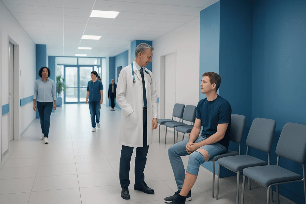
Most osteochondromas are benign and can be monitored if they do not cause symptoms or complications. Excision osteochondroma removes the bony growth and its cartilage cap while preserving healthy surrounding structures whenever possible.
Key Takeaways
- Most osteochondromas are benign and can be monitored if they do not cause symptoms or complications.
- Excision osteochondroma removes the bony growth and its cartilage cap while preserving healthy surrounding structures whenever possible.
- Recovery depends on the growth’s location, size, effects on muscles or joints, and whether bone reconstruction is needed.
- Many people can walk soon after surgery on the leg, but weight-bearing restrictions vary and should follow the surgeon’s plan.
- Persistent new pain, growth after skeletal maturity, or neurological or circulation symptoms should be assessed promptly.
Excision osteochondroma is a surgical procedure that removes an osteochondroma, a usually benign bone growth capped with cartilage. Surgery is not needed for every osteochondroma, but it may be recommended when the growth causes pain, limits movement, compresses nearby structures, changes in concerning ways, or creates uncertainty about the diagnosis.
Excision Osteochondroma: What the Procedure Involves
Excision osteochondroma is an operation to remove an osteochondroma: a bone growth that develops near the end of a bone, often around the knee, shoulder, hip, or ankle. These growths commonly arise during childhood or adolescence while bones are still growing. They are usually benign, meaning non-cancerous, and many are found incidentally on an X-ray taken for another reason.
An osteochondroma is continuous with the underlying bone and is covered by a cartilage cap. During surgery, the orthopedic surgeon removes the outward-growing bone and cartilage cap, aiming to protect nearby tendons, muscles, nerves, blood vessels, and the joint. The exact approach differs according to the location; for example, excision osteochondroma distal femur surgery requires careful planning because the growth is near the knee joint and important structures at the back and sides of the thigh.
The purpose of surgery is usually to relieve symptoms or prevent complications rather than to treat cancer. A complete removal of a symptomatic solitary osteochondroma generally provides lasting relief, although follow-up remains important, particularly for people with multiple hereditary exostoses, also called hereditary multiple osteochondromas.
Who May Benefit From Osteochondroma Removal Surgery?
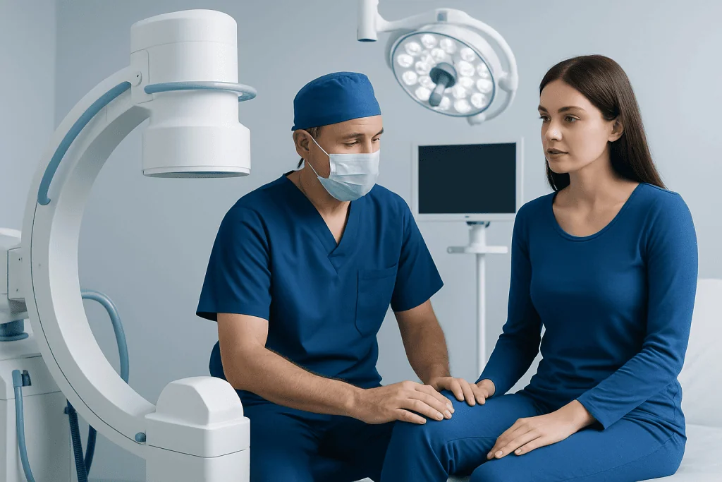
Observation is appropriate for many people with a painless, stable osteochondroma. A clinician may recommend periodic examination and imaging, especially in a growing child, to assess symptoms and development. The decision about osteochondroma removal surgery is individualized and should consider the person’s age, activity level, symptoms, imaging findings, and the growth’s relationship to nearby tissues.
Surgery may be considered when an osteochondroma causes ongoing pain, reduced joint motion, repeated irritation from tendons or muscles, visible deformity, limb-length or alignment concerns, or pressure on a nerve or blood vessel. A growth that fractures after an injury, forms a painful fluid-filled bursa over it, or interferes with everyday activities can also be a reason to discuss excision.
Further assessment is important if a known growth becomes painful or enlarges after skeletal maturity, or if imaging shows changes in the cartilage cap or surrounding tissue. These changes do not automatically mean cancer, but they need timely specialist review. People with multiple growths may benefit from coordinated orthopedic care because individual lesions can affect different joints and functions.
How Excision Osteochondroma Is Planned and Performed
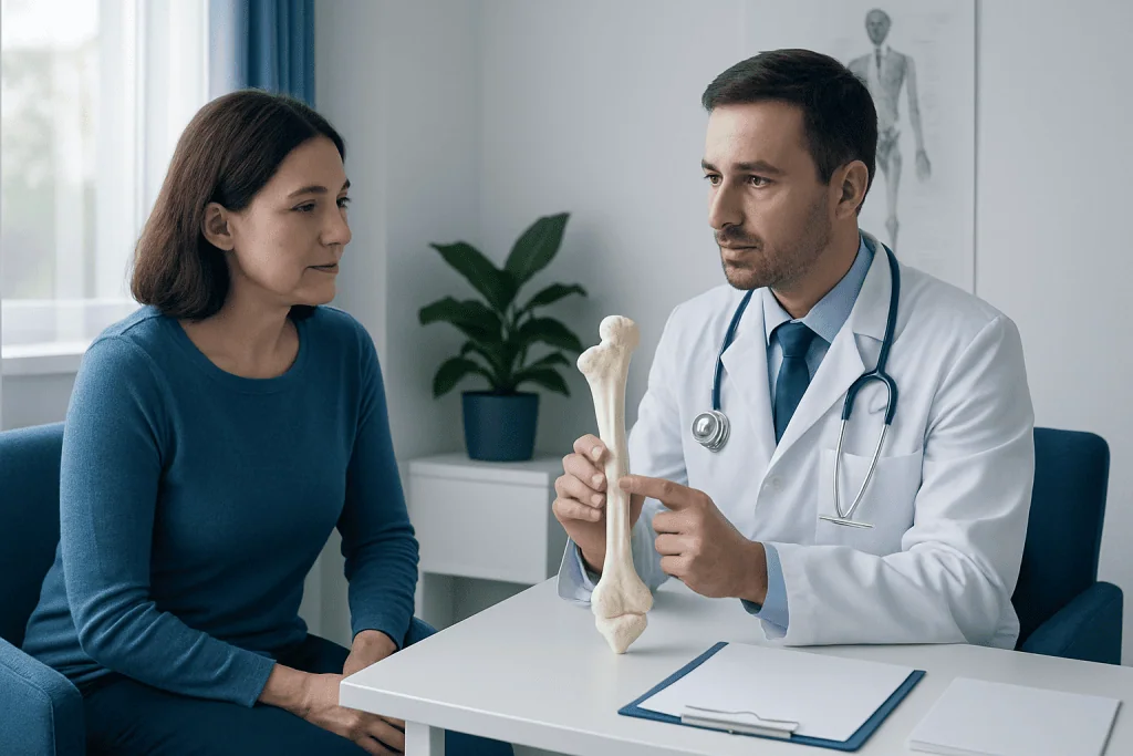
Before surgery, an orthopedic specialist reviews the person’s symptoms and medical history and performs a focused physical examination. Plain X-rays usually show the characteristic connection between the growth and the underlying bone. MRI may be used to assess the cartilage cap and the relationship to nerves, blood vessels, tendons, muscles, or the joint. CT may be useful for lesions in anatomically complex areas.
Excision is generally performed under general anesthesia. The surgeon makes an incision positioned to allow safe access to the lesion, identifies and protects important surrounding tissues, and removes the growth at its base. The cartilage cap is removed with the bony prominence. If the lesion is large, close to a joint, or has altered the bone significantly, the surgeon may smooth the bone or use additional stabilization or reconstruction where needed.
The removed tissue may be sent to a pathology laboratory for examination, particularly when imaging or symptoms have raised questions about the diagnosis. The incision is then closed and dressed. Some procedures are day surgery, while others require a short hospital stay depending on the site of surgery, expected pain control needs, mobility, and the complexity of the operation.
Documentation and billing terms can differ between healthcare systems. An excision osteochondroma CPT code is selected by the treating team or billing professionals based on the exact bone, surgical technique, and clinical circumstances. It is not a substitute for a surgical assessment or an indication of how complex an individual operation will be.
How Long Does It Take to Recover From Osteochondroma Removal Surgery?
Osteochondroma excision recovery varies considerably. Removal of a small, accessible lesion may allow a return to light daily activities within a few weeks, while surgery involving the pelvis, hip, spine, or a lesion near a major joint may require a longer rehabilitation period. Healing also depends on the incision, the amount of muscle and soft tissue involved, and whether other procedures were performed at the same time.
In the first days after surgery, the care team focuses on pain control, wound care, safe movement, and preventing stiffness. Swelling and tenderness around the surgical area are expected initially and usually improve gradually. Follow-up appointments allow the surgeon to inspect wound healing, discuss pathology if testing was performed, and adjust activity restrictions.
Many patients progress through guided exercises or physical therapy to restore range of motion, strength, balance, and confidence with movement. Returning to strenuous work, impact exercise, sports, or heavy lifting should wait until the surgeon confirms that healing and function are adequate. Children and adolescents may have different recovery goals and rehabilitation needs than adults.
It is helpful to ask the surgeon for a personalized recovery plan before the operation. This should cover bathing and dressing instructions, driving and work guidance, travel considerations, pain management, activity progression, and signs that should prompt contact with the healthcare team.
Can You Walk After Osteochondroma Surgery?
Many people can walk after osteochondroma surgery, but the timing and amount of weight they can place on the limb depend on where the growth was removed. For an uncomplicated excision osteochondroma femur procedure, some patients may begin protected walking soon after surgery, often with crutches or another walking aid. Others may need to limit weight-bearing for a period to protect healing tissues.
A lesion near the knee, such as one on the distal femur, can affect comfort, knee bending, and muscle control during early recovery. The surgeon and physiotherapist may recommend crutches, a brace, or specific exercises until walking is safe and comfortable. A growth removed from the upper body may not directly limit walking, although general anesthesia, pain medication, and discomfort can still affect balance during the first days.
Patients should not push through severe pain or change weight-bearing instructions without medical advice. Gradual increases in walking distance and activity help the body recover while lowering the chance of a fall, wound problem, or overuse of healing tissues. The care team can explain when it is safe to resume stairs, driving, work duties, and sports.
Benefits, Risks and Expected Results
The main potential benefits of excision osteochondroma are relief from pain or mechanical irritation, better joint movement, reduced pressure on nearby nerves or blood vessels, and improved ability to carry out work, school, exercise, or other daily activities. In cases where a growth causes visible prominence or deformity, surgery may also improve comfort with clothing or movement. Results depend on whether symptoms were caused directly by the osteochondroma and whether other joint or soft-tissue conditions are present.
As with any operation, there are possible risks. These include bleeding, infection, anesthesia-related problems, blood clots, delayed wound healing, scar sensitivity, stiffness, persistent pain, and injury to nearby nerves, blood vessels, tendons, or muscles. The likelihood and type of risk vary by location; lesions near major joints or deep structures can require more complex surgery.
Recurrence after complete removal of a solitary osteochondroma is uncommon, but it can occur, especially if cartilage-cap tissue remains or if surgery is performed before skeletal maturity. People with multiple hereditary exostoses may develop symptoms from other lesions over time, even when a treated growth does not return. Regular clinical review is appropriate when advised by the orthopedic team.
Orthopedic surgeons, radiologists, pathologists, anesthesiologists, and rehabilitation specialists may work together to plan care. Acibadem International’s multidisciplinary specialists and JCI-accredited hospitals diagnose and treat osteochondroma-related concerns for international patients.
What Is the Prognosis After Osteochondroma Treatment?
The prognosis after osteochondroma treatment is generally favorable. Most solitary osteochondromas remain benign, and people who do not have symptoms may never require an operation. When surgery is needed and the growth is fully removed, many people experience meaningful improvement in the symptoms caused by the lesion.
Prognosis is influenced by the lesion’s site, its effects on nearby structures, and any associated orthopedic issues. For example, pain caused by a tendon rubbing over a bony prominence may improve after excision, while long-standing joint stiffness or nerve symptoms can take more time to settle. Rehabilitation and follow-up are therefore important parts of the overall result.
A small proportion of cartilage-capped bone growths can undergo malignant transformation, most often to a cartilage cancer, but this is uncommon. The risk is higher in people with multiple hereditary exostoses than in those with a single lesion. New or increasing pain, growth after the bones have stopped growing, or other concerning changes should be evaluated rather than assumed to be part of normal recovery.
Should I Get My Osteochondroma Removed? When to Seek Medical Care
Whether an osteochondroma should be removed is a decision made with an orthopedic specialist after reviewing symptoms, examination findings, and imaging. Removal is often unnecessary for a painless, stable lesion that is not affecting movement or nearby structures. Surgery may be reasonable when symptoms are persistent, function is limited, complications are present, or the diagnosis needs clarification.
Medical assessment should be arranged for a newly noticed bony lump, persistent pain near a known osteochondroma, reduced range of motion, numbness, weakness, tingling, color or temperature changes in a limb, or swelling that does not settle. Prompt review is particularly important if a growth enlarges or becomes painful after skeletal maturity, or if pain is new and unexplained.
After surgery, patients should contact their clinical team urgently for fever, increasing redness or drainage from the incision, worsening rather than improving pain, marked swelling, new numbness or weakness, calf pain or swelling, chest pain, or shortness of breath. These symptoms have several possible causes and need professional assessment.
A qualified orthopedic team can explain whether monitoring, symptom management, or surgery is most appropriate. Shared decision-making helps ensure that the expected benefits of excision are balanced against recovery time and the specific risks of surgery for that individual.
Frequently asked questions
What is excision osteochondroma?
Excision osteochondroma is surgery to remove an osteochondroma, a typically benign bone growth with a cartilage cap. The procedure is usually considered when the growth causes symptoms, affects function, presses on nearby structures, or has features that need further evaluation.
How long does it take to recover from osteochondroma removal surgery?
Recovery can range from several weeks for a straightforward procedure to several months when surgery is more extensive or involves a major joint. The timeline depends on the growth’s location, the amount of soft tissue affected, weight-bearing restrictions, and rehabilitation needs.
Can you walk after osteochondroma surgery?
Many patients can walk after surgery, sometimes with crutches or another support. The amount of weight-bearing allowed depends on the surgical site and the surgeon’s instructions, especially after removal from the femur or near the knee, hip, or ankle.
What is the prognosis after osteochondroma treatment?
The outlook is generally very good, particularly for a solitary osteochondroma that is monitored or completely removed when needed. Symptoms often improve after successful excision, though recovery of strength, mobility, or nerve symptoms may take time.
Should I get my osteochondroma removed?
Not every osteochondroma needs removal. An orthopedic specialist may recommend observation for a painless, stable lesion and discuss surgery when pain, limited movement, nerve or blood vessel pressure, deformity, or concerning changes are present.
Can an osteochondroma grow back after surgery?
Recurrence after complete removal is uncommon, especially after skeletal maturity. It may be more likely if cartilage-cap tissue remains or when surgery is performed while a child is still growing; people with multiple hereditary exostoses may also develop symptoms from other lesions.
References
- American Academy of Orthopaedic Surgeons
- National Cancer Institute
- OrthoInfo
- MedlinePlus
- World Health Organization
This article is for general information only and is not a substitute for professional medical advice. Please consult a qualified doctor about your individual situation.
Joint and spine care in Turkey — expert assessment & treatment
JCI-accredited · board-certified surgeons · reply within 24h
Add us as a Preferred Source to see more of our trusted health content across Google Search, AI Overviews and Discover.

More from the Health Library



Related Specialists
Dr. Sibel Uysal
Gynecology & Obstetrics
Dr. Nazli Özcan
Radiology
Dr. Parısa Sadat Hosseını
Anesthesiology
Prof. Dr. Mehmet Veli Karaaltın
Aesthetic Plastic & Reconstructive Surgery