Fes PET Scan: Preparation, Procedure and Results
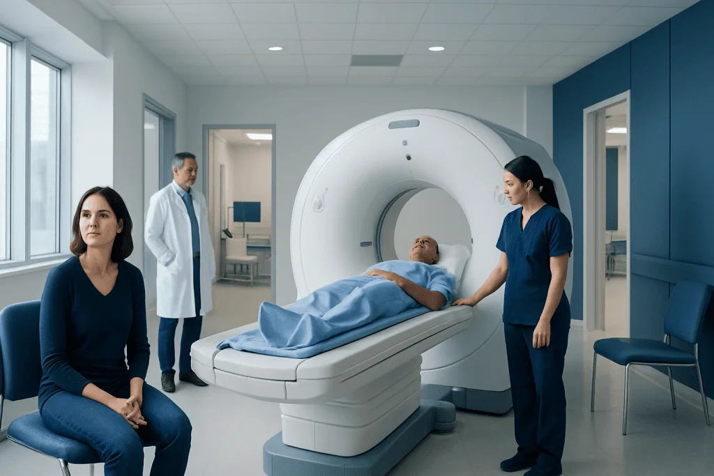
FES PET scanning is designed to visualize estrogen receptor activity, most often in breast cancer. Preparation is usually simpler than for FDG PET scans, but the imaging center’s instructions should always be followed.
Key Takeaways
- FES PET scanning is designed to visualize estrogen receptor activity, most often in breast cancer.
- Preparation is usually simpler than for FDG PET scans, but the imaging center’s instructions should always be followed.
- Certain estrogen-blocking medicines may affect results and may need to be reviewed before scheduling the scan.
- The scan is generally outpatient, and most people can return to normal activities afterward.
- Results are interpreted alongside pathology, other scans, symptoms and the person’s treatment history.
An FES PET scan is a specialized imaging test used mainly in some people with recurrent or metastatic breast cancer. It uses a tracer that attaches to estrogen receptors, helping clinicians assess whether cancer sites are likely to be estrogen receptor-positive.
Overview: What Is an FES PET Scan?
An FES PET scan is a type of positron emission tomography scan that helps show estrogen receptor activity in the body. It uses a small amount of a radioactive tracer called fluoroestradiol, or FES, which is chemically similar to estrogen. After injection, the tracer may collect in tissues containing estrogen receptors and can be detected by the PET scanner.
The test is used primarily for people with certain types of breast cancer, especially when recurrent or metastatic disease is suspected or known. It can help clinicians understand whether cancer in different areas of the body appears to have estrogen receptor-positive features. This information may support treatment planning, but it does not replace a biopsy when tissue sampling is needed or feasible.
FES PET scans differ from standard PET scans that use FDG, a glucose-like tracer. An FDG PET scan highlights areas with increased glucose use, which can occur with many cancers as well as inflammation or infection. In contrast, FES imaging focuses on estrogen receptor binding, providing a different biological view of a tumor.
How FES PET Imaging Works and Who May Benefit
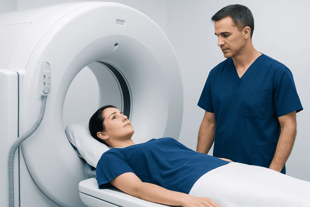
Before the scan, a healthcare professional injects the FES tracer into a vein, usually in the arm or hand. The tracer circulates through the bloodstream and binds to estrogen receptors where they are present. A PET/CT scanner then produces detailed images that show where tracer uptake is seen.
An FES PET scan may be considered for adults with estrogen receptor-positive breast cancer when there is concern about recurrence or spread, when lesions are difficult to biopsy, or when tumor receptor status may have changed over time. Cancer cells can sometimes differ between the original breast tumor and metastatic sites, so imaging may add useful information about the overall pattern of disease.
Not everyone with breast cancer needs this test. It is not generally used as a routine screening examination, and it is not a substitute for mammography, breast ultrasound, MRI, conventional CT or other cancer assessments. The oncology team decides whether FES imaging is appropriate based on the individual clinical question and prior test results. For broader assessment and planning, patients may also discuss breast cancer care with their specialist.
Pregnancy and breastfeeding require special consideration because the test involves radiation exposure. Anyone who may be pregnant, is breastfeeding, has allergies, or has kidney or liver concerns should tell the imaging team before the appointment.
FES PET Scan Preparation and Medication Review

The precise FES PET scan protocol can vary between imaging centers. Patients should follow the instructions from the nuclear medicine department, as those directions are tailored to the scanner, tracer and reason for testing. In many cases, normal eating and drinking are allowed before FES PET imaging, unlike FDG PET scans that often require fasting and carbohydrate restriction.
Medication review is particularly important. Some medicines that directly block or modulate estrogen receptors can reduce FES tracer binding and may affect how the images are interpreted. The oncology and imaging teams should know about all prescribed medicines, over-the-counter products, hormone treatments and supplements. Patients should not stop cancer treatment or any regular medicine on their own; only the treating clinician can advise whether timing needs to change.
What not to do 24 hours before a PET scan? The answer depends on the type of PET scan. For an FES PET scan, patients should not assume that the fasting, exercise or low-carbohydrate instructions used for an FDG PET scan apply. They should avoid making unapproved medication changes, attend well hydrated unless instructed otherwise, and follow the center’s directions about clothing, jewellery and arrival time. For any PET scan, informing staff about possible pregnancy, breastfeeding, diabetes and recent imaging is important.
What happens if I eat carbs the day before a PET scan? Carbohydrates can be relevant for FDG PET scans because FDG behaves like glucose and blood sugar levels can influence image quality. For FES PET scans, carbohydrate restriction is usually not the central preparation issue, and eating carbohydrates the day before is often not a problem. However, the patient should follow the instructions for their specific appointment, especially if FES PET is being performed alongside another imaging test.
Step-by-Step: What Happens During an FES PET Scan?
On arrival, the patient checks in and is asked about their medical history, current medicines and any possibility of pregnancy or breastfeeding. The team explains the procedure and obtains the required consent. A small intravenous line is placed, and the FES tracer is injected.
There is usually a waiting period after injection to allow the tracer to distribute through the body. During this time, the patient may be asked to rest quietly and drink water. The exact timing varies by protocol. The imaging team may also ask the patient to empty their bladder before scanning because the tracer is removed partly through urine.
For the scan itself, the patient lies on a narrow table that moves slowly through the PET/CT machine. Remaining still helps avoid blurred images. The examination is painless, although lying flat may be uncomfortable for some people. The scan commonly takes around 20 to 30 minutes, while the full visit is longer because of registration, injection and the uptake period.
Afterward, a radiologist or nuclear medicine physician reviews the images in the context of prior scans, biopsy findings and treatment history. In cancer care, PET imaging may be part of a wider assessment that includes medical oncology treatment planning and other targeted investigations.
After the Scan: Recovery, Toilet Instructions and Safety
Most people can leave after an FES PET scan and resume normal daily activities unless their clinician gives different advice. The injected tracer loses radioactivity over time and is cleared largely through urine. Drinking fluids, if medically appropriate, and urinating regularly can help the body eliminate it.
Why do you have to flush the toilet twice after a PET scan? Some centers recommend flushing twice after urinating for a limited period after a PET scan to reduce the small amount of radioactive tracer that may remain in the toilet. This is a simple radiation-safety measure, particularly in shared bathrooms. The imaging department will provide specific instructions on hygiene, toilet use and close contact with others when needed.
The radiation exposure from an FES PET scan is generally limited and is weighed against the expected clinical value of the information obtained. Allergic reactions to the tracer are uncommon, but patients should seek urgent medical help for symptoms such as trouble breathing, facial swelling or widespread hives after any injection. Mild soreness or bruising at the injection site can occur.
Recovery does not usually involve restrictions on driving, work or eating. People who are breastfeeding should receive individualized advice from the imaging team about whether, and for how long, milk expression or direct breastfeeding needs to be adjusted.
Benefits, Limits and Understanding FES PET Scan Results
The main benefit of FES PET scanning is that it can provide a whole-body picture of estrogen receptor availability without requiring multiple biopsies. This can be helpful when metastatic sites are hard to reach safely, when different lesions may behave differently, or when clinicians are considering endocrine therapy. It may also help clarify whether a suspicious finding is likely to have estrogen receptor-positive characteristics.
However, FES PET scan results require careful interpretation. Uptake does not by itself confirm cancer, and a lack of uptake does not always rule out cancer. Some tumors have low or mixed estrogen receptor expression, very small lesions may not be visible, and certain medicines can influence tracer uptake. The scan cannot determine every feature of a tumor or replace pathology in all situations.
Results may be described as FES-avid or showing uptake, meaning that an area has bound the tracer, or as having low or absent uptake. The treating oncologist explains what this means in the person’s situation and whether it affects treatment choices, further imaging or biopsy decisions. A multidisciplinary review can be valuable when imaging and pathology findings do not fully agree.
Questions about FES PET scan cost should be discussed directly with the imaging provider, insurer or hospital financial counseling team. Costs and coverage can vary by country, facility, insurance plan, clinical indication and whether other imaging is performed at the same visit.
Food Questions: Best Foods Before a PET Scan
What are the best foods to eat the day before a PET scan? For an FES PET scan, most people can eat their usual balanced meals unless the imaging center gives different instructions. Good hydration is often encouraged, provided there are no fluid restrictions for another medical condition. Patients should not begin a special diet solely because they have read general PET preparation advice online.
For an FDG PET scan, preparation often differs and may include specific instructions about fasting, blood glucose management and sometimes limiting carbohydrate intake before the test. This distinction is important when comparing an FES PET scan vs PET scan: FES is a specialized PET tracer examination, while FDG PET is the more common glucose-based PET study.
If a patient has diabetes, takes insulin or other glucose-lowering medicines, or is scheduled for multiple tests on the same day, individualized preparation is essential. The patient should contact the imaging department in advance rather than skipping meals or changing diabetes medication without guidance.
When to Seek Medical Care
Patients should contact their oncology or imaging team before the scan if they become ill, develop a fever, learn they may be pregnant, begin a new hormone-related medicine, or are unable to follow the preparation instructions. It is also sensible to call if there are questions about fasting, regular medicines, diabetes care or breastfeeding.
After the scan, urgent medical care is appropriate for signs of a severe allergic reaction, such as difficulty breathing, swelling of the lips or tongue, fainting or rapidly spreading rash. These reactions are rare, but prompt assessment is important. Persistent redness, pain or swelling at the injection site should also be reported to a healthcare professional.
FES PET imaging is one component of individualized cancer care. Acibadem International’s multidisciplinary specialists and JCI-accredited hospitals can assess imaging findings and treatment options for international patients, including breast cancer treatment.
Frequently asked questions
How is an FES PET scan different from a regular PET scan?
An FES PET scan uses a tracer that binds to estrogen receptors, while many regular PET scans use FDG, a glucose-like tracer. FDG imaging shows areas of increased glucose use, whereas FES imaging is designed to assess estrogen receptor activity. The tests answer different clinical questions and may sometimes be used alongside one another.
Do I need to fast before an FES PET scan?
Many FES PET protocols do not require fasting, but preparation instructions can vary by center and by other tests scheduled that day. The patient should follow the instructions provided by the imaging department. People with diabetes should ask for individualized advice rather than changing medication or food intake on their own.
Can hormone therapy affect an FES PET scan?
Some treatments that interact with estrogen receptors may affect FES tracer uptake and therefore influence scan interpretation. The oncology and nuclear medicine teams should review all current and recent cancer treatments before the scan. Patients should never stop a prescribed medicine unless their treating clinician tells them to do so.
How long does an FES PET scan take?
The scanning portion often takes about 20 to 30 minutes, but the entire appointment may take several hours. Time is needed for check-in, tracer injection, an uptake period and imaging. The center can provide the most accurate estimate for the individual protocol.
Is an FES PET scan painful?
The test is generally not painful apart from the brief needle stick for the intravenous injection. During imaging, the patient needs to lie still on a table, which may cause temporary discomfort for those with pain or mobility limitations. The imaging staff can help with positioning and support.
Can an FES PET scan diagnose breast cancer?
An FES PET scan is not usually used to make an initial diagnosis of breast cancer. It is mainly used to evaluate estrogen receptor-positive disease in selected clinical situations, often when recurrence or metastasis is being assessed. Diagnosis and treatment decisions are based on the full clinical picture, including pathology and other imaging.
References
- National Cancer Institute
- U.S. Food and Drug Administration
- Society of Nuclear Medicine and Molecular Imaging
- American Cancer Society
This article is for general information only and is not a substitute for professional medical advice. Please consult a qualified doctor about your individual situation.
Oncology care in Turkey — second opinion and treatment plan
JCI-accredited · board-certified surgeons · reply within 24h
Add us as a Preferred Source to see more of our trusted health content across Google Search, AI Overviews and Discover.

More from the Health Library


Related Specialists

Prof. Dr. Safa Gürsoy
Orthopedic Surgery & Traumatology
Dt. Hatice Ağan
Oral & Dental Health
Nazlıhan Bozdağ
Physical Medicine & Rehabilitation
Prof. Dr. Atakan Yeşil
Gastroenterology