Forced Vital Capacity — Explained by Medical Evidence, Not Myths

Forced vital capacity measures how much air can be forcefully exhaled after a full inhalation. It is usually assessed with spirometry, a common and noninvasive lung function test.
Key Takeaways
- Forced vital capacity measures how much air can be forcefully exhaled after a full inhalation.
- It is usually assessed with spirometry, a common and noninvasive lung function test.
- FVC results are interpreted together with values such as FEV1 rather than on their own.
- Low forced vital capacity can occur in several conditions, including restrictive lung disease and some obstructive disorders.
- Test quality matters, so proper technique and repeated efforts are important for accurate results.
Forced vital capacity is the total volume of air a person can blow out forcefully after taking the deepest breath possible. It is measured during spirometry and helps doctors understand how well the lungs and airways are working, especially when breathing symptoms need further evaluation.
What forced vital capacity means
Forced vital capacity, often shortened to FVC, is the total amount of air a person can exhale as forcefully and completely as possible after taking the deepest breath in. In simple terms, it shows how much air the lungs can move out during a strong, fast breath out. Doctors use it as part of a wider picture of lung function rather than as a stand-alone answer.
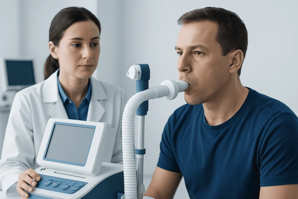
This measurement is most commonly taken during spirometry, a standard breathing test performed in clinics, hospitals, and pulmonary laboratories. During the test, the person inhales fully, seals their lips around a mouthpiece, and then blows out hard and long until no more air comes out. The machine records the volume and speed of the exhaled air.
Forced vital capacity is useful because many lung and airway conditions can change how much air the lungs hold or how easily air can move out. A lower-than-expected value may point toward a restrictive pattern, air trapping, weak breathing muscles, or a technical issue during testing. That is why medical evidence emphasizes interpretation by a qualified clinician, using age, sex, height, symptoms, and other test results.
Why doctors measure forced vital capacity
Doctors measure forced vital capacity to help evaluate symptoms such as shortness of breath, wheezing, chronic cough, chest tightness, or reduced exercise tolerance. It can also be used to follow known lung disease over time, check response to treatment, or assess lung function before some procedures.
On its own, FVC tells only part of the story. It is usually interpreted alongside forced expiratory volume in one second, called FEV1, and the FEV1/FVC ratio. This combination helps distinguish broad breathing patterns. For example, obstructive conditions often reduce airflow speed more than volume, while restrictive conditions may reduce the total volume exhaled.
Clinical context matters. A physically small person may naturally have a lower absolute FVC than a taller person, and this may still be normal for them. For that reason, spirometry reports compare measured values with predicted ranges based on personal characteristics rather than using one fixed number for everyone.
FVC may be part of the evaluation of conditions such as COPD, asthma, interstitial lung disease, chest wall disorders, and some neuromuscular conditions. In selected situations, doctors may also recommend broader pulmonary function tests to obtain more detail about lung volumes and gas transfer.
How the test is done and how to prepare

The forced vital capacity test is usually quick, noninvasive, and done while sitting. A healthcare professional may place a clip on the nose so that all air goes through the mouthpiece. The person is then asked to breathe in completely and blow out as hard, fast, and as long as possible. Because one effort can vary from another, the test is typically repeated several times to make sure the results are reliable.
Good technique is essential. A poor seal around the mouthpiece, coughing during the first second, stopping too early, or taking an incomplete breath in can all affect the result. Test staff coach the person through each attempt so the final numbers reflect true lung function as closely as possible.
Preparation instructions may vary. A doctor may advise avoiding smoking for a period before testing, wearing loose clothing, and not eating a very heavy meal immediately beforehand. Some patients are also told whether to take or temporarily withhold inhalers before the test, depending on the question being asked. It is important to follow the testing center’s instructions rather than making changes independently.
Sometimes spirometry is repeated after an inhaled bronchodilator. This helps show whether airflow improves with medication, which can be useful when symptoms suggest asthma or another reversible airway problem. If imaging or specialist evaluation is needed, a doctor may pair breathing tests with chest diseases assessment by a pulmonary team.
What results can show
FVC results are compared with predicted values and then interpreted as normal, low, or occasionally higher than expected. A normal FVC generally means the exhaled lung volume falls within the expected range for that person. However, a normal FVC does not always rule out all lung disease, especially if symptoms are ongoing or if other lung function values are abnormal.
A low forced vital capacity can happen for several reasons. One possibility is a restrictive pattern, where the lungs cannot fully expand. This may occur in interstitial lung disease, some chest wall conditions, obesity-related mechanical limitation, or certain neuromuscular disorders. Another possibility is air trapping in obstructive lung disease, where some air remains in the lungs and reduces the amount that can be forcefully exhaled.
The FEV1/FVC ratio helps doctors interpret these possibilities. In many obstructive conditions, the ratio is reduced because airflow in the first second is disproportionately limited. In restrictive patterns, the ratio may be normal or even high while FVC is low. Sometimes further testing is needed to confirm whether the problem is truly restrictive, especially because spirometry alone does not directly measure total lung capacity.
Results should always be read in context. Age, height, ethnicity used by the reference equation, effort during testing, recent illness, and underlying medical conditions can all influence the numbers. For that reason, patients should avoid self-diagnosing from a printed report without a clinician’s explanation.
Common causes of an abnormal forced vital capacity
An abnormal forced vital capacity does not point to one single disease. Instead, it signals that the lungs, airways, chest wall, or breathing muscles may need closer assessment. In everyday practice, doctors look at symptoms, examination findings, smoking history, environmental exposures, medications, and imaging alongside spirometry results.
Conditions linked with reduced FVC may include:
- Restrictive lung diseases, including some forms of pulmonary fibrosis and other interstitial lung disorders
- Obstructive diseases with air trapping, such as severe asthma or chronic obstructive pulmonary disease
- Obesity or chest wall problems that limit full lung expansion
- Neuromuscular disorders that weaken the breathing muscles
- Poor test effort or technical factors during spirometry
Temporary factors can also influence the result. A respiratory infection, pain after surgery, or difficulty following instructions may lower the measured value. In these situations, repeat testing after recovery may provide a clearer picture.
When doctors suspect a broader respiratory condition, they may combine spirometry with imaging, blood oxygen assessment, or specialist review. If there are symptoms such as recurrent wheeze or variable breathlessness, evaluation for asthma may also be considered.
How abnormal results are evaluated and managed
Treatment does not aim to change the FVC number alone; it focuses on the cause behind the result. If the pattern suggests asthma or another airway disorder, management may include inhaled medication, trigger avoidance, and follow-up lung function testing. If a restrictive process is suspected, the doctor may request more detailed tests to identify whether the issue comes from the lung tissue, chest wall, or breathing muscles.
Additional tests can include complete pulmonary function testing, imaging such as chest X-ray or CT, oxygen measurements, exercise assessment, or laboratory studies. In selected cases, specialist input from pulmonology, allergy, neurology, or rehabilitation may be appropriate. The goal is to move from a test result to a clear diagnosis and then to a treatment plan tailored to the person.
Self-care can also support lung health. Avoiding smoking and secondhand smoke, staying physically active within a doctor’s advice, keeping vaccinations up to date, and managing chronic conditions such as reflux or obesity may all help breathing symptoms. People with established lung disease may benefit from breathing exercises or structured pulmonary rehabilitation when recommended.
For patients needing specialist assessment, Acibadem International’s multidisciplinary specialists in JCI-accredited hospitals diagnose and treat respiratory conditions for international patients. Depending on the findings, care may involve bronchoscopy or other pulmonary evaluations when clinically indicated.
When to seek medical care
A person should seek medical advice if they have ongoing shortness of breath, frequent wheezing, a chronic cough, unexplained reduced stamina, or chest tightness that keeps returning. These symptoms do not always mean serious disease, but they deserve proper assessment, especially if they interfere with daily life or sleep.
More urgent medical attention is needed for severe trouble breathing, bluish lips, confusion, chest pain, coughing up blood, or sudden worsening of known lung disease. These symptoms require prompt evaluation rather than waiting for routine testing.
Anyone who has received an abnormal spirometry report should discuss the result with a qualified doctor rather than trying to interpret the numbers alone. A single FVC value is most useful when paired with the rest of the breathing test, the person’s medical history, and a professional examination.
Frequently asked questions
Is forced vital capacity the same as lung capacity?
Not exactly. Forced vital capacity measures the amount of air a person can forcefully exhale after a full inhalation, while total lung capacity refers to the total amount of air the lungs can hold. FVC is an important part of lung assessment, but it does not measure every aspect of lung volume.
What is a normal forced vital capacity?
A normal forced vital capacity depends on a person's age, sex, height, and the reference equation used by the laboratory. Because of this, doctors compare the measured result with a predicted range instead of using one number for everyone. A clinician can explain whether the result is normal for that individual.
Can low forced vital capacity improve?
It can improve in some situations, especially if the cause is treatable or temporary. For example, better control of airway disease, recovery from infection, improved test technique, or treatment of an underlying condition may change the result. In other cases, the value may remain reduced and be used mainly to monitor the condition over time.
Does a low FVC always mean restrictive lung disease?
No. Although a low FVC can suggest a restrictive pattern, it can also happen because of air trapping in obstructive lung disease, obesity, muscle weakness, or suboptimal effort during the test. Doctors often need the full spirometry pattern and sometimes additional lung function tests to clarify the cause.
How long does an FVC test take?
The spirometry part is usually brief, often completed within minutes, although preparation and repeated efforts can make the appointment longer. The person may need to perform the maneuver several times so the results are accurate and reproducible. If bronchodilator testing is added, the visit may take more time.
Should inhalers be used before a forced vital capacity test?
This depends on why the test is being done. Some patients are asked to continue their inhalers, while others may be told to pause certain medications for a short period before testing. The safest approach is to follow the exact instructions given by the doctor or testing center.
References
- American Thoracic Society
- European Respiratory Society
- National Heart, Lung, and Blood Institute
- Centers for Disease Control and Prevention
- World Health Organization
This article is for general information only and is not a substitute for professional medical advice. Please consult a qualified doctor about your individual situation.
Explore treatments in Turkey — costs, top hospitals & a free quote
JCI-accredited · board-certified surgeons · reply within 24h
Add us as a Preferred Source to see more of our trusted health content across Google Search, AI Overviews and Discover.

More from the Health Library

Liver Spots — Explained by Medical Evidence, Not Myths

Ingrown Hair Cyst Bikini: An Evidence-Based Guide for Patients

Ureteral Stent — Explained by Medical Evidence, Not Myths

Microbiome: A Complete Medical Overview

Strawberry Tongue: A Complete Medical Overview