Hand Xray: A Complete Medical Overview
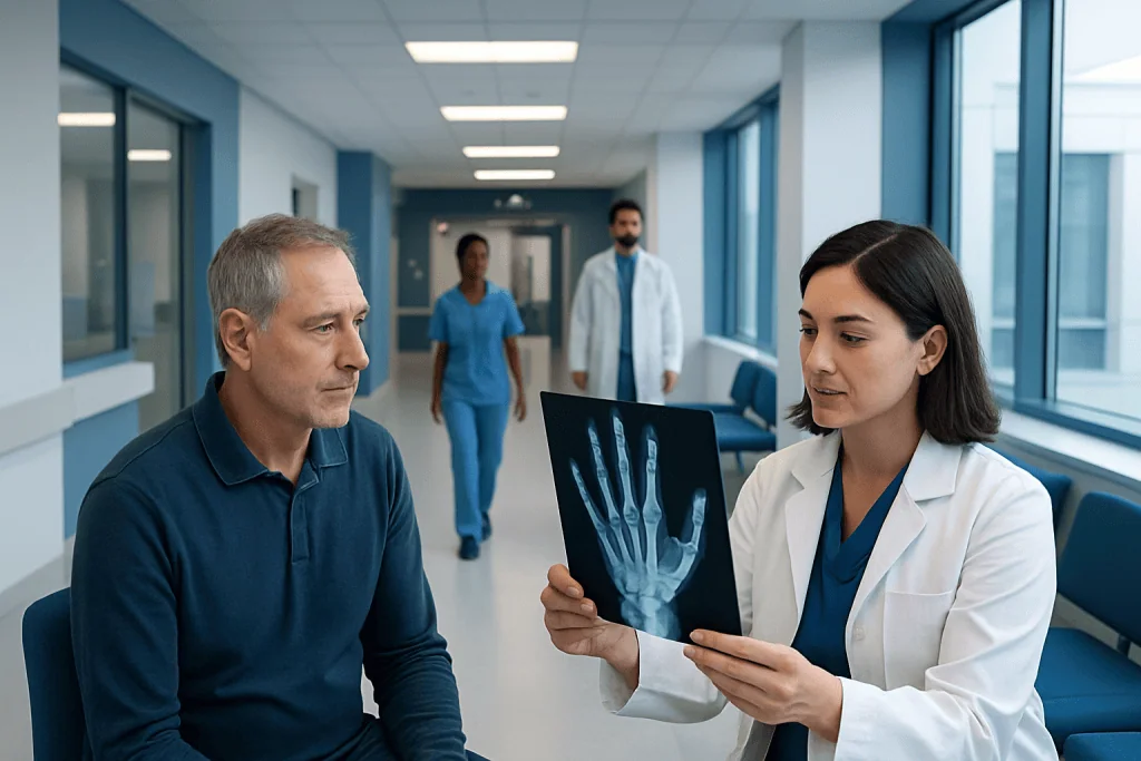
A hand xray uses a small amount of ionizing radiation to create images of the hand’s bones and joints. It is commonly used to assess suspected fractures, dislocations, arthritis, and unexplained pain or swelling.
Key Takeaways
- A hand xray uses a small amount of ionizing radiation to create images of the hand’s bones and joints.
- It is commonly used to assess suspected fractures, dislocations, arthritis, and unexplained pain or swelling.
- The examination is painless, usually takes only a few minutes, and generally requires no special preparation.
- More than one image is often taken from different angles because some injuries are easier to see in one view than another.
- An xray may not show every soft-tissue injury, such as a tendon or ligament tear, so further imaging can sometimes be needed.
A hand xray is a commonly used imaging examination that produces detailed pictures of the bones and joints in the hand. It can help clinicians identify fractures, dislocations, arthritis-related changes, infections, and certain bone abnormalities, often after an injury or when pain and stiffness persist.
What Is a Hand Xray?
A hand xray, also called a hand X-ray or radiograph, is an imaging test that uses a controlled, low dose of radiation to create pictures of the bones and joints in the hand. It is a fast, painless examination that can help a clinician look for a broken bone, dislocation, joint changes, or other structural causes of hand symptoms. Images may include the fingers, thumb, palm, wrist, or a combination of these areas depending on the concern.
The hand contains many small bones and joints that work closely together. Because injuries can be subtle, xray images are typically taken from two or more directions, such as from above, the side, and an angled view. Looking at several views helps the radiologist and treating clinician assess bone alignment and identify findings that may not be visible on a single image.
A hand xray is often the first imaging test used after a fall, sports injury, direct blow, crush injury, or sudden pain. It can also be useful when symptoms develop gradually, including stiffness, swelling, reduced movement, or changes in finger shape. The test provides important information, but it is interpreted alongside symptoms, the physical examination, and the person’s medical history.
What Can a Hand Xray Show?
The main role of a hand xray is to show bone and joint structure. It can identify many types of fractures, ranging from a clear break after trauma to a small crack caused by repetitive stress. It may also show whether broken bone pieces have shifted out of their usual position, which can influence decisions about splinting, reduction, or surgical treatment.
Hand xrays can also help detect dislocations and subluxations, where a joint is fully or partly out of alignment. In addition, they may reveal signs of arthritis, including narrowing of joint spaces, bone spurs, changes in bone shape, or erosions around a joint. These findings can support an evaluation for osteoarthritis, inflammatory arthritis, or other joint conditions, although an xray alone does not always establish a specific diagnosis.
In selected situations, an xray may show a retained foreign object, such as some types of glass or metal, after a penetrating injury. It may also reveal changes associated with bone infection, cysts, tumors, or congenital differences in bone development. Not all materials are visible on xray, and many soft-tissue concerns require other tests.
- Fractures, stress fractures, and bone healing after a previous fracture
- Joint dislocation, malalignment, or instability-related bone changes
- Arthritis-related joint damage and degenerative changes
- Some foreign bodies and bone abnormalities
- Changes that may prompt further assessment for infection or other bone disorders
When Is a Hand Xray Recommended?

A clinician may recommend a hand xray when an injury causes pain, swelling, bruising, tenderness over a bone, deformity, or difficulty moving the fingers or thumb. Even when a person can still use the hand, an xray may be appropriate if pain is localized, especially after a fall onto an outstretched hand, a twisting injury, or impact during sport or work.
The test may also be requested for symptoms that are not linked to a single injury. Examples include persistent hand pain, morning stiffness, recurrent swelling of the finger joints, gradually reduced grip, or visible changes in joint shape. In children and adolescents, hand xrays may sometimes be used as part of an assessment of bone age or growth, depending on the clinical situation.
Follow-up xrays can be used after a known fracture to check alignment and healing. However, the timing and need for repeat imaging depend on the location and pattern of the fracture, the person’s symptoms, and the treatment plan. A clinician will decide whether repeat images are helpful rather than performing them routinely in every case.
How the Hand Xray Procedure Works
No special preparation is usually needed for a hand xray. Before the examination, the patient may be asked to remove rings, watches, bracelets, or other objects that could obscure the images. It is important to tell the radiology team about any possible pregnancy, although extremity xrays expose the reproductive organs to very little scattered radiation and appropriate precautions can be considered.
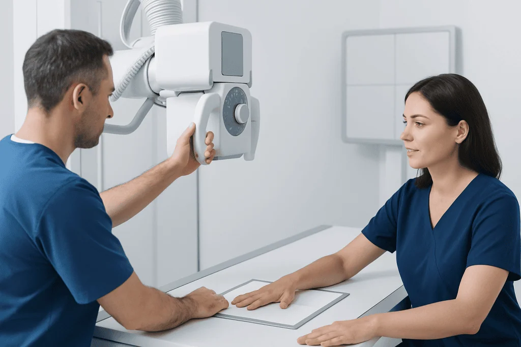
During the test, the patient sits or stands beside the xray equipment and places the hand on a flat imaging detector. The radiographer will gently guide the hand and fingers into different positions. The hand needs to remain still briefly for each image; movement can make an image blurred and may require it to be repeated.
The xray itself does not hurt. If the hand is injured, positioning may be uncomfortable, but the radiographer can work carefully and adjust the examination where possible. The imaging portion commonly takes only a few minutes, after which the patient can usually return to normal activities unless the injury itself requires support, rest, or urgent treatment.
A radiologist, a doctor trained to interpret medical images, reviews the images and issues a report. The requesting clinician discusses the findings in the context of the patient’s symptoms and examination. In urgent injury settings, an initial result may be available promptly, while formal reporting processes can vary by healthcare setting.
Understanding Hand Xray Results and Their Limits
A normal hand xray means that no clear abnormality was seen in the bones or joints on the images obtained. This is reassuring, but it does not always rule out injury. Very small fractures, early stress fractures, and some fractures around complex joints can be difficult to see immediately, particularly soon after the injury occurred.
Hand xrays are also limited in their ability to assess soft tissues. Tendons, ligaments, nerves, cartilage, and many muscle injuries are not directly shown in detail. If symptoms and examination findings suggest a soft-tissue injury despite a normal xray, a clinician may recommend a period of protection and reassessment, repeat xray imaging, ultrasound, CT, or MRI depending on the suspected problem.
If a fracture is identified, the report may describe its location, whether it extends into a joint, whether the bone pieces are displaced, and whether the fracture appears stable. These details help guide treatment. A simple stable fracture may be managed with a splint or cast, while displaced, unstable, or joint-involving injuries may require assessment by an orthopedic or hand specialist.
For joint disease, the xray report may describe findings such as joint-space narrowing, bone spurs, erosions, or swelling around a joint. These observations are useful but should not be interpreted in isolation. Symptoms, blood tests, examination findings, and sometimes further imaging may be necessary to clarify the cause of hand joint pain.
Safety, Radiation and Aftercare
Hand xrays use a relatively low amount of radiation, and modern imaging services follow the principle of using the lowest exposure needed to obtain a useful image. Because the hand is away from the chest, abdomen, and pelvis, the radiation dose to other parts of the body is very limited. For most people, the clinical benefit of appropriately requested imaging outweighs the small radiation risk.
There are usually no side effects from a standard xray and no recovery period. The patient can eat, drink, drive, and continue ordinary activities after the test unless their clinician gives instructions related to the underlying injury. If a splint, bandage, or cast has been applied, it should be kept in place as advised.
After an injury, it is sensible to follow the care plan while waiting for or reviewing results. This may include resting the hand, avoiding activities that worsen pain, elevating it to help reduce swelling, and using pain relief recommended by a qualified clinician or pharmacist. A person should not attempt to straighten a visibly deformed finger or hand without medical guidance.
People who are pregnant or think they could be pregnant should inform the imaging team before the examination. The team can confirm whether the test is necessary, discuss precautions, and ensure imaging is performed safely. It is also helpful to mention previous hand injuries, surgery, implanted hardware, or recent imaging when relevant.
When to Seek Medical Care
Prompt medical assessment is advisable after a hand injury if there is severe pain, visible deformity, marked swelling, inability to move a finger, numbness, tingling, or a finger that looks pale, blue, or feels cold. These features can indicate an injury that needs timely treatment to protect circulation, nerve function, and long-term hand movement.
Medical care is also important for an open wound near a painful area, a suspected foreign body, or signs of infection such as increasing redness, warmth, swelling, drainage, fever, or worsening pain. A hand injury caused by a bite should be assessed promptly because bites can introduce infection and may damage deeper structures.
For symptoms without a recent injury, a doctor should evaluate persistent hand pain, recurrent swelling, stiffness that affects daily tasks, reduced grip strength, or gradually changing finger shape. Early assessment can help identify treatable causes and support an appropriate management plan.
Acibadem International’s multidisciplinary specialists and JCI-accredited hospitals provide diagnostic assessment and treatment planning for international patients with hand injuries and hand-related conditions. The appropriate next step depends on the individual findings, symptoms, and imaging results.
Frequently asked questions
How long does a hand xray take?
The imaging portion of a hand xray usually takes only a few minutes. Allowing time for registration, positioning, and preparation, the overall appointment may take longer depending on the healthcare setting and whether urgent care is needed.
Does a hand xray hurt?
The xray radiation itself is painless and cannot be felt. Positioning an injured hand may be uncomfortable, especially if there is swelling or a fracture, but the radiographer will aim to move the hand as gently as possible.
Can a hand xray show a ligament or tendon injury?
A hand xray primarily shows bones and joint alignment rather than tendons and ligaments. It can sometimes show indirect signs of a soft-tissue injury, but ultrasound or MRI may be more useful when a tendon or ligament problem is suspected.
Can a fracture be missed on a hand xray?
Yes, some small, nondisplaced, stress-related, or very early fractures may not be clearly visible on initial images. If symptoms strongly suggest a fracture despite a normal xray, a clinician may recommend immobilization, repeat imaging, or another scan.
Do rings need to be removed for a hand xray?
Rings, watches, bracelets, and similar items are usually removed because they can block part of the image or create artifacts. If a ring cannot be removed because of swelling, inform the radiographer rather than forcing it off.
What happens if the hand xray shows a fracture?
Treatment depends on the bone involved, the fracture pattern, alignment, and whether a joint is affected. Management may include a splint, cast, reduction to improve alignment, hand therapy, or referral to an orthopedic or hand specialist when needed.
References
- Radiological Society of North America
- American College of Radiology
- National Institute of Arthritis and Musculoskeletal and Skin Diseases
- NHS
- Mayo Clinic
This article is for general information only and is not a substitute for professional medical advice. Please consult a qualified doctor about your individual situation.
Joint and spine care in Turkey — expert assessment & treatment
JCI-accredited · board-certified surgeons · reply within 24h
Add us as a Preferred Source to see more of our trusted health content across Google Search, AI Overviews and Discover.