Hip Ultrasound for Breech Baby: Preparation, Procedure and Results

Breech positioning is an important risk factor for developmental dysplasia of the hip (DDH). Hip ultrasound uses sound waves, not radiation, and is usually most useful during the first months of life.
Key Takeaways
- Breech positioning is an important risk factor for developmental dysplasia of the hip (DDH).
- Hip ultrasound uses sound waves, not radiation, and is usually most useful during the first months of life.
- The scan commonly takes about 15 to 30 minutes and does not require sedation or special preparation.
- An abnormal or unclear result does not always mean a baby needs treatment; repeat imaging or specialist assessment may be recommended.
- Early identification of DDH can allow treatment while the hip is still developing.
A hip ultrasound for a breech baby is a painless imaging test that checks whether the hip joints are developing normally. Because breech positioning increases the chance of developmental dysplasia of the hip, many babies are referred for screening even when the newborn examination appears normal.
Hip Ultrasound for Breech Baby: Overview
A hip ultrasound for breech baby is a screening examination used to look at the shape, position and stability of a baby’s hip joints. It is commonly recommended because babies who were breech late in pregnancy, particularly during the third trimester, have a higher likelihood of developmental dysplasia of the hip (DDH). This is a condition in which the ball-and-socket hip joint has not formed or fitted together as expected.
The test is safe and comfortable. A sonographer moves a small probe over the baby’s hip area while ultrasound images are displayed on a screen. There is no radiation, no needles and usually no need for sedation. Screening is often arranged at around 4 to 6 weeks of age, although the timing may differ according to local guidance, the baby’s medical history and the findings of the newborn examination.
DDH can range from a mildly shallow socket to a hip that is unstable or dislocated. Many babies have no obvious symptoms, which is why appropriate screening and follow-up are valuable. A normal ultrasound can provide reassurance, while an abnormal result helps the care team decide whether observation, repeat imaging or referral to a pediatric orthopedic specialist is appropriate.
Who May Need Screening and Why
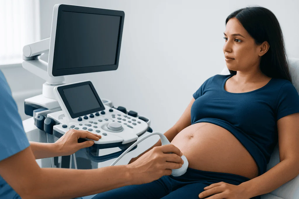
Not every newborn needs a hip ultrasound. Doctors usually begin with a physical hip examination soon after birth and again at routine infant visits. Screening ultrasound is often advised for babies with specific risk factors, even if that examination is normal. Breech presentation is one of the most important of these factors.
A baby may be considered breech if the buttocks or feet were positioned to be delivered first, or if breech positioning occurred during the later weeks of pregnancy. Risk may remain relevant even when a baby turns head-down before birth. Other factors that can influence screening decisions include a close family history of DDH, a previous sibling with DDH, female sex, restricted space in the uterus, or certain foot and neck positioning concerns.
- Breech position in late pregnancy or at delivery
- A parent or sibling with developmental dysplasia of the hip
- An unusual, unstable or difficult-to-assess hip examination
- Associated musculoskeletal findings identified by the clinician
The purpose of screening is not to label a baby with a condition unnecessarily. It is to identify hips that may benefit from monitoring while the bones, socket and surrounding soft tissues are still maturing. Families should follow the timing recommended by their pediatrician or maternity team.
How the Hip Ultrasound Works
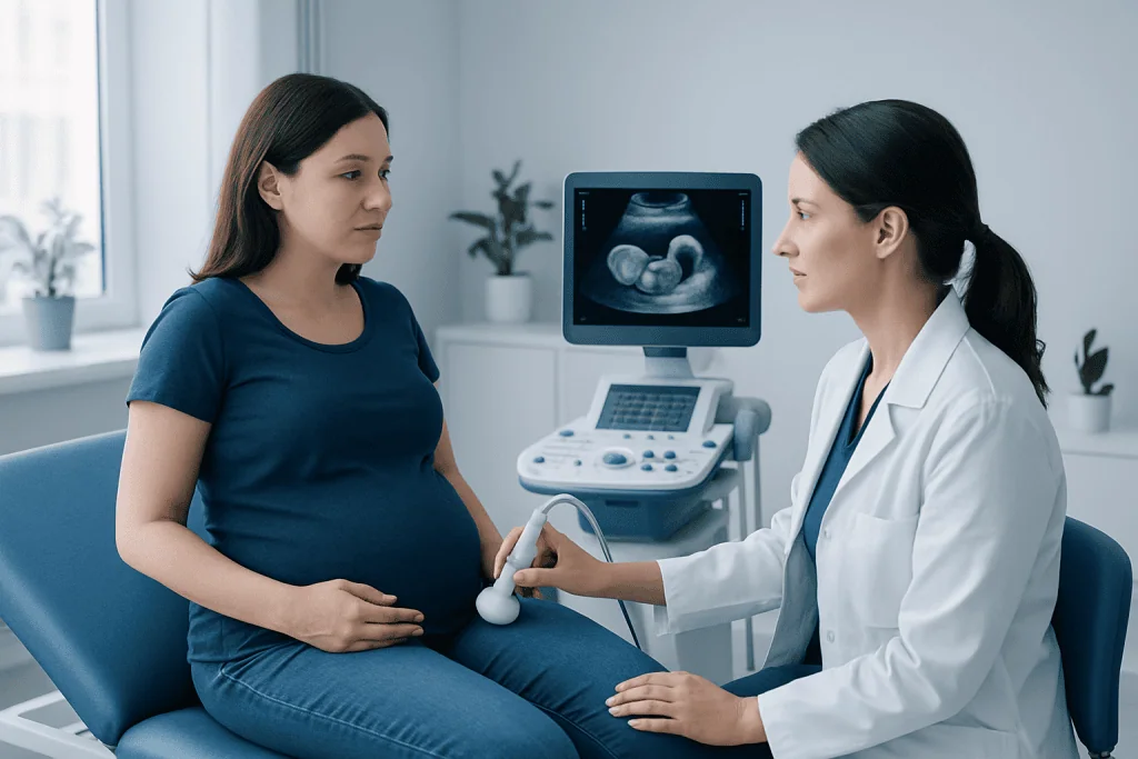
Ultrasound creates images using high-frequency sound waves. A handheld device called a transducer sends sound waves into the body and receives returning signals. A computer turns these signals into moving images of the hip joint, including the rounded upper end of the thigh bone and the cartilage-rich hip socket.
This technique is particularly useful in young babies because much of the hip is still made of cartilage and is not fully visible on an X-ray. Ultrasound allows the clinician to assess the hip’s structure and, when needed, observe how it sits in the socket during gentle positioning.
The report may describe the depth and shape of the socket, the position of the femoral head and whether the hip appears stable. Measurements and classifications can help specialists interpret the images, but these details must be considered alongside the baby’s age, examination findings and overall development. A hip that looks immature very early in life may mature normally on follow-up.
Step-by-Step: What Happens During the Procedure
No special preparation is usually needed for a baby hip ultrasound. Families may find it helpful to feed the baby shortly before the appointment, bring familiar comfort items and choose clothing that is easy to remove around the diaper area. A calm or sleeping baby can make positioning easier, but crying does not make the scan unsafe.
During the examination, the baby is generally placed on their side in a padded support. Warm gel is applied to the skin over the outer hip. The sonographer gently places the transducer against the area and captures images from each hip. The baby may be repositioned so that both hips can be examined carefully.
The sonographer may use gentle, controlled movement to observe the hip joint, but the procedure should not be painful. Parents can usually remain with their baby throughout the scan and may be able to soothe or feed the baby if needed. Images are then reviewed by a radiologist, pediatrician or orthopedic specialist, depending on the care pathway.
Most appointments take around 15 to 30 minutes, although the imaging portion may be shorter. The family may receive preliminary information on the day, while the formal report is often sent to the referring clinician for discussion.
Results, the 50% Rule and Possible Next Steps
A result may be described as normal, immature, borderline, dysplastic, unstable or dislocated. The wording can vary between imaging centers and specialists. A normal result generally means the hips look appropriately positioned and developed for the baby’s age. If the result is unclear or shows mild immaturity, a repeat ultrasound may be advised after a period of growth.
The “50% rule” is an informal way some clinicians describe femoral head coverage on an ultrasound image. In simple terms, it refers to whether at least about half of the femoral head appears covered by the hip socket. Lower coverage can suggest that the socket is shallow or that the hip is not centered as expected. However, this measurement is not interpreted alone; specialists also evaluate hip angles, stability and the baby’s age.
If DDH is confirmed, early care may include monitoring or use of a soft positioning brace, often called a Pavlik harness, to help keep the hip in a stable position while it develops. The exact approach depends on the severity, the child’s age and specialist assessment. Some babies require further imaging, and less commonly, procedures may be considered when dysplasia is detected later or does not respond to early treatment.
Parents should avoid trying to position the hips themselves unless a specialist has given clear instructions. Healthy hip positioning generally allows the legs to bend and move freely. When swaddling, the hips and knees should have room to flex and spread naturally rather than being held tightly straight together.
Benefits, Risks and Recovery After the Scan
The main benefit of hip ultrasound for a breech baby is early assessment of a condition that may not be visible from the outside. Detecting hip instability or dysplasia early can support timely follow-up and may reduce the likelihood of more complex treatment later in childhood.
Ultrasound does not use ionizing radiation, and the procedure is not expected to cause harm when performed appropriately. The gel may feel cool, and a baby may become unsettled during positioning, but there is no recovery period. Babies can feed, sleep, travel and continue normal routines immediately after the appointment.
A limitation of screening is that hip development changes quickly during the first weeks of life. An examination performed very early may show temporary immaturity that resolves naturally, while waiting too long may mean ultrasound is less useful as the bones become more visible on X-ray. The referring clinician will recommend the most suitable timing.
A result suggesting DDH can understandably worry parents, but it does not mean a child will have long-term mobility problems. With appropriate assessment and follow-up, many children with early-detected hip dysplasia develop well. The care team can explain what the individual result means and whether any treatment is needed.
When to Seek Medical Care
Parents should attend recommended hip screening appointments for a breech baby, even if the baby appears comfortable and moves both legs normally. DDH is often not painful in infancy and may not produce visible signs. Routine checkups give clinicians repeated opportunities to assess hip movement and development.
Medical advice should be sought promptly if one leg seems harder to move outward during diaper changes, the legs appear different in length, one thigh or buttock crease appears noticeably different together with limited movement, or the baby consistently seems uncomfortable when one hip is moved. These signs do not confirm DDH, but they deserve assessment.
As a child begins standing or walking, families should discuss a limp, toe-walking on one side, a waddling gait, reduced hip movement or ongoing hip discomfort with a doctor. These findings can have different causes, and early professional evaluation is the safest way to determine whether imaging or specialist referral is needed.
Acibadem International’s multidisciplinary pediatric, radiology and orthopedic teams in JCI-accredited hospitals can assess hip concerns and coordinate appropriate care for international patients.
Frequently asked questions
Do all breech babies need hip ultrasound?
Many healthcare systems recommend hip ultrasound for babies who were breech in late pregnancy or at birth, even if the newborn hip examination is normal. The exact recommendation and timing can differ by country, hospital protocol and individual risk factors. A pediatrician can confirm whether screening is appropriate for a particular baby.
How long does a hip ultrasound take for a baby?
The scan itself often takes about 15 to 30 minutes. Additional time may be needed for registration, settling or feeding the baby, and for discussing the next steps. Sedation is not usually needed.
What does the 50% rule mean on a hip ultrasound for developmental dysplasia?
The 50% rule usually refers to how much of the femoral head, or ball of the hip joint, is covered by the hip socket on ultrasound. Coverage of about half or more is generally considered reassuring, while lower coverage may suggest a shallow socket or reduced containment. Specialists interpret this alongside other ultrasound measurements, the physical examination and the baby’s age.
How long do you have to worry about hip dysplasia in babies?
Hip development is monitored most closely during infancy, especially in babies with breech presentation, family history or an abnormal examination. Even after a normal early ultrasound, the pediatrician continues to check hip movement during routine visits as the child grows. Concerns such as a limp or limited hip movement after walking begins should also be assessed.
Is hip ultrasound painful for a baby?
Hip ultrasound is not considered painful. The baby may dislike being held in one position or feel the cool gel on the skin, but the test does not involve injections, radiation or surgery. Parents can usually stay with the baby and provide comfort throughout.
What happens if a breech baby’s hip ultrasound is abnormal?
An abnormal result may lead to repeat imaging, referral to a pediatric orthopedic specialist or treatment depending on the degree of hip immaturity or instability. Mild findings can sometimes improve as the baby grows, while more significant DDH may need early support such as a harness. The specialist will explain the result and provide an individualized follow-up plan.
References
- American Academy of Pediatrics
- American Academy of Orthopaedic Surgeons
- International Hip Dysplasia Institute
- National Institute for Health and Care Excellence
- Radiological Society of North America
This article is for general information only and is not a substitute for professional medical advice. Please consult a qualified doctor about your individual situation.
Ultrasound in Turkey — costs, top hospitals & a free quote
JCI-accredited · board-certified surgeons · reply within 24h
Add us as a Preferred Source to see more of our trusted health content across Google Search, AI Overviews and Discover.

Check-up & Preventive Medicine
Comprehensive health screenings designed for early detection and prevention.
3 specialists in this unitMore from the Health Library



Related Specialists

Dr. Seval Karakaya Atınçer
Anesthesiology
Prof. Dr. A. Sibel Erdamar Çetin
Gastrointestinal Oncology Unit Clinical Service
Assoc. Prof. Dr. Mustafa Emir Tavşanlı
Neurology
Prof. Dr. Ece Aydoğ
Physical Medicine & Rehabilitation