How Much Radiation in a CT Scan: How It Works, Results and What to Expect
CT radiation exposure varies widely, so there is no single dose for every CT examination. CT scans generally use more radiation than standard X-rays but can provide important information quickly and accurately.
Key Takeaways
- CT radiation exposure varies widely, so there is no single dose for every CT examination.
- CT scans generally use more radiation than standard X-rays but can provide important information quickly and accurately.
- The possible long-term cancer risk from one medically necessary CT scan is considered small, while the benefit may be immediate and significant.
- Contrast material is not radioactive and is usually cleared through the kidneys, mainly in urine.
- Patients should tell the imaging team about pregnancy, possible pregnancy, kidney disease, prior contrast reactions and relevant medications.
A CT scan uses ionizing radiation to create detailed cross-sectional images. The amount of radiation varies by the part of the body scanned, the clinical question, the scanner and the imaging protocol; clinicians use the lowest dose that can provide a useful answer.
Overview: how much radiation is in a CT scan?
How much radiation in a CT scan depends on what part of the body is being examined and why. A CT scan may deliver an effective dose ranging from relatively low levels for some targeted examinations to higher levels for studies of the chest, abdomen and pelvis, especially when several image series are needed. The radiology team adjusts the examination to answer the clinical question while keeping radiation exposure as low as reasonably achievable.
Computed tomography, or CT, combines multiple X-ray measurements taken around the body to create detailed cross-sectional images. It can show internal organs, blood vessels, bones and tissues more clearly than many standard X-rays. This can be particularly valuable when doctors need to assess injuries, identify infection, investigate symptoms or plan treatment promptly.
Radiation dose is often described as an effective dose in millisieverts (mSv), but this number is an estimate rather than a personal prediction of harm. Dose can differ according to body size, age, equipment, scan length, use of multiple phases and the settings selected by the radiologist and radiographer.
How CT scanning works and why it is used
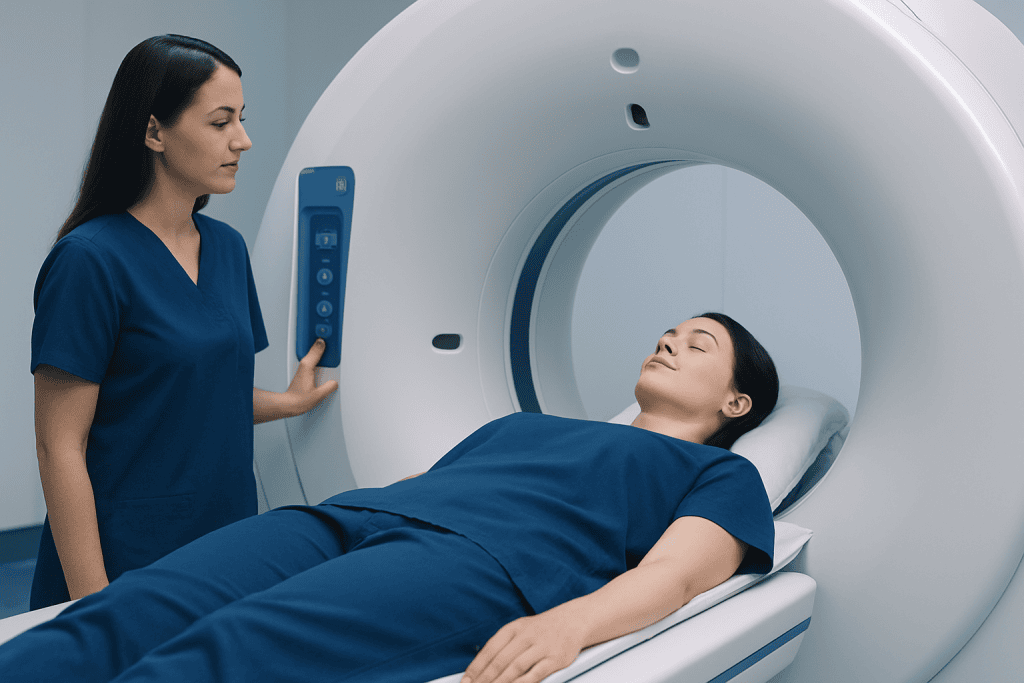
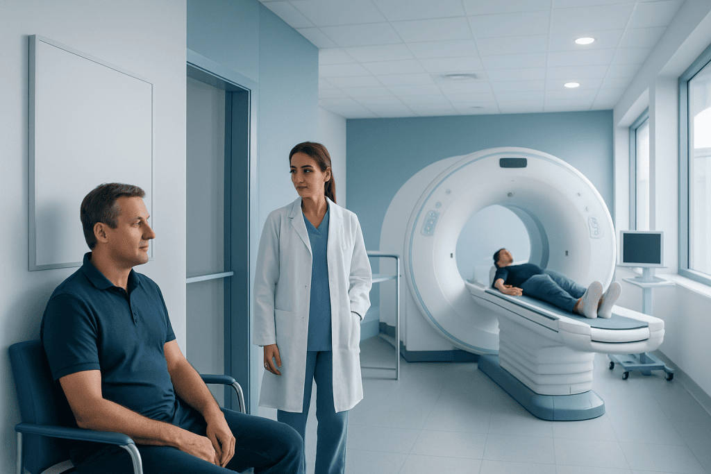
During a CT scan, the patient lies on a table that moves slowly through a ring-shaped scanner. An X-ray tube rotates around the body while detectors measure how much radiation passes through different tissues. A computer then reconstructs these measurements into detailed images, which a radiologist reviews and reports.
CT may be used to evaluate sudden symptoms such as severe pain, breathing problems, suspected stroke or trauma. It is also used to assess conditions involving the lungs, abdomen, urinary tract, blood vessels, bones and brain. In some situations, CT helps guide biopsies, drainage procedures or treatment planning.
Many scans are performed without contrast material. For others, an iodine-based contrast agent helps make blood vessels, inflammation, tumors or organs easier to distinguish. Contrast improves image detail in selected cases but does not add radiation; the radiation comes from the X-rays used by the scanner.
Candidacy and preparing for a CT scan
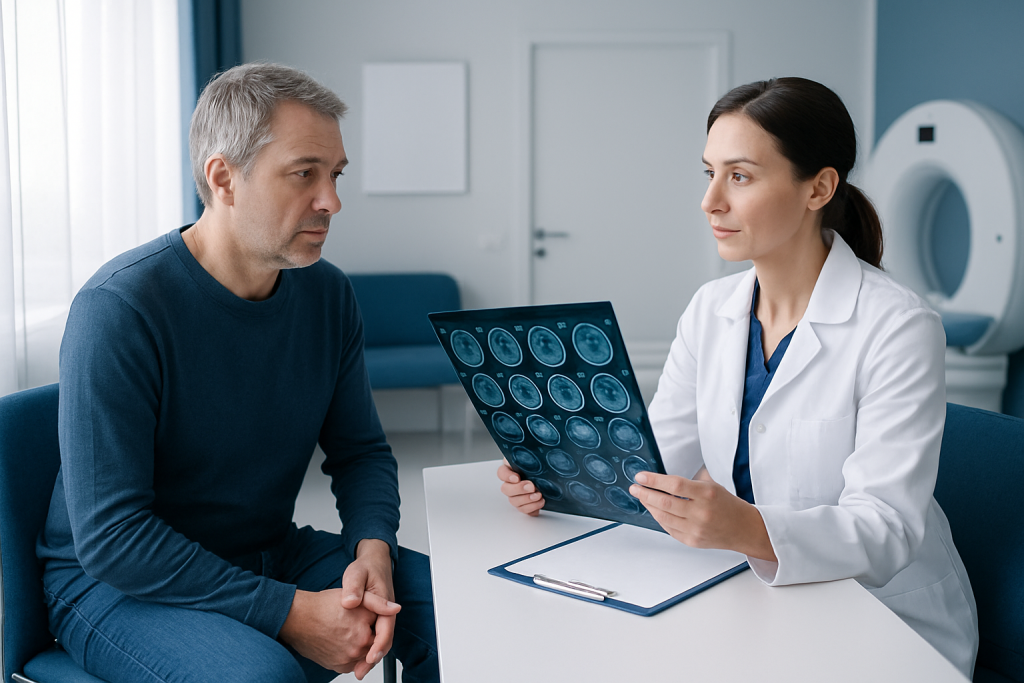
A doctor may recommend CT when its detailed images are likely to answer an important medical question better or faster than another test. Ultrasound and MRI do not use ionizing radiation and may be preferred in some circumstances. However, they do not always provide the same information, may take longer or may not be suitable in urgent settings.
Before the scan, patients should tell the imaging team if they are pregnant, think they may be pregnant, are breastfeeding, have kidney disease, diabetes, thyroid disease, asthma or previous reactions to contrast material. The team should also know about any medicines, allergies and previous imaging studies. This information helps them select the safest and most appropriate protocol.
Preparation varies by examination. Some CT scans require no special preparation, while scans using contrast may involve temporary fasting or a kidney blood test for selected patients. Patients should follow the instructions provided by their healthcare team rather than stopping medication independently.
What happens during the procedure and recovery
On arrival, the patient may be asked to change into a gown and remove metal objects or jewelry from the area being scanned. The radiographer positions the patient on the table and may use cushions or straps to support a comfortable, still position. Remaining still is important because movement can blur the images.
If intravenous contrast is needed, a small cannula is placed in a vein. Some people notice a brief warm sensation, a metallic taste or a feeling of needing to urinate when contrast is injected. These sensations usually pass within moments. The scanner is open at both ends, and the radiographer can communicate with the patient throughout the examination.
The image acquisition itself often takes only seconds to minutes, although the overall appointment may be longer because of preparation and positioning. Most people can return to normal activities straight away. After contrast, drinking fluids may be advised if medically appropriate, and the body generally removes the contrast through urine.
CT findings are interpreted alongside symptoms, examination findings and other tests. A result may be available quickly in emergency care, while non-urgent reports can take longer depending on the clinical setting. A CT result is not always a final diagnosis; further assessment may sometimes be needed.
Radiation risks, benefits and dose reduction
Ionizing radiation can slightly increase the lifetime chance of cancer, particularly when exposures are high or repeated. For an individual, the additional risk from one appropriately justified CT scan is generally considered small. The exact level cannot be calculated precisely for a particular person because it depends on age, sex, body area, dose and other lifetime exposures.
The potential benefit of CT can be substantial. It may detect serious bleeding after an injury, identify a blood clot, reveal a complication requiring treatment or help avoid unnecessary surgery. A scan that is medically necessary should not be delayed solely because of radiation concerns; instead, patients can discuss why it is needed and whether another test could provide the required information.
Radiology departments use dose-optimization methods, including automated exposure controls, patient-size adjustments and focused scan ranges. Modern protocols can also use lower-dose techniques for appropriate indications. Keeping prior imaging records available may help clinicians avoid unnecessary repeat examinations.
- Ask what clinical question the CT scan is intended to answer.
- Tell the team about recent imaging performed elsewhere.
- Ask whether a low-dose protocol, ultrasound or MRI is appropriate for the situation.
- Do not skip a recommended urgent scan without discussing it with the treating clinician.
What percentage of people get cancer from a CT scan?
There is no reliable single percentage that applies to every person or every CT scan. Research estimates are based largely on population data and models of radiation exposure, rather than on a test that can determine whether one particular scan will cause cancer in one person. The estimated added lifetime risk from a single standard CT examination is small, but it varies with the scan type and the person’s age and sex.
Children and younger adults are generally more sensitive to radiation than older adults because their tissues are still developing and they have more years ahead in which a radiation-related cancer could occur. For this reason, pediatric CT should be carefully justified and use child-sized protocols. Repeated examinations can add to cumulative exposure, which is another reason clinicians review whether each scan is necessary.
It is important to put this small potential risk in context. When CT is recommended for a significant health concern, the information it provides may lead to timely treatment or rule out a dangerous condition. Patients can ask their doctor or radiologist about the expected benefit in their individual situation.
How long does contrast stay in your body after a CT scan?
Most iodine-based CT contrast is removed from the bloodstream by the kidneys and leaves the body in urine. In people with normal kidney function, the majority is usually cleared within about 24 hours, although the precise timing can vary. Contrast material is not radioactive, so it does not remain in the body as radiation.
People with significantly reduced kidney function may clear contrast more slowly. The clinical team may check kidney function or adapt the imaging plan when this is relevant. A previous contrast reaction also deserves attention, as it may influence the preparation or choice of examination.
After a contrast-enhanced CT, patients should seek prompt medical advice if they develop symptoms such as difficulty breathing, swelling of the face or throat, widespread hives, severe dizziness or rapidly worsening illness. Mild, short-lived warmth or an unusual taste during injection is common and is not usually an allergic reaction.
Which scan has the highest radiation, and which CT scan has the lowest radiation?
Radiation dose varies more by protocol than by the name of the scan alone. CT examinations covering a larger body area, scans performed in several phases before and after contrast, and certain specialized studies can have higher radiation doses because more images are needed. CT of the abdomen and pelvis, multiphase liver studies and some cardiac or vascular CT protocols may involve higher exposure than a single limited series.
Among CT examinations, targeted scans using optimized low-dose protocols may have lower radiation exposure. Examples can include low-dose CT protocols used for selected lung cancer screening programs, some sinus examinations and certain focused bone or urinary-tract protocols. However, the lowest-dose scan is not automatically the best scan; image quality must still be sufficient to answer the medical question.
A standard chest X-ray generally uses substantially less radiation than a CT scan, while MRI and ultrasound do not use ionizing radiation. The best imaging test depends on the suspected condition, urgency, body area and the information needed. The imaging specialist can tailor the test to minimize exposure without compromising necessary diagnostic quality.
When to seek medical care
Patients should seek urgent medical care for sudden or severe symptoms such as chest pain, severe shortness of breath, signs of stroke, major injury, fainting, uncontrolled bleeding, severe abdominal pain or a rapidly worsening condition. In these situations, CT may be recommended because it can provide critical information quickly.
Following a CT with contrast, urgent assessment is appropriate for breathing difficulty, swelling, severe rash, fainting or other signs of a serious allergic-type reaction. Contact the healthcare team promptly for persistent vomiting, marked reduction in urine output or concerning symptoms after the examination, particularly in people with known kidney disease.
For non-urgent questions about radiation exposure, patients can speak with the referring doctor or radiology department before the appointment. Acibadem International’s multidisciplinary specialists and JCI-accredited hospitals can assess imaging needs and coordinate diagnostic care for international patients.
Frequently asked questions
Is the radiation from one CT scan dangerous?
A single CT scan performed for a clear medical reason is generally considered to carry a small potential long-term radiation risk. Its diagnostic benefit may be much more important, especially in urgent situations. The imaging team aims to use the lowest dose that can provide useful images.
Is CT radiation higher than an X-ray?
Yes. A CT scan usually uses more radiation than a standard X-ray because it takes many X-ray measurements to create detailed cross-sectional images. The exact difference depends on the body part and the imaging protocol.
Can a CT scan cause cancer years later?
Ionizing radiation can slightly increase lifetime cancer risk, but it is not possible to determine whether a future cancer in an individual was caused by one scan. The added risk from one necessary CT examination is generally small. Risk can increase with higher and repeated exposures, which is why imaging is carefully justified.
Should children have CT scans?
Children can have CT scans when the expected medical benefit outweighs the potential risk. Because children are more sensitive to radiation, clinicians should use pediatric protocols and consider ultrasound or MRI when those tests can answer the question adequately. Parents can ask why CT is recommended and how the dose will be optimized.
Do I need to drink extra water after CT contrast?
Many people are encouraged to drink fluids after contrast if they are able to do so and have not been advised to limit fluids for another health reason. This supports normal kidney clearance of contrast. Patients with heart failure, kidney disease or fluid restrictions should follow their clinician’s specific advice.
Can I have a CT scan if I am pregnant?
Pregnancy or possible pregnancy should always be discussed with the referring clinician and imaging team before the scan. They can assess the urgency, consider alternatives when appropriate and modify the examination if CT is necessary. CT should not be withheld when it is needed to diagnose a potentially serious condition.
References
- U.S. Food and Drug Administration
- RadiologyInfo.org
- International Atomic Energy Agency
- National Cancer Institute
- American College of Radiology
This article is for general information only and is not a substitute for professional medical advice. Please consult a qualified doctor about your individual situation.
Explore treatments in Turkey — costs, top hospitals & a free quote
JCI-accredited · board-certified surgeons · reply within 24h
Add us as a Preferred Source to see more of our trusted health content across Google Search, AI Overviews and Discover.