Hypodermis: An Evidence-Based Guide for Patients

The hypodermis lies below the dermis and is also called subcutaneous tissue or subcutis. It contains fat, connective tissue, blood vessels, and nerves that protect and support the body.
Key Takeaways
- The hypodermis lies below the dermis and is also called subcutaneous tissue or subcutis.
- It contains fat, connective tissue, blood vessels, and nerves that protect and support the body.
- Changes in the hypodermis can occur with aging, weight change, injury, infection, inflammation, and some diseases.
- Doctors assess the hypodermis through physical examination and, when needed, imaging or biopsy.
- Medical care is important for painful lumps, spreading redness, skin breakdown, or sudden swelling.
The hypodermis is the deepest layer beneath the skin. It helps cushion the body, store energy, regulate temperature, and support blood vessels and nerves, while also playing a role in how some medical conditions and treatments affect the skin.
What the hypodermis is
The hypodermis is the deepest layer beneath the skin. In simple terms, it is the soft layer under the dermis that contains fat, connective tissue, blood vessels, and nerves. Although it is often discussed together with the skin, many anatomy references describe it as a supporting layer below the skin rather than part of the skin itself.
This layer matters because it does much more than store fat. The hypodermis cushions underlying muscles and bones, helps the body conserve heat, and creates a flexible connection between the skin and the structures underneath. It also provides a pathway for blood vessels and nerves that help keep tissues healthy.
For patients, understanding the hypodermis can make it easier to understand common issues such as bruising, injections, pressure-related skin damage, swelling, lumps under the skin, and changes linked with weight loss, aging, or inflammation. In clinical care, doctors often evaluate this layer when a person has skin pain, tenderness, thickening, or a new mass.
How the hypodermis differs from other skin layers
The body has three main layers in this region: the epidermis on the surface, the dermis underneath it, and the hypodermis below both. The epidermis acts as the outer barrier. The dermis contains collagen, elastin, hair follicles, sweat glands, and many small blood vessels. The hypodermis sits deepest and contains larger fat lobules separated by bands of connective tissue.
This difference in structure explains why each layer behaves differently. Surface problems such as dryness or mild irritation affect the epidermis. Scarring and many rashes involve the dermis. Deeper tenderness, pressure injury, and some types of swelling or infection can involve the hypodermis, sometimes making the skin feel warm, firm, or painful.
The thickness of the hypodermis varies from one part of the body to another and from person to person. It is influenced by age, sex, genetics, body composition, hormones, and general health. A thinner hypodermis can mean less cushioning over bony areas, while a thicker one may affect how injections are given or how some conditions appear on examination.
What the hypodermis does

The hypodermis has several essential roles in day-to-day health. One of its main jobs is protection. By absorbing mechanical stress, it helps reduce the effect of pressure and minor trauma on deeper tissues. This cushioning role is especially important over the hips, heels, buttocks, elbows, and other areas exposed to friction or prolonged pressure.
It also supports temperature regulation. Subcutaneous fat helps slow heat loss, acting as a form of insulation. In addition, the hypodermis stores energy that the body can use when needed. This storage function is part of normal metabolism and does not simply reflect body weight alone.
Another important function is tissue support. The hypodermis allows the skin to move over muscles and bones without excessive tension. It also contains larger blood vessels and nerves that help nourish the skin and communicate sensations such as pressure and deep pain. Because of this, problems in the hypodermis can affect comfort, wound healing, and mobility.
- Cushions the body against pressure and impact
- Helps retain body heat
- Stores energy in fat cells
- Supports blood vessels, lymphatic channels, and nerves
- Anchors the skin to deeper tissues while allowing movement
Common changes and conditions affecting the hypodermis
The hypodermis can change for many reasons, including normal aging. With age, this layer may become thinner in some areas, reducing padding and making bruising or pressure injury more likely. Weight loss can also reduce subcutaneous fat, while weight gain may expand it. Hormonal changes, chronic illness, and reduced mobility may further affect tissue quality.
Inflammation and infection can involve the hypodermis. Cellulitis may spread into deeper soft tissue and cause redness, warmth, swelling, and tenderness. Collections of infected material such as abscesses can form beneath the skin and may need drainage. Patients with recurrent infections, diabetes, poor circulation, or weakened immunity may need closer evaluation. In related cases, doctors may also assess for skin cancer if a lesion appears persistent, unusual, or does not heal as expected.
Some people develop tender nodules or thickened areas within subcutaneous fat, a pattern seen in panniculitis and other inflammatory disorders. Benign growths such as lipomas can arise in fatty tissue and often feel soft and movable. Trauma may lead to bruising, hematoma, or fat necrosis. Swelling due to lymphatic or venous problems can also alter the feel of the hypodermis. A new lump, severe pain, rapidly spreading redness, or skin breakdown should always be reviewed by a qualified clinician.
How doctors evaluate the hypodermis
Assessment usually begins with a medical history and physical examination. A doctor may ask when a lump, swelling, or painful area began, whether it is growing, and whether there are symptoms such as fever, drainage, itching, numbness, or recent injury. They may also ask about medications, diabetes, circulation problems, autoimmune disease, or past skin conditions.
During the examination, the clinician looks at color, warmth, tenderness, firmness, mobility, and the borders of the affected area. This helps distinguish possibilities such as infection, inflammation, fluid retention, scar tissue, or a benign fatty lump. If the concern relates to pressure injury or wound healing, the surrounding skin and circulation are also assessed.
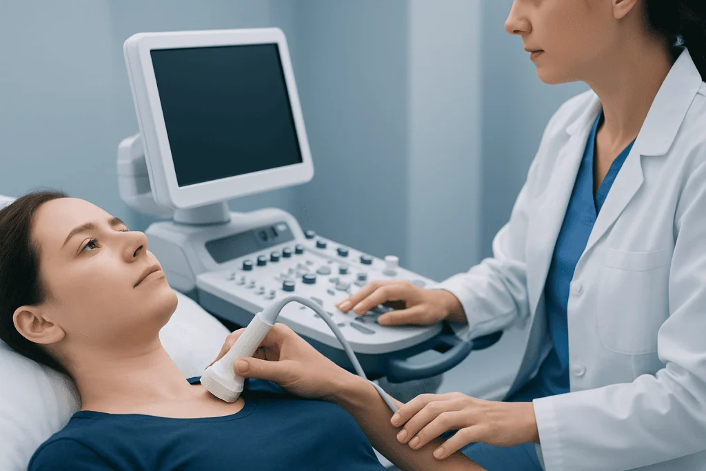
Further testing depends on the problem. Ultrasound can help evaluate a lump, fluid collection, or soft tissue swelling. In selected cases, MRI or CT may provide a deeper view. If there is concern about an unclear mass or an inflammatory skin disorder, a biopsy may be recommended. Diagnostic imaging such as MRI may be helpful when a lesion extends deeper or when the diagnosis remains uncertain after examination.
Treatment and medical care
Treatment depends on the cause. Normal anatomical variation or age-related thinning of the hypodermis may not need specific treatment, but it may call for preventive skin care and pressure protection. Infections are treated with appropriate medical therapy, and some abscesses need drainage. Inflammatory conditions may require medicines aimed at the underlying cause rather than the skin alone.
Benign soft lumps such as lipomas are often monitored unless they are painful, growing, or bothersome. If removal is needed, doctors may recommend a minor procedure. When a lesion seems suspicious, additional evaluation is important rather than assuming it is harmless. In some cases, specialists may use biopsy to clarify the diagnosis and guide the next steps.
Care for injuries affecting the hypodermis may include rest, wound care, pressure relief, and monitoring for infection. If circulation problems, nerve symptoms, or chronic swelling are present, treatment may involve several specialties. Near the end of a patient journey, multidisciplinary teams can be valuable; Acibadem International’s specialists in JCI-accredited hospitals diagnose and treat skin and soft-tissue conditions for international patients when more advanced assessment is needed.
Prevention and self-care for healthy deep skin tissue
Protecting the hypodermis starts with general skin health and pressure awareness. Staying active, changing position regularly, and using supportive footwear or cushioning can reduce stress on vulnerable areas. This is particularly important for older adults, people with limited mobility, and those with diabetes or circulation problems.
Good nutrition and hydration support tissue integrity and healing. Maintaining a stable, healthy weight can help reduce extremes that affect subcutaneous tissue, although individual body composition naturally varies. Gentle skin care, prompt cleansing of cuts, and avoiding repeated friction may also lower the risk of deeper irritation or infection.
People who self-inject medicines should follow clinician instructions carefully, including rotation of injection sites when advised. If the skin repeatedly becomes hard, tender, or lumpy after injections, a doctor should review technique and the area involved. When there is concern about recurring inflammation, deep infection, or a wound that is slow to heal, further evaluation may include imaging such as ultrasound or review of related skin disorders such as eczema if the surface skin barrier is also affected.
When to seek medical care
Medical review is advisable if a person notices a new lump under the skin, an area that is becoming larger, or persistent deep tenderness without a clear reason. A doctor should also assess skin that is red, hot, swollen, draining, or associated with fever, as these may suggest infection or inflammation involving the hypodermis.
Prompt care is important for skin breakdown over pressure areas, sudden swelling of a limb, severe pain after injury, or bruising that is unusually extensive. People with diabetes, poor circulation, reduced sensation, or weakened immunity should seek help earlier because deeper skin and soft-tissue problems may progress more quickly or heal more slowly.
Even when symptoms seem mild, it is sensible to arrange a medical appointment if a problem is not improving, keeps returning, or is interfering with movement, sleep, or daily comfort. Early assessment can help identify whether the issue is a benign change, a treatable infection, an inflammatory condition, or another soft-tissue problem that needs targeted care.
Frequently asked questions
Is the hypodermis part of the skin?
The hypodermis lies below the dermis and is often described as a supporting layer beneath the skin. Some sources discuss it together with the skin because it works closely with the upper layers, but anatomically it is usually considered subcutaneous tissue.
What is the main function of the hypodermis?
Its main functions are cushioning, insulation, energy storage, and support for blood vessels and nerves. It also helps attach the skin to deeper tissues while allowing the skin to move smoothly over the body.
Can the hypodermis cause lumps under the skin?
Yes. Benign fatty growths such as lipomas, fluid collections, inflammation, bruising, or infection can all create lumps or thickened areas within the hypodermis. Any new, enlarging, painful, or persistent lump should be examined by a doctor.
Does the hypodermis change with age?
Yes, age can change the thickness and quality of subcutaneous tissue. In some areas it becomes thinner, which may reduce cushioning and increase the risk of pressure-related discomfort, bruising, or slower healing.
How do doctors check problems in the hypodermis?
Doctors usually begin with a physical examination and review of symptoms. If needed, they may use ultrasound, MRI, or a biopsy to understand whether the problem is related to fat tissue, fluid, infection, inflammation, or a mass.
When should someone worry about symptoms involving the hypodermis?
It is important to seek medical attention for spreading redness, warmth, severe pain, fever, drainage, sudden swelling, or skin breakdown. A lump that keeps growing or does not improve should also be checked, even if it is not very painful.
References
- American Academy of Dermatology
- Merck Manual Professional Edition
- Mayo Clinic
- National Institutes of Health
- Cleveland Clinic
This article is for general information only and is not a substitute for professional medical advice. Please consult a qualified doctor about your individual situation.
Explore treatments in Turkey — costs, top hospitals & a free quote
JCI-accredited · board-certified surgeons · reply within 24h
Add us as a Preferred Source to see more of our trusted health content across Google Search, AI Overviews and Discover.

More from the Health Library

Sinus Arrhythmia: A Complete Medical Overview

Lamictal: A Complete Medical Overview

Iron Bisglycinate: A Complete Medical Overview

Squamous Epithelium: An Evidence-Based Guide for Patients

Healthy BMI for Women: A Complete Medical Overview