

Intraoperative MRI: Real-Time Imaging Guidance During Brain Surgery
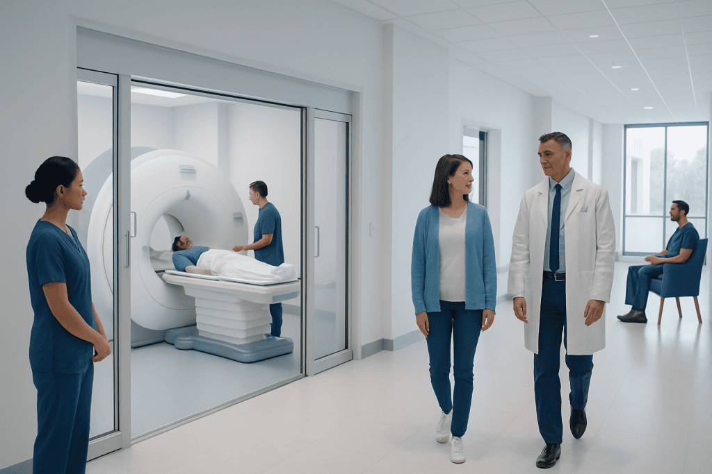
Intraoperative MRI provides updated images during brain surgery, helping surgeons see changes that occur after the operation begins. It is commonly used in selected brain tumor surgeries, especially when safe and complete tumor removal is an important goal.
Key Takeaways
- Intraoperative MRI provides updated images during brain surgery, helping surgeons see changes that occur after the operation begins.
- It is commonly used in selected brain tumor surgeries, especially when safe and complete tumor removal is an important goal.
- The technology may help identify remaining tumor tissue, adjust surgical planning, and support protection of important brain areas.
- Not every patient or surgery requires intraoperative MRI; the decision depends on the diagnosis, tumor location, patient factors, and hospital resources.
- Patients should discuss the expected benefits, limitations, and safety considerations with a qualified neurosurgical team.
Intraoperative MRI is an advanced imaging technology that allows neurosurgeons to obtain MRI scans during an operation, most often during brain tumor surgery. It can help the surgical team assess progress, update navigation, and make careful decisions while the patient is still in the operating room.
Overview
Intraoperative MRI, often called iMRI, is magnetic resonance imaging performed during surgery. In neurosurgery, it is most often used during operations on the brain, where millimeters can matter. Instead of relying only on MRI images taken before the operation, the surgical team can obtain updated images while the procedure is still underway.
This matters because the brain can shift during surgery. Once the skull is opened, cerebrospinal fluid may drain, tissue may relax, and parts of the brain may move slightly. This natural movement, called brain shift, can make preoperative navigation less accurate as the operation progresses. Intraoperative MRI helps the team update the visual map and reassess the surgical field.
The technology does not replace the experience of the neurosurgeon. Rather, it is one of several tools that may be combined with surgical microscopes, neuronavigation, neurophysiological monitoring, endoscopy, and functional imaging. The goal is to support precise decision-making, especially when operating close to important areas involved in movement, speech, vision, memory, or other functions.
How Intraoperative MRI Works
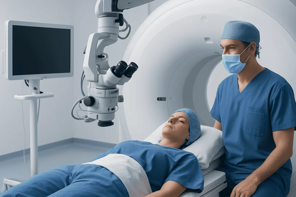
Standard MRI uses a strong magnetic field and radiofrequency signals to create detailed images of soft tissues. Intraoperative MRI applies the same imaging principle inside or adjacent to a specially designed operating environment. The operating room must be planned carefully because MRI magnets require strict safety controls for equipment, instruments, anesthesia devices, and monitoring systems.
Different hospital systems use different setups. Some operating rooms contain a fixed MRI scanner that can be brought into position during the procedure. Others move the patient, while still safely positioned and monitored, to an MRI scanner connected to the operating room. In each model, the patient remains under the care of the anesthesia and surgical teams throughout the imaging process.
During surgery, the neurosurgeon may pause at a planned point and request an intraoperative scan. The images can show the current location of the surgical cavity, nearby brain structures, swelling, blood products, and whether visible tumor tissue may remain. The updated images can then be integrated into navigation systems to guide the next stage of surgery, if further removal is appropriate and safe.
When Intraoperative MRI May Be Used

Intraoperative MRI is most commonly associated with brain tumor surgery. It may be considered for low-grade gliomas, high-grade gliomas, pituitary-region tumors, deep-seated lesions, pediatric brain tumors, and selected metastases, depending on the case. Its value is often greatest when the tumor borders are difficult to distinguish from normal tissue or when the surgical goal includes removing as much tumor as safely possible.
It can also be helpful when the tumor is near critical brain areas. In such cases, the surgical team must balance tumor removal with protection of neurological function. iMRI may assist this balance by showing what has already been removed and what remains in relation to important structures. However, the decision to continue removing tissue is always individualized and guided by safety.
Possible uses include:
- Checking for residual tumor during the same operation.
- Updating navigation after brain shift occurs.
- Confirming the relationship between the surgical cavity and nearby structures.
- Supporting complex procedures in which anatomy may change during surgery.
- Reducing the need for an early second operation in selected cases, when additional safe removal can be performed immediately.
Not all brain surgeries require intraoperative MRI. Some lesions are visible and accessible without it, while others may be better managed with different technologies. The neurosurgical team considers the diagnosis, imaging appearance, patient health, expected benefit, and available expertise before recommending iMRI.
Potential Benefits and Limitations
The main potential benefit of intraoperative MRI is real-time reassessment. Surgeons begin with a preoperative plan, but surgery is a dynamic process. By obtaining updated imaging during the operation, the team can better understand whether the surgical goal has been reached or whether further work may be possible. In tumor surgery, this may support a greater extent of safe removal in appropriately selected patients.
iMRI may also improve confidence in decision-making. If the scan shows that the target has been adequately addressed, the surgeon may avoid unnecessary exploration. If it shows remaining tissue that appears surgically reachable, the team can consider additional removal during the same anesthetic. This can be especially useful when residual tissue is not easily distinguished by the naked eye or microscope.
However, intraoperative MRI has limitations. It cannot make surgery risk-free, and it cannot always distinguish tumor from inflammation, blood products, treatment-related change, or normal tissue with complete certainty. It also requires additional time, specialized staff, MRI-compatible equipment, and a carefully controlled environment. In some cases, the safest choice may be to stop surgery even if imaging shows residual abnormal tissue, particularly when the remaining area is close to essential brain function.
Patients should also understand that iMRI is a decision-support tool, not a guarantee of outcome. Recovery, tumor control, neurological function, and long-term treatment plans depend on many factors, including tumor biology, location, pathology results, the patient’s overall health, and whether additional therapies such as radiotherapy, chemotherapy, targeted therapy, or rehabilitation are needed.
What Patients Can Expect Before and During Surgery
Before surgery, patients usually undergo detailed imaging and consultation with the neurosurgical team. The doctor explains the reason for surgery, expected goals, possible alternatives, and whether intraoperative MRI is likely to be useful. Additional tests may include functional MRI, diffusion tensor imaging, blood tests, anesthesia evaluation, and sometimes neuropsychological or speech assessments, depending on the tumor location.
Patients should inform the medical team about implanted devices, previous surgeries, allergies, kidney disease, pregnancy, and any metal fragments or implants. MRI safety screening is very important because the magnet can interact with certain devices or metal objects. Many modern implants are MRI conditional, meaning they may be safe under specific conditions, but this must be verified before scanning.
During the operation, the patient is usually under general anesthesia, although selected brain surgeries may involve awake mapping when speech or movement areas need testing. If intraoperative MRI is used, the surgical team pauses the operation at the appropriate stage. The sterile field, anesthesia lines, monitoring, and patient position are managed according to strict protocols. MRI-compatible equipment is used in the scanner area.
After the scan, the surgeon reviews the images and decides whether the operation should continue, be adjusted, or proceed to closure. Family members are typically updated after surgery according to hospital practice. The final diagnosis often depends on pathology examination of the tissue, which may take additional time and may influence the next steps in treatment.
Safety Considerations
Intraoperative MRI is performed within a highly controlled safety environment. The MRI magnet is always taken seriously because it can strongly attract ferromagnetic objects. For this reason, operating rooms equipped for iMRI use special MRI-compatible instruments, anesthesia machines, monitors, and positioning systems. Staff members follow detailed checklists before anyone or anything enters the MRI zone.
From the patient’s perspective, safety planning includes MRI screening, anesthesia monitoring, infection-control measures, and careful management of body temperature, blood pressure, breathing, and fluids. If contrast material is needed, the team considers kidney function, allergy history, and the clinical reason for contrast use. Not every intraoperative scan requires contrast.
As with any brain surgery, risks depend on the condition being treated and the location of the operation. These may include bleeding, infection, seizures, swelling, weakness, speech changes, vision changes, memory or cognitive effects, cerebrospinal fluid leakage, and anesthesia-related complications. iMRI itself is not the main source of these surgical risks, but it adds procedural complexity and time, so it should be used when the expected benefit justifies its use.
Patients can support safety by providing a complete medical history and asking questions before surgery. They should bring implant cards when available and tell the team about pacemakers, cochlear implants, aneurysm clips, stents, shunts, medication pumps, spinal stimulators, dental or orthopedic metalwork, and any prior metal injury to the eyes or body.
Recovery and Follow-Up After MRI-Guided Neurosurgery
Recovery after brain surgery depends on the type of operation, tumor location, neurological status before surgery, and whether additional treatment is needed. Some patients spend time in an intensive care or high-dependency unit for close monitoring. Nurses and doctors check alertness, strength, speech, pupils, pain level, wound condition, and vital signs. Postoperative imaging may still be performed after surgery, even if intraoperative MRI was used.
Pathology results are an important part of follow-up. They help determine the exact tumor type, grade, molecular features when relevant, and whether further treatment is recommended. A multidisciplinary team may include neurosurgeons, neurologists, neuroradiologists, pathologists, radiation oncologists, medical oncologists, rehabilitation specialists, nurses, and speech or physical therapists.
Self-care after discharge usually includes wound care, medication guidance, activity restrictions, and follow-up appointments. Patients should not drive, fly, return to work, or restart strenuous exercise until their doctor confirms it is safe. If seizures are a concern, the medical team will give specific advice about safety precautions and medication use.
At Acibadem International, multidisciplinary specialists and JCI-accredited hospitals diagnose and treat complex neurosurgical conditions, including selected cases where intraoperative MRI may be part of the surgical plan for international patients. Individual recommendations are made after careful review of medical records, imaging, and patient-specific risks and goals.
When to Discuss Intraoperative MRI With a Doctor
Patients diagnosed with a brain tumor or another complex brain lesion may ask their neurosurgeon whether intraoperative MRI could be helpful in their case. The answer may be yes, no, or uncertain depending on the anatomy, suspected diagnosis, planned surgical approach, and available alternatives. A clear discussion can help patients understand how iMRI may or may not change the surgical plan.
Useful questions include: What is the main goal of surgery? Is the lesion close to critical brain areas? How will the team monitor brain function? Could intraoperative MRI help identify residual tissue? What are the limitations of the technology in this case? What other imaging or mapping tools will be used? What follow-up treatments may be needed after pathology results?
Medical attention should be sought promptly if a person develops concerning neurological symptoms such as a first seizure, new or worsening weakness, persistent severe headache, speech difficulty, vision changes, confusion, or loss of balance. These symptoms do not always mean a brain tumor is present, but they deserve timely evaluation by a qualified healthcare professional.
Frequently asked questions
What is intraoperative MRI?
Intraoperative MRI is MRI scanning performed during an operation, most often during brain surgery. It provides updated images while the patient is still in the operating room. These images can help the surgical team assess progress and adjust navigation if needed.
Why is intraoperative MRI useful in brain tumor surgery?
Brain tissue can shift during surgery, which may reduce the accuracy of images taken before the operation. Intraoperative MRI gives the surgeon a current view of the surgical area. It may help show whether tumor tissue remains and whether further removal appears safe.
Does intraoperative MRI make brain surgery safer?
It can support safer decision-making in selected cases by improving visualization during surgery. However, it does not remove all surgical risks and does not guarantee a specific outcome. Safety depends on many factors, including tumor location, patient health, surgical technique, and brain function monitoring.
Is the patient awake during intraoperative MRI?
Most patients are under general anesthesia during brain surgery with intraoperative MRI. In selected cases involving speech or movement mapping, parts of the operation may be performed with the patient awake, but this is planned carefully and explained in advance. The approach depends on the condition and the brain area involved.
Can everyone have intraoperative MRI?
No. Some patients may not be suitable because of certain implants, metal fragments, medical devices, or clinical factors. In other cases, the technology may not add enough benefit to justify its use. The neurosurgical and radiology teams assess suitability before surgery.
Does intraoperative MRI replace postoperative MRI?
Not always. Even when intraoperative MRI is used, doctors may still request postoperative imaging to document the surgical result and guide follow-up care. The timing and type of imaging depend on the diagnosis, surgery, and treatment plan.
What should patients ask their surgeon before MRI-guided brain surgery?
Patients may ask why intraoperative MRI is being recommended, what information it is expected to provide, and how it could change the operation. They should also ask about risks, alternatives, brain function monitoring, recovery, and whether additional treatment may be needed after pathology results are available.
References
- American Association of Neurological Surgeons
- Radiological Society of North America
- European Association of Neurosurgical Societies
- National Institute for Health and Care Excellence
- World Federation of Neurosurgical Societies
This article is for general information only and is not a substitute for professional medical advice. Please consult a qualified doctor about your individual situation.
Oncology care in Turkey — second opinion and treatment plan
JCI-accredited · board-certified surgeons · reply within 24h
Add us as a Preferred Source to see more of our trusted health content across Google Search, AI Overviews and Discover.

More from the Health Library



Related Specialists

Dr. Mehmet Tayfun Osmanağaoğlu
Gynecology & Obstetrics
Dr. Faruk Batur
Inpatient Clinic Physicians Clinical Service
Assoc. Prof. Dr. Nilüfer Aykaç
Pulmonology
Dr. Savaş Beyaztaş
Cardiology