Kidney Xray: Preparation, Procedure and Results

A kidney xray is commonly called a KUB X-ray, meaning kidneys, ureters and bladder. It can detect some stones, especially those containing calcium, but it cannot reliably rule out all kidney or urinary tract conditions.
Key Takeaways
- A kidney xray is commonly called a KUB X-ray, meaning kidneys, ureters and bladder.
- It can detect some stones, especially those containing calcium, but it cannot reliably rule out all kidney or urinary tract conditions.
- Most people need little or no preparation; instructions may differ if the X-ray is part of a larger imaging examination.
- The examination uses a small amount of ionizing radiation and is generally completed within minutes.
- Pregnancy, possible pregnancy and previous imaging should be discussed with the imaging team before the test.
A kidney xray is a plain abdominal radiograph, often called a KUB X-ray, that can help identify certain urinary stones and provide a general view of the kidneys, ureters and bladder area. Preparation is usually simple, and the test is painless, fast and performed without needles or recovery time.
Kidney Xray Overview: What the Test Shows
A kidney xray is a plain X-ray image of the abdomen that includes the area of the kidneys, ureters and bladder. Its medical term is commonly a KUB radiograph, from “kidneys, ureters and bladder.” It uses a small amount of ionizing radiation to create a two-dimensional image of structures inside the abdomen.
The test is most often used when a clinician is checking for or monitoring certain urinary stones. It may show stones that are dense enough to be visible on X-ray, including many calcium-containing stones. It can also help assess the general position of the kidneys and identify some patterns of bowel gas or calcification that may be relevant to symptoms.
A kidney xray has important limits. It does not measure how well the kidneys work, and many stones are not visible on plain X-rays. Ultrasound or computed tomography (CT) may be more appropriate when a clearer look at the kidneys, urinary blockage, infection-related complications or non-visible stones is needed.
How a Kidney Xray Works and Who May Need One
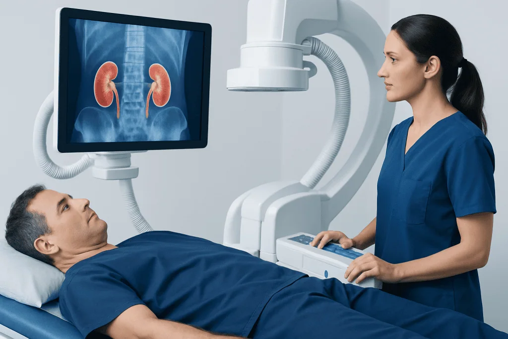
X-rays pass through the body at different rates. Dense materials, such as bone and some stones, absorb more radiation and appear lighter on the final image. Softer tissues are less distinct on a plain X-ray, which is why a kidney xray provides useful but limited information about the urinary tract.
A clinician may request a KUB X-ray for a person with known kidney stones, symptoms that could be related to a visible stone, or follow-up after treatment for stones. It may also be used alongside urine testing, blood tests and other imaging to help build a fuller clinical picture. For a broader discussion of stone disease, see kidney stones.
Not every person with flank pain, blood in the urine or urinary symptoms needs a kidney xray. The most suitable scan depends on the symptom pattern, medical history, pregnancy status, prior imaging and the clinical question. The referring clinician or radiologist can decide whether a KUB X-ray, ultrasound, CT or another examination is most useful.
- A KUB X-ray may be useful for following stones already known to be visible on X-ray.
- Ultrasound may be preferred when radiation should be avoided or a clinician needs to assess swelling of the kidney.
- CT can offer more detailed information when the diagnosis is uncertain or complications are suspected.
Kidney Xray Preparation and Candidacy

Kidney x ray preparation is usually straightforward. For many routine KUB X-rays, people can eat, drink and take regular medicines as usual. However, the imaging center’s instructions should always take priority because preparation can vary when the X-ray is paired with another test, such as contrast imaging of the urinary tract or a bowel examination.
For kidney ureter bladder x ray preparation, a clinician may occasionally ask a person to avoid certain foods, use a bowel preparation or arrive with a particular bladder status. These steps are not needed for every patient; they are intended to reduce bowel contents or gas that can obscure part of the image. The preparation of patient for x ray of kidney should therefore be confirmed when the appointment is booked.
Before the examination, patients should tell the team if they are pregnant, might be pregnant, have had recent abdominal imaging, or have concerns about radiation exposure. They should also mention any medical devices or recent procedures. A plain kidney xray does not usually involve contrast dye, injections or sedation.
Comfortable clothing without metal is helpful. Belts, jewelry, coins, zippers and other metal objects around the abdomen or pelvis may need to be removed because they can interfere with the image.
Kidney Xray Procedure: Step by Step
The procedure to take x ray of kidney medical term is a KUB radiograph. On arrival, a radiographer confirms the person’s identity and reviews relevant safety information. The patient may be asked to change into a gown or remove metal objects from the waist and pelvic area.
For the main image, the person usually lies flat on an X-ray table while the radiographer positions the X-ray equipment above the abdomen. In some cases, additional images may be taken while standing or from a different angle. The radiographer gives clear instructions on keeping still and may ask the person to briefly hold their breath.
The exposure itself lasts only a fraction of a second. The radiographer steps behind a protective screen or out of the room to take the image, while remaining able to see and communicate with the patient. The entire appointment often takes around 10 to 20 minutes, depending on the number of images needed.
There are no needles, incisions or anesthesia for a standard kidney xray. After the images are checked for quality, the patient can generally leave immediately and return to normal daily activities.
Can an X-ray Show if Something Is Wrong with Your Kidneys?
Yes, an X-ray can sometimes show findings related to the kidneys or urinary tract, particularly some kidney stones. It may also show the outline or location of the kidneys in some people and can be useful for tracking a stone that was visible on previous images. However, a normal kidney xray does not prove that the kidneys are healthy.
Plain X-rays do not show kidney tissue in enough detail to diagnose many conditions. They cannot reliably identify all stones, and they do not directly show kidney function, early infection, small tumors, scarring or every type of blockage. Uric acid stones, for example, are often not seen on a KUB X-ray.
When more information is needed, clinicians may recommend ultrasound, CT, urine tests or blood tests. An ultrasound can show whether urine is backing up into the kidney and can assess structures without radiation. CT may be selected when detailed imaging is necessary, particularly for suspected stones that are not visible on X-ray.
Results should be interpreted in context. A radiologist reviews the images and sends a report to the clinician who ordered the test, who then explains what the findings mean for the individual’s symptoms and next steps.
Benefits, Risks and Recovery After a Kidney Xray
The main benefit of a kidney xray is that it is quick, widely available and noninvasive. It can be helpful for monitoring specific known stones without requiring contrast dye or a lengthy scan. It may also help clinicians decide whether further imaging is needed.
The principal risk is exposure to a low dose of ionizing radiation. The dose from a single plain X-ray is generally small, but imaging should be used only when medically appropriate. Radiation exposure is particularly important to discuss during pregnancy or when pregnancy is possible.
There is no physical recovery period after a standard KUB X-ray. Patients can usually eat, drink, drive and continue normal activities right away. If an imaging study includes additional procedures or contrast material, the recovery advice may be different and will be provided by the care team.
The result may be available the same day or within a few days, depending on the setting and urgency. If the report identifies a stone, the clinician may discuss observation, pain management, prevention planning or a urology referral. Some stones may be managed with kidney stone treatment when intervention is appropriate.
Do I Need to Drink Lots of Water Before a Kidney Ultrasound?
Not usually, but instructions for a kidney ultrasound vary according to what area is being examined. A bladder that is comfortably full can improve views of the bladder and lower ureters, so some patients are asked to drink water before a renal or urinary tract ultrasound and avoid urinating until after the scan.
Drinking excessive amounts of water is not necessary and can be uncomfortable. The imaging provider generally gives a specific amount and timing, if bladder filling is required. People with conditions that require fluid restriction, such as some heart or kidney conditions, should ask their clinician before increasing fluid intake.
A renal ultrasound is different from a kidney xray: it uses sound waves rather than radiation. It can provide information about kidney size, cysts, swelling and urinary drainage that a plain X-ray may not show. The best test depends on the reason for imaging.
What Should You Not Do Before a Kidney Ultrasound?
Patients should not assume that preparation is identical for every kidney ultrasound. They should follow the instructions provided by the imaging center, as a scan of the kidneys alone may require little preparation, while an abdominal or bladder-focused ultrasound may involve fasting or arriving with a full bladder.
Unless specifically instructed, people should not drink an unusually large volume of water, stop prescribed medicines or begin laxatives before the test. They should also avoid emptying the bladder immediately before the appointment if they were asked to arrive with a full bladder. If instructions are unclear, calling the imaging department before the appointment is the safest approach.
It is also helpful to wear clothing that allows easy access to the abdomen. Informing the sonographer about pregnancy, recent surgery, pain or difficulty lying flat can help them adapt the examination for comfort and safety.
Do I Have to Undress for a Renal Ultrasound?
For a renal ultrasound, patients may need to lift a shirt or change into a gown so the sonographer can place gel and a handheld probe on the abdomen and sides of the back. In many cases, only the area being scanned needs to be exposed, and privacy is maintained with drapes or clothing adjustments.
Jewelry or clothing around the abdomen may need to be moved out of the way. The examination is usually painless, though gentle pressure from the probe can feel uncomfortable if the area is already tender. The sonographer can adjust positioning when possible.
Unlike a kidney xray, ultrasound does not use ionizing radiation. It is commonly used for people who are pregnant or for those who need evaluation of kidney structure and urine drainage. The referring clinician can explain why one test is recommended over another.
When to Seek Medical Care
Prompt medical assessment is important for severe or worsening pain in the side, back or lower abdomen; fever or chills with urinary symptoms; vomiting that prevents fluid intake; visible blood in the urine; or difficulty passing urine. These symptoms can have several causes and may require timely testing and treatment.
People should also contact a clinician for persistent urinary burning, repeated urinary infections, new swelling, changes in urine output or recurrent episodes of suspected stones. A kidney xray may be one part of the evaluation, but it should not delay medical care when symptoms are severe.
Acibadem International’s multidisciplinary specialists and JCI-accredited hospitals assess kidney and urinary tract concerns for international patients, coordinating appropriate imaging and urology care when needed.
Frequently asked questions
What is a kidney xray called?
A kidney xray is often called a KUB X-ray. KUB stands for kidneys, ureters and bladder, the parts of the urinary tract included in the image. It is a type of plain abdominal radiograph.
How should I prepare for a kidney xray?
Many people need no special preparation for a routine kidney xray and can eat, drink and take usual medicines normally. The imaging center may give different instructions if the X-ray is combined with another examination. Patients should ask in advance whether fasting, bowel preparation or bladder instructions apply.
Can a kidney xray detect kidney stones?
A kidney xray can detect some kidney stones, especially stones that contain calcium and are large enough to be seen. However, it can miss stones that are small, hidden by bowel contents or composed of materials that do not appear on plain X-rays. Ultrasound or CT may be recommended if a stone is still suspected.
Is a kidney xray painful?
No, the X-ray itself is painless and does not involve injections or instruments entering the body. Some people may find lying still on the table uncomfortable if they already have abdominal or back pain. The radiographer can help with safe positioning.
How long does a kidney xray take?
The image exposure takes less than a second, while the full appointment commonly takes around 10 to 20 minutes. Extra views can extend the appointment slightly. There is no usual recovery time afterward.
Is a kidney xray safe during pregnancy?
Pregnant patients or those who may be pregnant should tell the imaging team before any X-ray. The care team will consider whether the examination is necessary and whether ultrasound or another method is more suitable. Imaging decisions are individualized to protect both the patient and pregnancy while addressing urgent health needs.
References
- Radiological Society of North America
- American College of Radiology
- National Institute of Diabetes and Digestive and Kidney Diseases
- National Health Service
- Urology Care Foundation
This article is for general information only and is not a substitute for professional medical advice. Please consult a qualified doctor about your individual situation.
Explore treatments in Turkey — costs, top hospitals & a free quote
JCI-accredited · board-certified surgeons · reply within 24h
Add us as a Preferred Source to see more of our trusted health content across Google Search, AI Overviews and Discover.