Lipoma Ultrasound: Preparation, Procedure and Results

Ultrasound uses sound waves to assess the size, depth, texture, and blood flow of a suspected lipoma. Most superficial ultrasounds need little or no preparation and are completed within a short outpatient appointment.
Key Takeaways
- Ultrasound uses sound waves to assess the size, depth, texture, and blood flow of a suspected lipoma.
- Most superficial ultrasounds need little or no preparation and are completed within a short outpatient appointment.
- Typical lipomas are soft, movable, slow-growing, and usually painless, but imaging findings must be interpreted with a clinical examination.
- An ultrasound can support a lipoma diagnosis but cannot reliably rule out every other type of soft-tissue mass.
- A lump that is growing quickly, painful, firm, deep, fixed, or unusually large should be assessed promptly by a qualified clinician.
A lipoma ultrasound is a quick, radiation-free imaging test used to examine a lump beneath the skin. It can often identify features consistent with a lipoma, while also helping clinicians decide whether monitoring, further imaging, biopsy, or removal is appropriate.
Lipoma Ultrasound: What It Shows
A lipoma ultrasound is a non-invasive scan used to evaluate a lump that may be a lipoma. Lipomas are common benign growths made of mature fat cells. They often develop just under the skin, feel soft or rubbery, move slightly when pressed, and grow slowly over time.
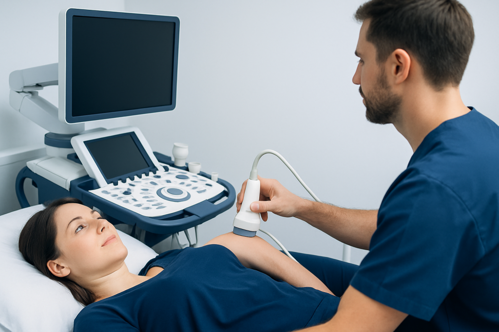
During the scan, a sonographer or radiologist places a handheld probe over the area of concern. The probe sends sound waves into the tissues and receives returning echoes, which create real-time images. Unlike X-rays or CT scans, ultrasound does not use ionising radiation.
For a superficial lump, ultrasound can help show whether it is mainly fatty, whether it sits above or below the muscle layer, and whether it has features that look typical or atypical. Results are considered alongside the person’s symptoms, medical history, and physical examination rather than in isolation.
If a lump is confirmed or strongly suspected to be a lipoma and is causing discomfort, limiting movement, or causing cosmetic concerns, a clinician may discuss lipoma removal as an option.
How a Lipoma Ultrasound Works and Who May Need One
Ultrasound works because different tissues reflect sound waves differently. Fatty tissue, fluid-filled cysts, muscle, and solid masses can produce different appearances on the image. Doppler ultrasound may also be used to assess blood flow within or around a lump, which can provide additional information.
A clinician may recommend an ultrasound when a lump cannot be confidently identified during an examination, when its size or depth is uncertain, or when it has changed. It is particularly useful for lumps close to the skin surface, including those on the trunk, shoulders, arms, thighs, neck, or forehead.
Not every suspected lipoma requires imaging. A small, stable, soft, mobile lump with a typical examination may be monitored clinically. However, ultrasound is commonly chosen when reassurance is needed or when the clinician wants to distinguish a possible lipoma from a cyst, enlarged lymph node, vascular lesion, or another soft-tissue mass.
People with a deep lump, a mass near a joint or nerve, multiple lumps, unexplained pain, or a personal history that raises concern for another condition may need more detailed assessment. In these situations, MRI, specialist review, or tissue sampling may be recommended after the initial scan.
Preparation and Step-by-Step Procedure

Most people do not need special preparation for a lipoma ultrasound. They can usually eat, drink, and take regular prescribed medicines as normal unless their healthcare team gives different instructions. Wearing loose clothing or clothing that makes the affected area easy to access can make the appointment more comfortable.
At the appointment, the person may be asked questions about when the lump appeared, whether it has grown, and whether it is painful or associated with skin changes. The examiner may first feel the area and compare it with nearby tissues. The person is then positioned so the site can be examined easily.
A clear water-based gel is applied to the skin. The ultrasound probe is moved gently over the lump and surrounding area while images are captured. Pressure is usually mild, though an already tender lump may be uncomfortable when touched. The scan itself commonly takes only a short time, depending on the lump’s location and complexity.
Afterward, the gel is wiped away and normal activities can be resumed immediately. A radiologist may review the images before a report is sent to the referring clinician. Some centres discuss preliminary findings on the day, while others arrange a follow-up conversation once the report is complete.
How Accurate Is Ultrasound for Lipoma Diagnosis?
Ultrasound is a useful first-line test for many superficial soft-tissue lumps and can often identify a lesion with features typical of a lipoma. A typical lipoma may appear as a well-defined, elongated or oval mass within the subcutaneous fat layer, often with an appearance similar to surrounding fatty tissue and little internal blood flow.
However, no imaging test should be treated as a complete substitute for clinical judgement. Lipomas can vary in appearance depending on their depth, internal fibrous tissue, location, and size. Some lesions that are not lipomas can also look partly fatty or resemble a benign lump on ultrasound.
Accuracy is generally strongest when the lump is superficial and has classic imaging and examination features. Ultrasound may be less definitive for deep masses, very large masses, lesions in anatomically complex areas, or lumps with mixed or unusual internal features. MRI gives more detailed soft-tissue information and may be the preferred next test in those settings.
If the ultrasound report is indeterminate or identifies atypical features, the clinician may recommend further imaging, referral to a surgical or musculoskeletal specialist, or biopsy. This does not mean that a serious diagnosis is likely; it means the lump needs a more certain assessment before a management decision is made.
What Is the Next Step After a Lipoma Ultrasound?
The next step after a lipoma ultrasound depends on the scan findings, symptoms, and examination. When the lump has typical benign features and is not troublesome, the clinician may recommend observation. People may be advised to note whether it changes in size, texture, mobility, or tenderness.
If the finding is consistent with a lipoma but it is painful, repeatedly irritated, affecting movement, pressing on nearby structures, or causing cosmetic concern, a surgical consultation can help discuss treatment choices. Complete excision is commonly considered when removal is appropriate, and the removed tissue may be examined in a laboratory to confirm the diagnosis.
If a scan does not clearly show a typical lipoma, the next step may be MRI or, less often, image-guided biopsy. These tests help characterize the tissue more fully. The choice depends on the lesion’s location, size, depth, and any features that need clarification.
Acibadem International’s multidisciplinary specialists and JCI-accredited hospitals assess soft-tissue lumps and coordinate imaging, pathology, and surgical care for international patients when needed.
What Are the Concerning Features of a Lipoma?
Most lipomas are harmless, but a new or changing lump deserves medical assessment. Features that may prompt more timely investigation include rapid or continuing growth, persistent pain, a firm or fixed feel, a lump that seems deep rather than just under the skin, or a mass that affects movement or sensation.
A lump that is large, irregular, difficult to move, associated with weakness or numbness, or accompanied by changes in the overlying skin should also be reviewed. These features do not diagnose cancer and often have non-serious explanations, but they should not be assumed to be due to a simple lipoma without assessment.
On imaging, features such as an unclear boundary, marked internal blood flow, mixed tissue components, thick internal partitions, or extension into deeper structures may lead the radiologist to recommend MRI or specialist input. The significance of any one feature depends on the full clinical and imaging context.
People should avoid repeatedly squeezing, puncturing, or attempting to drain a lump at home. A lipoma is not a fluid-filled spot, and home procedures can cause infection, bleeding, scarring, and delay an accurate diagnosis.
What Can Be Mistaken as a Lipoma on Ultrasound?
Several benign conditions can resemble a lipoma before or during an ultrasound assessment. Epidermoid cysts, for example, are common lumps under the skin and may feel mobile. They usually contain keratin rather than fat and can become inflamed or infected, which may make them painful or red.
Other possibilities include enlarged lymph nodes, benign nerve-related growths, fibrous masses, vascular malformations, haematomas after an injury, and hernias in certain body areas. A deeper fatty mass may also need to be distinguished from other lipomatous tumours, which is one reason imaging reports sometimes recommend MRI.
The ultrasound appearance, location, physical examination, and symptoms all help narrow the possibilities. If uncertainty remains, a clinician may arrange additional tests rather than making an assumption from a single image. This careful approach is intended to ensure that the management plan matches the diagnosis.
In some cases, a lump may be part of a different skin or soft-tissue condition rather than a lipoma. A qualified doctor can explain what the report means in the context of the individual’s health and whether monitoring or further assessment is sensible.
When to Seek Medical Care
Medical care should be sought for any new lump that persists, enlarges, or causes concern. It is especially important to arrange an appointment if a lump grows quickly, becomes painful, feels hard or fixed, lies deep in the tissue, measures noticeably larger over time, or is associated with numbness, weakness, or reduced movement.
Urgent assessment is appropriate if there are signs of infection around a lump, such as increasing redness, warmth, swelling, drainage, fever, or severe pain. A lump that appears after an injury but does not improve should also be evaluated, as it may not simply be a bruise.
For a known lipoma, regular self-awareness is usually enough when it remains stable and symptom-free. If its character changes, a clinician can reassess it and decide whether repeat ultrasound or a different test is needed. Early review can provide reassurance and support appropriate care.
After removal, most people return home the same day and follow wound-care guidance from their surgical team. Recovery varies with the location and size of the lipoma, but temporary soreness, bruising, and a small scar are possible. The main benefits of removal are relief of symptoms and diagnostic certainty; risks can include bleeding, infection, scarring, numbness, and recurrence, although recurrence is uncommon after complete excision.
Frequently asked questions
Is a lipoma ultrasound painful?
A lipoma ultrasound is usually painless. The probe is moved over gel on the skin, and only light pressure is normally needed. If the lump is already sore, pressing on the area may cause brief discomfort.
Do people need to fast before a lipoma ultrasound?
Fasting is not usually needed for an ultrasound of a superficial lipoma. People can generally eat, drink, and take regular medicines as usual. The imaging centre should be contacted in advance if there are any specific instructions.
Can ultrasound tell if a lipoma is cancerous?
Ultrasound can identify features that appear typical or atypical, but it cannot confirm every diagnosis on its own. If a mass has unclear or concerning features, a clinician may recommend MRI, biopsy, or specialist assessment. Most soft, superficial lipomas are benign.
What is the next step after a lipoma ultrasound?
If the scan shows a typical, symptom-free lipoma, observation may be all that is needed. If the lump is painful, enlarging, cosmetically bothersome, or uncertain on imaging, the next step may be specialist review, MRI, biopsy, or removal. The referring clinician should explain the report and the most appropriate plan.
How accurate is ultrasound for lipoma diagnosis?
Ultrasound is helpful for evaluating many superficial lumps and can often support a diagnosis of lipoma when the appearance is typical. Its accuracy is lower for deep, large, or unusual masses, and other conditions can occasionally look similar. MRI may be used when ultrasound does not provide enough certainty.
What are the concerning features of a lipoma?
Features needing assessment include rapid growth, persistent pain, a hard or fixed lump, a deep location, irregular shape, numbness, weakness, or reduced movement. These signs do not necessarily indicate a serious condition, but they warrant medical review. Any new or changing lump should be checked by a qualified clinician.
References
- American College of Radiology
- Radiological Society of North America
- National Health Service
- Mayo Clinic
- Merck Manual Consumer Version
This article is for general information only and is not a substitute for professional medical advice. Please consult a qualified doctor about your individual situation.
Ultrasound in Turkey — costs, top hospitals & a free quote
JCI-accredited · board-certified surgeons · reply within 24h
Add us as a Preferred Source to see more of our trusted health content across Google Search, AI Overviews and Discover.

Check-up & Preventive Medicine
Comprehensive health screenings designed for early detection and prevention.
2 specialists in this unit