Mass on Liver CT Scan: Preparation, Procedure and Results
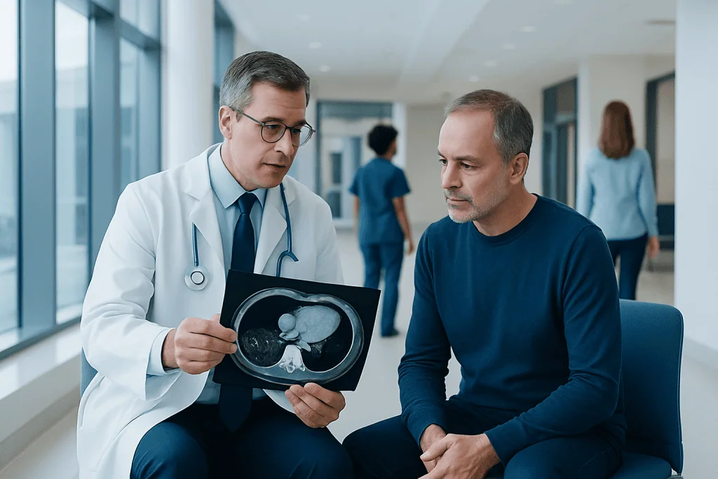
A liver mass seen on CT is a finding, not a diagnosis; many lesions are benign. Contrast-enhanced CT can often characterize common liver lesions by how they take up contrast over time.
Key Takeaways
- A liver mass seen on CT is a finding, not a diagnosis; many lesions are benign.
- Contrast-enhanced CT can often characterize common liver lesions by how they take up contrast over time.
- Preparation may include fasting and reviewing kidney function, allergies and medicines before contrast is used.
- A CT-guided liver biopsy is considered when imaging cannot provide a confident diagnosis or when tissue results will change care.
- Urgent assessment is important for severe abdominal pain, jaundice, fever, fainting or unexplained weight loss.
A mass on liver CT scan is an area that looks different from surrounding liver tissue. Many liver masses are noncancerous, but the scan findings, medical history, blood tests and, in selected cases, a biopsy are used to determine what the finding means.
Overview: What Does a Mass on Liver CT Scan Mean?
A mass on liver CT scan means that computed tomography has identified a spot, lump or area in the liver that has a different appearance from the surrounding tissue. It may be found while investigating symptoms, monitoring an existing condition or incidentally during a scan performed for another reason. A CT finding alone does not determine whether the mass is harmless or serious.
Common noncancerous findings include simple cysts, haemangiomas (clusters of blood vessels), focal nodular hyperplasia and fatty changes. Other possibilities include infections, inflammatory conditions, primary liver cancer or cancer that has spread to the liver from another part of the body. The significance depends on the lesion’s size, shape, enhancement pattern after contrast, number of lesions, liver health and the person’s medical history.
Radiologists assess the scan carefully and provide a report for the clinician who requested it. Further evaluation may include a multiphase CT scan, MRI, ultrasound, blood tests or tissue sampling. This structured approach helps avoid unnecessary procedures while ensuring that findings needing attention are investigated appropriately.
How a Liver CT Scan Works and Who May Need One
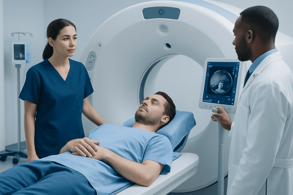
A CT scan uses rotating X-ray equipment and computer processing to create detailed cross-sectional images of the liver and nearby organs. For many liver assessments, a contrast agent containing iodine is injected into a vein. Images are taken at specific stages as contrast travels through the bloodstream, because different liver lesions show characteristic patterns of contrast uptake and washout.
A liver CT may be recommended after an abnormal ultrasound or blood test, to investigate abdominal symptoms, to assess known liver disease, or to monitor a previously identified lesion. It may also be used to evaluate injury, infection or possible spread of a known cancer. The test is not routinely necessary for every person with mild, nonspecific abdominal symptoms.
Before scheduling contrast CT, the care team should know about previous reactions to contrast, kidney disease, pregnancy or possible pregnancy, diabetes medicines and all prescribed or over-the-counter medicines. In some situations, ultrasound or MRI may be preferred, especially when avoiding radiation or iodinated contrast is important.
Preparation and Step-by-Step CT Procedure
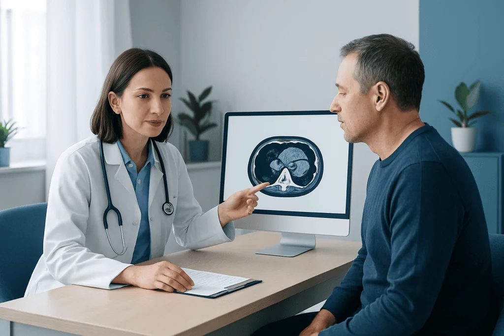
Preparation instructions vary by facility and the reason for the scan. Many patients are asked not to eat for several hours before a contrast-enhanced liver CT, although they may usually drink clear fluids unless told otherwise. They should follow the imaging department’s instructions rather than stopping food, fluids or medicines on their own.
What should you not do before a liver scan? Patients should not ignore instructions about fasting, fail to report a prior contrast reaction, or take unapproved supplements or medicines without discussing them with the care team. They should also avoid arriving dehydrated if fluids are permitted, because good hydration can support kidney clearance of contrast. Those taking diabetes medication, blood thinners or other regular medicines should ask for individualized guidance.
At the appointment, the patient may change into a gown and remove metal items from the scan area. A radiographer places an intravenous line if contrast is needed. The patient lies on a table that moves through the CT scanner; the procedure is painless, and staff may ask for brief breath-holds to reduce motion blur. Contrast can cause a short-lived warm sensation, metallic taste or feeling of needing to urinate, which usually passes quickly.
The imaging itself commonly takes only minutes, although the overall visit may be longer for registration, preparation and observation. The radiologist then reviews the images, sometimes alongside older scans, to decide whether the finding has features typical of a benign lesion or needs further assessment.
Can You Tell if a Liver Lesion Is Cancerous From a CT Scan?
Can you tell if a liver lesion is cancerous from a CT scan? CT can often strongly suggest the nature of a liver lesion, particularly when multiphase contrast images show a classic pattern. For example, simple cysts and many haemangiomas can often be identified confidently by imaging. In people with cirrhosis or chronic hepatitis, certain imaging patterns may also support a diagnosis of hepatocellular carcinoma without a biopsy when accepted clinical criteria are met.
However, CT cannot always confirm whether a lesion is cancerous. Small lesions, atypical appearances, fatty liver changes, prior treatment effects and certain benign or malignant tumours may overlap on imaging. MRI with liver-specific contrast, contrast-enhanced ultrasound, repeat imaging after an interval, blood tests and multidisciplinary review can provide additional information.
If uncertainty remains and the result would affect management, a clinician may recommend CT-guided biopsy. During this procedure, imaging helps guide a needle into the target area so a pathologist can examine tissue. A biopsy is not automatically required for every liver lesion because it has risks and may not add useful information when imaging findings are already clear.
How Serious Is It to Have a Mass on Your Liver?
How serious is it to have a mass on your liver? The answer varies widely. A small simple cyst or typical haemangioma may require no treatment and cause no health problems. Some lesions only need follow-up imaging to confirm stability. Other masses need prompt assessment because they may be related to liver disease, infection or cancer.
Factors that influence concern include whether the person has cirrhosis, chronic hepatitis B or C, a previous or current cancer, unexplained weight loss, abnormal liver blood tests or symptoms such as jaundice. The lesion’s appearance, growth over time and whether there is one lesion or several are also important. A healthcare professional should interpret these details in context rather than relying on the word “mass” alone.
When cancer is diagnosed, treatment is individualized according to the type of tumour, its location and extent, liver function and overall health. Options may include surgery, local image-guided therapy, radiation-based treatments, systemic medicines or transplantation evaluation in appropriate circumstances. Related conditions such as liver cancer are best assessed by a team that includes hepatology, gastroenterology, radiology, pathology, surgery and oncology specialists.
CT-Guided Liver Biopsy: Benefits, Risks and Recovery
A liver biopsy may be recommended when imaging does not provide a reliable answer, when infection or inflammatory disease is suspected, or when tumour tissue is needed to guide treatment. CT guidance enables the radiologist to plan a safe needle path and confirm that the sample is taken from the intended area. Ultrasound guidance may be used instead when the lesion is well seen on ultrasound.
How painful is a CT-guided liver biopsy? The skin and deeper tissues are numbed with local anaesthetic, so most people feel pressure, brief stinging during numbing and a short sensation as the biopsy needle is used rather than severe pain. Some people receive medication to help them relax if appropriate. They should tell the team immediately if they feel significant pain, dizziness or discomfort during the procedure.
After the biopsy, patients are observed for several hours while staff check for pain, bleeding or changes in blood pressure. Mild soreness at the needle site or referred discomfort in the right shoulder can occur and usually improves within a day or two. Patients typically need someone to take them home and may be advised to avoid strenuous activity and heavy lifting for a short period.
Bleeding is the main uncommon but important risk, along with infection, injury to nearby structures, bile leakage and an inconclusive sample. The team checks platelet counts, blood clotting and medicines such as anticoagulants before the procedure to reduce risk. Patients should follow all aftercare directions and contact the team promptly if concerning symptoms develop.
Results, Follow-Up and When to Seek Medical Care
CT results may be available quickly, but the final report and care plan can take longer, particularly if prior images must be compared or further tests are arranged. A report may describe the lesion’s location, size, density and contrast behaviour, then recommend no follow-up, repeat imaging, MRI, specialist review or biopsy. Patients may find it helpful to ask what the lesion is most likely to represent, whether it requires monitoring and what symptoms should prompt earlier review.
When to seek medical care: Medical assessment should be sought promptly for yellowing of the skin or eyes, severe or worsening upper abdominal pain, fever with abdominal symptoms, persistent vomiting, confusion, fainting, black stools, vomiting blood, or new abdominal swelling. Following a biopsy, urgent advice is needed for worsening pain, fever, dizziness, shortness of breath, bleeding or signs of an allergic reaction.
Most incidental liver lesions do not result from anything a person has done and cannot be prevented. Supporting liver health remains worthwhile: limiting alcohol, maintaining a weight suitable for the individual, managing diabetes and cholesterol where relevant, avoiding unnecessary herbal or medication products that may affect the liver, and discussing hepatitis vaccination or testing with a clinician when indicated.
For international patients, Acibadem International’s multidisciplinary specialists and JCI-accredited hospitals can evaluate liver findings with coordinated imaging, pathology and treatment planning. The appropriate next step should always be decided with a qualified clinician who has access to the full scan report and medical history.
Frequently asked questions
Is a mass on the liver usually cancer?
No. Many liver masses are benign, including cysts and haemangiomas, particularly when they are found incidentally in people without chronic liver disease. The likelihood of cancer depends on imaging features, liver health, prior cancer history and other clinical information.
What happens if a liver lesion is found incidentally on CT?
The clinician reviews the CT report, previous scans and medical history to determine whether the lesion has a typical harmless appearance. If it is indeterminate, MRI, ultrasound, follow-up CT or occasionally a biopsy may be recommended.
Do all liver masses need a biopsy?
No. A biopsy is not needed when imaging clearly identifies a benign lesion or when accepted imaging criteria establish a diagnosis. It is generally considered when tissue information is necessary to clarify the diagnosis or guide treatment.
Can a CT scan show the difference between a cyst and a tumour in the liver?
CT can often distinguish a simple cyst from a solid lesion because a cyst has fluid characteristics and does not enhance like tissue after contrast. Some lesions remain uncertain, and MRI or another test may then be used for clarification.
How long does it take to recover from a CT scan with contrast?
Most people can return to normal activities immediately after a CT scan, unless they received sedating medicine or were given different instructions. Drinking fluids may be advised when appropriate, and patients should seek advice if they develop symptoms of a delayed contrast reaction.
Should a person be worried while waiting for liver CT results?
Waiting can be stressful, but a liver finding does not by itself mean cancer or another serious illness. Asking when and how results will be communicated can help, and urgent symptoms such as jaundice, severe pain or fever should be assessed without waiting for a routine follow-up.
References
- American College of Radiology
- National Institute of Diabetes and Digestive and Kidney Diseases
- European Association for the Study of the Liver
- Radiological Society of North America
- World Health Organization
This article is for general information only and is not a substitute for professional medical advice. Please consult a qualified doctor about your individual situation.
Oncology care in Turkey — second opinion and treatment plan
JCI-accredited · board-certified surgeons · reply within 24h
Add us as a Preferred Source to see more of our trusted health content across Google Search, AI Overviews and Discover.