MR Angiography: Imaging Blood Vessels in the Brain, Neck, and Body

MR angiography uses magnetic fields and radio waves to visualize blood vessels in the brain, neck, chest, abdomen, pelvis, arms, and legs. Some MRA scans use a gadolinium-based contrast agent, while others can be performed without contrast depending on the clinical question.
Key Takeaways
- MR angiography uses magnetic fields and radio waves to visualize blood vessels in the brain, neck, chest, abdomen, pelvis, arms, and legs.
- Some MRA scans use a gadolinium-based contrast agent, while others can be performed without contrast depending on the clinical question.
- MRA can help detect narrowed arteries, aneurysms, vascular malformations, blood clots, and blood vessel injury or inflammation.
- Patients should tell the imaging team about implants, kidney disease, pregnancy, allergies, or claustrophobia before the scan.
- Results are interpreted by a radiologist and shared with the referring doctor to guide diagnosis, monitoring, or treatment planning.
MR angiography, also called MRA, is a specialized MRI technique used to create detailed images of arteries and veins throughout the body. It helps doctors evaluate blood vessel narrowing, blockages, aneurysms, and other vascular conditions, often without using ionizing radiation.
Overview
MR angiography, commonly shortened to MRA, is a type of magnetic resonance imaging designed to show blood vessels. Instead of focusing mainly on organs or soft tissues, the scan highlights the flow of blood through arteries and veins. This allows doctors to examine vessels in the brain, neck, chest, abdomen, pelvis, arms, and legs without using X-rays.
MRA can be performed with or without an injected contrast agent. Contrast-enhanced MRA often uses a gadolinium-based agent to make vessels appear clearer on the images. Non-contrast MRA techniques use the natural movement of blood and specific MRI settings to create vascular images, which can be helpful for people who should avoid contrast.
The test is usually requested when a doctor needs more information about circulation, a suspected narrowing or blockage, an aneurysm, or an unusual blood vessel pattern. It may also be used for follow-up after vascular treatment or to help plan surgery or minimally invasive procedures. The best imaging method depends on the body area, the urgency of the condition, and the patient’s overall health.
What MR Angiography Can Show
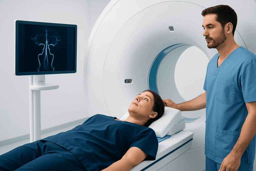
MR angiography can provide detailed information about the size, shape, and course of blood vessels. In the brain, it may help identify aneurysms, vessel narrowing, vascular malformations, or changes related to stroke evaluation. In the neck, MRA is often used to assess the carotid and vertebral arteries, which supply blood to the brain.
In the chest, abdomen, and pelvis, MRA can help evaluate the aorta and major branches, including concerns such as aneurysm, dissection, congenital vessel differences, or blood supply to organs. In the arms and legs, it may be used to assess peripheral artery disease, vessel injury, or circulation before certain treatments.
Doctors may recommend MRA for a range of clinical questions, including:
- Suspected narrowing or blockage of an artery
- Possible aneurysm or abnormal bulging of a vessel wall
- Evaluation of blood vessels after surgery, stenting, or other treatment
- Assessment of vascular malformations or unusual vessel connections
- Investigation of blood vessel inflammation or injury
- Planning for selected surgical or endovascular procedures
How the Scan Is Performed

During an MRA scan, the patient lies on a movable table that slides into the MRI scanner. A special coil, which helps receive the MRI signal, may be placed around the head, neck, chest, abdomen, or limb being examined. The scanner makes loud tapping or thumping sounds while images are being acquired, so ear protection is provided.
The patient needs to remain as still as possible because movement can blur the images. Some scans require brief breath-holding, particularly when imaging blood vessels in the chest or abdomen. A technologist monitors the patient throughout the examination and can communicate through an intercom.
If contrast is needed, an intravenous line is placed in a vein, usually in the arm or hand. The contrast agent is injected during part of the scan, and images are taken as it passes through the blood vessels. Many people feel no unusual sensation, although some may notice a cool feeling at the injection site or a brief metallic taste.
The total appointment time varies depending on the area being examined and whether contrast is used. The imaging portion may take several minutes to over half an hour, and additional time is needed for preparation and safety screening. After the scan, most patients can return to normal activities unless they have been given a sedative.
Preparation and Safety
Before MRA, patients complete a safety questionnaire because MRI uses a strong magnetic field. The imaging team needs to know about pacemakers, defibrillators, cochlear implants, aneurysm clips, implanted pumps, neurostimulators, metal fragments, or any prior surgery involving metal devices. Many modern implants are MRI-conditional, but they must be checked carefully before scanning.
Patients should also tell the team about kidney disease, dialysis, pregnancy, breastfeeding, previous reactions to contrast agents, or severe allergies. Gadolinium-based contrast agents are generally well tolerated, but kidney function may be assessed before contrast is used in people at risk of kidney problems. The radiology team will decide whether contrast is appropriate and which protocol is safest.
Metal objects must be removed before entering the MRI room, including jewelry, watches, hairpins, hearing aids, removable dental work, and some clothing items with metal fasteners. People who feel anxious in enclosed spaces should mention this when scheduling the scan. Practical options may include reassurance, music, a wider-bore scanner when available, or medication prescribed by a doctor if needed.
Benefits and Limitations
One of the main benefits of MR angiography is that it can show blood vessels without ionizing radiation. This is different from CT angiography and conventional X-ray angiography, which use X-rays. MRA also provides excellent soft tissue detail, allowing doctors to evaluate vessels in relation to nearby structures.
MRA is noninvasive in most cases and does not require a catheter placed into an artery. For many patients, this makes it a useful diagnostic or follow-up test. Non-contrast options are also valuable for selected patients, especially when contrast is not preferred.
However, MRA is not ideal for every situation. Very small vessels, heavily calcified arteries, certain stents or metal implants, and patient movement can affect image quality. In urgent cases, CT angiography may be faster and more widely available, while catheter angiography may still be needed when treatment is planned during the same procedure.
Because each imaging test has strengths and limitations, doctors choose the method that best answers the clinical question. The decision may depend on symptoms, kidney function, implant safety, the body region being studied, and whether immediate treatment might be required.
Understanding the Results
After the scan, a radiologist reviews the images and prepares a report for the referring doctor. The report may describe whether blood vessels are normal in size and shape, whether there is narrowing or blockage, and whether aneurysms, clots, malformations, dissections, or other abnormalities are present. It may also compare the findings with previous imaging if available.
Results are interpreted together with the patient’s symptoms, physical examination, medical history, and other tests. A finding on MRA does not always mean treatment is needed right away. Some vascular changes are monitored over time, while others may require medication, lifestyle management, further imaging, or referral to a specialist.
Patients are encouraged to discuss the report with their doctor rather than trying to interpret the images alone. Helpful questions include what the finding means, whether further tests are needed, what treatment options exist, and when follow-up should occur. Clear communication helps ensure that the scan results are used appropriately in the overall care plan.
When to See a Doctor
A doctor may consider MR angiography when symptoms suggest a possible blood vessel problem. These symptoms can vary depending on the body area and may include recurrent neurological symptoms, severe or unusual headaches, dizziness related to blood flow concerns, limb pain with walking, unexplained swelling, or signs of reduced circulation. MRA may also be recommended when previous tests suggest a vascular abnormality.
People with known vascular conditions, such as aneurysm, carotid artery disease, peripheral artery disease, aortic disease, or vascular malformation, may need imaging follow-up at intervals determined by their specialist. The timing depends on the size and location of the finding, symptoms, and treatment history.
Anyone scheduled for MRA should tell the healthcare team about new symptoms, implanted devices, kidney problems, pregnancy, or concerns about lying still in the scanner. In appropriate cases, multidisciplinary specialists and JCI-accredited hospitals at Acibadem International diagnose and treat vascular conditions for international patients, coordinating imaging results with individualized medical care.
Frequently asked questions
Is MR angiography the same as a regular MRI?
MR angiography is a specialized type of MRI focused on blood vessels. It uses MRI technology but applies specific imaging techniques to highlight arteries or veins and blood flow. A regular MRI may show organs, joints, the brain, or soft tissues in more general detail.
Does MR angiography use radiation?
No, MR angiography does not use ionizing radiation. It relies on a strong magnetic field and radio waves to create images. This is one reason it may be preferred for certain patients or for follow-up imaging when appropriate.
Will contrast be needed for an MRA scan?
Some MRA scans use a gadolinium-based contrast agent, while others can be done without contrast. The choice depends on the body area, the medical question, image quality needs, and the patient’s kidney function or other health factors. The radiology team will explain the plan before the scan.
Is gadolinium contrast safe?
Gadolinium-based contrast agents are widely used and are generally well tolerated. The imaging team checks for risk factors, especially significant kidney disease or previous contrast reactions, before giving contrast. Patients should always share their medical history so the safest imaging approach can be chosen.
How long does MR angiography take?
The imaging time varies by the area being examined and whether contrast is used. Many scans take from several minutes to over half an hour, with additional time for preparation, screening, and positioning. The scheduling team can provide a more specific estimate for the planned examination.
Can patients with implants have MR angiography?
Many patients with implants can have MRI or MRA, but this must be checked carefully. Some devices are MRI-safe or MRI-conditional, while others may not be compatible. Patients should bring implant cards or device information whenever possible.
What happens after the MRA scan?
A radiologist reviews the images and sends a report to the referring doctor. The doctor then explains the findings in the context of symptoms and medical history. Depending on the results, the next step may be reassurance, follow-up imaging, medication, specialist referral, or treatment planning.
This article is for general information only and is not a substitute for professional medical advice. Please consult a qualified doctor about your individual situation.
Explore treatments in Turkey — costs, top hospitals & a free quote
JCI-accredited · board-certified surgeons · reply within 24h
Add us as a Preferred Source to see more of our trusted health content across Google Search, AI Overviews and Discover.

More from the Health Library

Diagnostic Medical Ultrasound: How It Works and What It Can Show

Diagnostic Criteria for PTSD: How F43.1 Is Diagnosed

Common Reasons Doctors Order Lab Tests: Screening, Diagnosis, and Monitoring

Neuroradiology Tests Explained: MRI, CT, and Angiography for Brain Disorders

Urine Test Results Explained: Infection, Protein, Blood, and Other Common Findings