MRI Adrenal Gland: Preparation, Procedure and Results
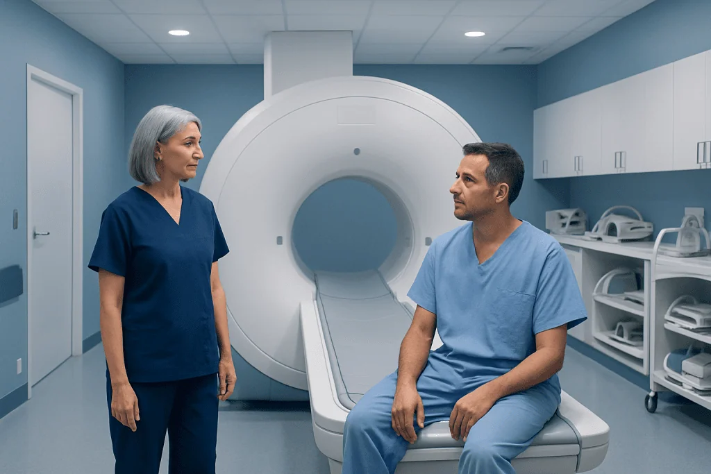
Adrenal MRI uses magnetic fields and radio waves, not X-rays or radiation. It can help distinguish many benign adrenal lesions from lesions that need further assessment.
Key Takeaways
- Adrenal MRI uses magnetic fields and radio waves, not X-rays or radiation.
- It can help distinguish many benign adrenal lesions from lesions that need further assessment.
- Contrast is not required for every examination; the choice depends on the clinical question and prior imaging.
- MRI findings are interpreted alongside hormone blood or urine tests, symptoms and medical history.
- Most people return to normal activities immediately after the scan, unless sedation has been used.
An MRI adrenal gland examination is a noninvasive scan that produces detailed images of the small hormone-producing glands above the kidneys. It is commonly used to evaluate an adrenal nodule or mass, clarify findings from another scan, and support treatment planning without using ionizing radiation.
Overview: What Is an MRI Adrenal Gland Scan?
An MRI adrenal gland scan is a magnetic resonance imaging examination focused on the adrenal glands. These two small glands sit above the kidneys and produce hormones involved in blood pressure, stress responses, salt balance and metabolism. MRI creates detailed pictures of their size, shape and internal tissue characteristics without exposing the person to ionizing radiation.
Doctors may request an adrenal MRI after an ultrasound, CT scan or other test identifies an adrenal nodule, also called an adrenal incidentaloma. The scan may help determine whether a lesion has features commonly associated with a benign adenoma, cyst, bleeding or another condition. It may also be used when a person has symptoms or laboratory findings suggesting excess adrenal hormone production.
Importantly, imaging is only one part of adrenal assessment. A radiologist’s report is considered together with medical history, physical examination and, when appropriate, blood or urine tests for adrenal hormones. This combined approach helps clinicians decide whether observation, additional testing or treatment is appropriate.
How Adrenal MRI Works and Who May Need It
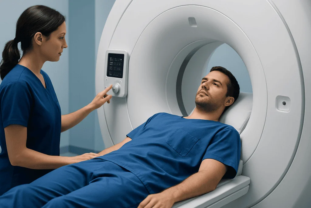
MRI uses a powerful magnet and radiofrequency waves to generate images of the body. For adrenal imaging, the examination often includes sequences obtained while the person holds their breath briefly. Special chemical-shift sequences compare the amount of fat within an adrenal lesion, which can help identify many lipid-rich benign adenomas.
An MRI adrenal protocol is tailored to the reason for the scan. The radiology team may use several image sequences before and after contrast, when contrast is appropriate. This individualized MRI adrenal gland planning helps provide useful images while avoiding unnecessary steps.
Doctors may recommend adrenal MRI for an indeterminate adrenal mass seen on CT, an adrenal lesion in a younger person, a lesion requiring radiation-free follow-up, or suspected bleeding or cystic change. MRI can also be useful when CT contrast is unsuitable. People with certain implanted devices, metallic fragments or severe claustrophobia may need additional screening or an alternative imaging approach.
- Known or newly found adrenal nodule or mass
- Possible hormone-producing adrenal lesion
- Unclear result on ultrasound or CT
- Need for follow-up imaging without ionizing radiation
- Assessment of tissue characteristics before specialist management
MRI Adrenal Gland Preparation and Step-by-Step Procedure

MRI adrenal gland preparation usually begins with a safety questionnaire. Patients should tell the imaging team about pacemakers, neurostimulators, cochlear implants, surgical clips, artificial joints, metal fragments, kidney disease, pregnancy, allergies and previous reactions to MRI contrast. Many modern implants are MRI-compatible under specific conditions, but this must be checked in advance.
Instructions vary by facility and by whether contrast is planned. Some people may be asked not to eat for a few hours beforehand, particularly if contrast or sedation is expected. Regular medicines should not be stopped unless the prescribing clinician gives specific instructions. Comfortable clothing without metal is helpful, and jewelry, watches, hearing aids, removable dental appliances and other metal items must be removed before entering the scan room.
During the examination, the person lies on a movable table that slides into the MRI scanner. A coil is positioned around the abdomen to receive images. The machine makes repeated knocking or humming sounds, and ear protection is provided. The person should remain still and may be asked to hold their breath for short periods. An adrenal MRI commonly takes about 30 to 60 minutes, depending on the protocol and whether contrast is used.
If contrast is needed, a gadolinium-based contrast agent is injected through a small intravenous line during part of the scan. The radiographer can communicate throughout the procedure, and an alarm device is usually available for the patient to use if they need assistance. An MRI technologist performs the scan, while a radiologist interprets the images.
Should I Get an MRI of My Adrenal Gland With or Without Contrast?
Whether an adrenal MRI should be performed with or without contrast depends on the clinical question, the appearance of a lesion on prior imaging and the person’s health history. Many adrenal lesions can be evaluated with non-contrast MRI sequences, especially chemical-shift imaging, which may identify features typical of a benign adenoma.
Contrast-enhanced images can provide additional information about blood flow and enhancement patterns within a lesion. They may be useful when a finding remains unclear, when there is concern for a more complex mass, or when clinicians need more detail for treatment planning. However, contrast does not automatically need to be used for every adrenal MRI.
Gadolinium contrast agents are generally well tolerated, but the care team reviews kidney function and prior contrast reactions before use. People with severe kidney impairment require individualized assessment. The referring doctor and radiologist can determine the most appropriate MRI adrenal protocol based on the available information.
What Are the First Signs of Adrenal Problems?
Adrenal problems can cause different symptoms depending on whether the glands produce too much or too little hormone, and some adrenal masses cause no symptoms at all. Early signs are often nonspecific and can overlap with many common health conditions, so symptoms alone cannot confirm an adrenal disorder.
Possible signs of excess cortisol include unexplained weight gain around the trunk, easy bruising, muscle weakness, high blood pressure, raised blood sugar, mood changes or irregular menstrual periods. Excess aldosterone may contribute to difficult-to-control high blood pressure and low potassium. Excess adrenaline-type hormones may cause episodes of palpitations, sweating, headache, tremor or sudden blood pressure elevation.
Low adrenal hormone production may be associated with persistent fatigue, weakness, nausea, abdominal discomfort, dizziness when standing, low blood pressure, salt craving or skin darkening in some conditions. A clinician may arrange blood tests and other investigations when symptoms, examination findings or family history raise concern. For broader information about hormone-related conditions, clinicians may also consider assessment for adrenal gland diseases.
What Are the First Signs of an Adrenal Tumor?
Most adrenal tumors are discovered incidentally during imaging performed for another reason. Many are benign and do not produce hormones, meaning there may be no noticeable signs. When symptoms do occur, they are more often related to hormone overproduction than to the tumor itself.
Depending on the hormone involved, possible symptoms include persistent or episodic high blood pressure, recurrent headaches, palpitations, sweating, unexplained low potassium, muscle weakness, changes in body fat distribution, easy bruising, new diabetes or changes in menstrual patterns. Rarely, a larger mass may cause a feeling of fullness or discomfort in the side or upper abdomen, although this is not specific to an adrenal tumor.
These symptoms have many possible explanations, including common medical conditions. Prompt but calm assessment by a qualified clinician is appropriate when symptoms are recurrent, severe, associated with significant blood pressure changes, or accompanied by an adrenal mass found on imaging. Hormone testing helps determine whether a lesion is functioning, while imaging helps assess its anatomy.
Will an MRI Rule Out if an Adrenal Tumor Is Cancerous?
No imaging test, including MRI, can always rule out cancer with complete certainty. MRI can identify imaging features that make a benign lesion more likely and can also show characteristics that require closer evaluation, such as an irregular appearance, internal complexity, local invasion or growth over time. These findings help guide the next steps but do not replace clinical judgment.
Radiologists assess the lesion’s size, margins, signal characteristics and behavior on different MRI sequences. They also compare the scan with previous imaging whenever available. Hormone tests, the patient’s history, the presence of cancer elsewhere in the body and follow-up imaging may all influence the interpretation and management plan.
If a lesion remains uncertain or has concerning features, the care team may recommend consultation with endocrinology, endocrine surgery or oncology. Biopsy is not routinely used for every adrenal mass and may be unsafe in some circumstances, particularly until a hormone-producing pheochromocytoma has been excluded. Decisions about surgery or surveillance should be made by the treating specialists.
Results, Recovery, Benefits and When to Seek Medical Care
After an MRI, most people can eat, drink, drive and return to usual activities immediately. If sedative medication was used for anxiety or claustrophobia, someone else should accompany the patient home and driving should be avoided for the period advised by the care team. The radiologist prepares a report for the referring clinician, who explains what the findings mean in the context of other test results.
The benefits of adrenal MRI include detailed soft-tissue imaging, no ionizing radiation and the ability to characterize many adrenal lesions. The main limitations are that scans can be noisy, require lying still and may be difficult for people with severe claustrophobia. Rare risks include contrast reactions or concerns related to certain implants; screening before the examination is designed to reduce these risks.
Medical care should be sought promptly for severe or recurring episodes of headache, palpitations, sweating and markedly elevated blood pressure; fainting; severe weakness; persistent vomiting; or symptoms of very low blood pressure. A person should also contact their doctor after an MRI if they develop signs of an allergic reaction after contrast, such as widespread rash, swelling or breathing difficulty.
For international patients requiring coordinated evaluation, Acibadem International’s multidisciplinary specialists and JCI-accredited hospitals can assess adrenal findings and discuss appropriate diagnostic and treatment pathways.
Frequently asked questions
How long does an MRI adrenal gland scan take?
The scan commonly takes around 30 to 60 minutes. The exact duration depends on the imaging protocol, whether contrast is needed and whether additional sequences are required. Patients should allow extra time for registration and safety screening.
Do I need to fast before an adrenal MRI?
Some imaging centers request fasting for a few hours, especially when contrast or sedation is planned, while others do not. The patient should follow the instructions provided by the imaging facility. Regular medications should usually be continued unless a clinician advises otherwise.
Is MRI better than CT for adrenal glands?
MRI and CT provide different types of useful information. CT is often the first scan that detects an adrenal mass, while MRI may further characterize lesions or avoid radiation during follow-up. The best test depends on the clinical question, prior results and individual health factors.
Can an adrenal MRI show a hormone-producing tumor?
MRI can show the size and features of a lesion, but it cannot reliably determine hormone production on its own. Blood and urine tests are needed to assess cortisol, aldosterone, catecholamines or other hormones when clinically indicated. Imaging and hormone testing are interpreted together.
Is contrast MRI safe for adrenal imaging?
Gadolinium-based MRI contrast is generally well tolerated and is used only when it is likely to add helpful diagnostic information. The care team checks for kidney problems, previous contrast reactions and other relevant risks before administering it. Patients should report any past reactions or serious kidney disease.
What happens if an adrenal mass is found on MRI?
The next step depends on the scan appearance, size, growth history and whether hormone tests are abnormal. Many benign, nonfunctioning lesions can be monitored, while others need specialist review or treatment. A clinician will explain the recommended follow-up plan for the individual situation.
References
- American College of Radiology
- Endocrine Society
- European Society of Endocrinology
- Radiological Society of North America
- National Institute of Diabetes and Digestive and Kidney Diseases
This article is for general information only and is not a substitute for professional medical advice. Please consult a qualified doctor about your individual situation.
Medical weight-loss options in Turkey — costs & eligibility
JCI-accredited · board-certified surgeons · reply within 24h
Add us as a Preferred Source to see more of our trusted health content across Google Search, AI Overviews and Discover.
Check your numbers in seconds
BMI, calories, due date, blood pressure and 30+ more clinical calculators — free, instant, doctor-reviewed ranges.