MRI and Prostate Cancer: Preparation, Procedure and Results

A prostate MRI uses magnetic fields rather than ionizing radiation to create detailed images of the prostate. Multiparametric MRI combines several image types to assess suspicious areas and estimate their likelihood of clinically significant cancer.
Key Takeaways
- A prostate MRI uses magnetic fields rather than ionizing radiation to create detailed images of the prostate.
- Multiparametric MRI combines several image types to assess suspicious areas and estimate their likelihood of clinically significant cancer.
- MRI findings are commonly reported with a PI-RADS score, which helps guide next steps but is not a diagnosis by itself.
- Some people may avoid an immediate biopsy after a reassuring MRI, depending on PSA level, examination findings and individual risk.
- Preparation may include screening for metal implants, discussing kidney function and following local instructions about food, bowel preparation or contrast.
- New urinary symptoms, blood in urine, persistent bone pain or concerning test results should be assessed by a qualified clinician.
MRI and prostate cancer evaluation often involves a multiparametric MRI, a detailed scan that can show areas of the prostate that may need closer assessment. It does not diagnose cancer alone, but it can help clinicians decide whether a biopsy is needed and, if cancer is found, support treatment planning.
MRI and Prostate Cancer: What the Scan Can Show
MRI and prostate cancer assessment are closely linked because magnetic resonance imaging can provide detailed pictures of the prostate gland and nearby tissues. A prostate MRI is often requested after an elevated prostate-specific antigen (PSA) result, an abnormal digital rectal examination, a previous negative biopsy with continuing concern, or after a cancer diagnosis to help understand its local extent.
Most modern examinations are multiparametric MRI (mpMRI). This combines anatomical images with sequences that assess how water moves within tissue and how tissue takes up contrast material when contrast is used. Prostate cancers can have imaging features that differ from normal prostate tissue, although inflammation, benign enlargement and prior treatment can sometimes look similar.
The scan is an important decision-support tool, not a stand-alone cancer test. A normal or low-suspicion MRI reduces the chance of clinically significant cancer but does not entirely exclude it. Doctors interpret the scan alongside PSA trends, prostate size, family history, examination findings, age and overall health. For broader information on the condition, readers may review prostate cancer.
How a Prostate MRI Works and Who May Benefit
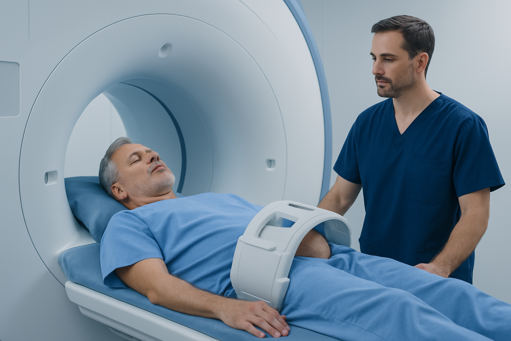
MRI uses a strong magnetic field and radio waves to create cross-sectional images. Unlike CT scans and X-rays, it does not use ionizing radiation. The examination is usually performed in a standard MRI scanner, with the person lying still on a padded table while images are taken. Modern prostate MRI is commonly performed without an internal coil, although the exact technique varies between imaging centers.
A prostate MRI may be especially helpful before a first biopsy, because it can identify a target for sampling and may help some men with low clinical risk avoid an unnecessary immediate biopsy. It can also be useful after a negative biopsy when PSA remains elevated, when planning active surveillance, and when assessing known cancer before treatment. It is one part of a wider urology and oncology assessment.
Not everyone can have MRI in the usual way. Certain implanted devices, metal fragments, some older aneurysm clips and severe claustrophobia may require special review. People with reduced kidney function, previous reactions to MRI contrast or a possible need for sedation should tell the care team in advance. The radiology department can determine whether MRI is suitable and whether contrast is appropriate.
Preparing for the Scan: What Should You Not Do Before a Prostate MRI?

Before a prostate MRI, a person should not assume that all centers use identical instructions. The imaging team’s guidance should take priority, since preparation may differ according to scanner protocols, contrast use and medical history. Patients should disclose all implants, previous surgery, allergies, kidney disease and medicines, including anticoagulants and diabetes medicines.
People are commonly asked not to bring metal objects into the MRI room. Watches, jewelry, hearing aids, removable dental devices, belts, bank cards and clothing with metallic components may need to be removed. It is also important not to conceal any metal exposure, including a possible workplace eye injury involving metal, as this can create a safety risk in the magnetic field.
Some centers ask patients to avoid a large meal shortly before the scan or to use a simple bowel preparation to reduce gas and motion in the rectum. Others may give different advice. Patients should not start laxatives, stop prescribed medication or fast for long periods unless their imaging team specifically instructs them to do so. If anxiety in confined spaces is a concern, discussing this before the appointment allows the team to consider supportive options.
- Bring previous PSA results, biopsy reports and earlier prostate imaging if available.
- Arrive early enough to complete MRI safety screening and change clothing if needed.
- Ask whether contrast is planned and whether recent kidney blood tests are required.
- Arrange transport if sedation has been prescribed for anxiety.
Step by Step: What Happens During and After a Prostate MRI
On arrival, the patient completes a safety questionnaire and changes into MRI-safe clothing if needed. A radiographer reviews the process, checks for implants or metal and may place an intravenous line if contrast is planned. The patient then lies on the scanning table, usually on the back, and a receiving device is positioned over the pelvis.
The table moves slowly into the scanner. The machine makes loud tapping or knocking sounds, so ear protection is provided. The patient can communicate with the radiographer throughout the scan and is asked to remain as still as possible. Image acquisition often takes around 30 to 45 minutes, though the total visit may be longer because of preparation and safety checks.
If gadolinium-based contrast is used, it is injected through the intravenous line during part of the examination. Most people feel no significant sensation, although a brief cool feeling at the injection site may occur. After the scan, the patient can generally return to normal activities and eat and drink as usual, unless sedation was used or the care team gives other instructions.
There is usually no recovery period after MRI itself. If contrast was given, normal hydration is generally encouraged unless a clinician has advised fluid restriction. If sedation was used, the person should follow discharge instructions, avoid driving and arrange for a responsible adult to accompany them home.
Understanding Results and Next Steps
A radiologist with prostate MRI expertise reviews the images and sends a report to the referring clinician. Reports often use the Prostate Imaging Reporting and Data System, known as PI-RADS, to describe the likelihood that a visible area represents clinically significant prostate cancer. Scores range from 1, very low likelihood, to 5, very high likelihood. The score is not a final diagnosis.
How long does it take to get results from an MRI for prostate cancer?
Timing varies by hospital, radiologist availability and whether the images need multidisciplinary review. Many patients receive results within several days to one or two weeks, but the referring urologist should confirm the local timeframe. If a result is needed for a planned consultation, patients can ask when and how they will be contacted.
Do you need a biopsy after an MRI for prostate cancer?
Not always. If MRI shows a suspicious area, a doctor may recommend a targeted biopsy, often combined with systematic sampling, to confirm whether cancer is present and determine its grade. If MRI is reassuring, some people may be monitored with PSA testing and clinical review rather than having an immediate biopsy. This choice depends on the full risk picture, because a negative MRI cannot rule out every cancer.
When biopsy is appropriate, MRI information can improve targeting of the area of concern. Options may include prostate biopsy techniques selected by the urology team according to the patient’s anatomy, risk factors and local practice.
Benefits, Limitations and Possible Risks
The main benefit of prostate MRI is detailed, noninvasive imaging that can help identify lesions more likely to need biopsy. It may help guide targeted sampling, reduce detection of some low-risk cancers that may never cause harm, and contribute to treatment planning. MRI can also show whether a known tumor may extend near the prostate capsule, seminal vesicles or adjacent structures, although it cannot determine every detail with certainty.
Its limitations are equally important. Small cancers and some clinically significant cancers may not be visible. Benign prostatic enlargement, infection, inflammation, bleeding after a recent biopsy and motion during the scan can affect image interpretation. Results should therefore be discussed with a urologist rather than viewed in isolation.
MRI is generally safe, but the strong magnet can be hazardous for people with incompatible metallic implants or fragments. Contrast reactions are uncommon but possible, and gadolinium contrast requires particular care in people with severe kidney impairment. Claustrophobia and difficulty remaining still can also make the examination challenging. The imaging team can explain practical adjustments and alternatives where appropriate.
When to worry about prostate MRI results?
A higher PI-RADS score, a lesion described as suspicious, or wording suggesting possible extension outside the prostate should prompt a timely conversation with the referring urologist. These terms do not confirm cancer, and even a high-suspicion MRI typically requires biopsy for diagnosis. Concern is also appropriate when a low-suspicion MRI does not fit with rapidly rising PSA, an abnormal examination or strong inherited risk; the doctor may recommend further assessment despite the scan.
When to Seek Medical Care
A person should arrange medical review for an elevated or rising PSA result, an abnormal prostate examination, or ongoing urinary symptoms such as difficulty starting urination, a weak stream, frequent urination, urgency or waking often at night. These symptoms are common and are frequently caused by noncancerous conditions, including benign prostate enlargement, but they deserve appropriate assessment.
Prompt medical advice is important for blood in the urine, inability to pass urine, fever with urinary symptoms, unexplained weight loss, persistent pain in the back, hips or pelvis, or new leg weakness. These symptoms have many possible causes and do not by themselves mean prostate cancer, but they should not be ignored.
People with a close relative affected by prostate cancer, particularly at a younger age, or those with known inherited cancer-related gene changes may benefit from discussing individualized screening and risk assessment earlier with a doctor. Acibadem International’s multidisciplinary specialists and JCI-accredited hospitals support international patients with prostate assessment, imaging and coordinated treatment planning, including prostate cancer treatment.
After any MRI result, the most useful next step is a follow-up discussion with the clinician who ordered it. They can explain what the report means in the context of the person’s PSA, symptoms, medical history and preferences, and can recommend surveillance, biopsy or other care when appropriate.
Frequently asked questions
Is MRI accurate for detecting prostate cancer?
A multiparametric prostate MRI is useful for identifying areas that may contain clinically significant prostate cancer, but it is not perfect. It can miss some cancers and can also show changes caused by inflammation or benign enlargement. Doctors use it together with PSA, examination findings and, when needed, biopsy results.
Does a prostate MRI use radiation?
No. MRI uses magnetic fields and radio waves rather than ionizing radiation. This differs from CT scans and X-rays. The MRI safety screening is still essential because the magnetic field can interact with certain implants or metal fragments.
Can I eat before a prostate MRI?
Many people can eat normally, but some imaging centers ask patients to avoid a large meal for a period before the appointment. Instructions may also address bowel preparation to improve image quality. The patient should follow the specific advice from the radiology department rather than changing diet or medication independently.
What does a PI-RADS 4 or 5 result mean?
PI-RADS 4 or 5 indicates that an MRI-visible area has a high or very high likelihood of clinically significant prostate cancer. It does not confirm a diagnosis. A urologist will usually discuss biopsy and other clinical information to determine the next step.
Can a normal prostate MRI rule out cancer?
A reassuring MRI lowers the likelihood of clinically significant prostate cancer, but it cannot exclude all cancer. If PSA levels, physical examination findings or family history remain concerning, a doctor may recommend monitoring, repeat assessment or biopsy. Decisions are individualized.
What happens if a prostate MRI finds a suspicious lesion?
The urologist will review the MRI report alongside PSA and other risk factors. In many cases, a targeted biopsy is recommended to sample the lesion and establish whether cancer is present. If cancer is confirmed, the biopsy grade and imaging findings help guide treatment or active surveillance discussions.
References
- European Association of Urology
- American Cancer Society
- National Cancer Institute
- Radiological Society of North America
- Prostate Imaging Reporting and Data System (PI-RADS) Steering Committee
This article is for general information only and is not a substitute for professional medical advice. Please consult a qualified doctor about your individual situation.
Prostate Cancer in Turkey — costs, top hospitals & a free quote
JCI-accredited · board-certified surgeons · reply within 24h
Add us as a Preferred Source to see more of our trusted health content across Google Search, AI Overviews and Discover.

More from the Health Library



Related Specialists

Dr. Yakup Sabri Arıker
Pediatrics
Dr. Zeynep Ekici Ljama
Anesthesiology
Prof. Dr. İletişim | 444 55 44
Otorhinolaryngology
Prof. Dr. Safa Gürsoy
Orthopedic Surgery & Traumatology