MRI Cerebral Aneurysm: Preparation, Procedure and Results
MRA is a specialized MRI technique that shows arteries in and around the brain. It may be used to investigate symptoms, screen selected higher-risk people, or monitor a known aneurysm.
Key Takeaways
- MRA is a specialized MRI technique that shows arteries in and around the brain.
- It may be used to investigate symptoms, screen selected higher-risk people, or monitor a known aneurysm.
- Most examinations are painless, but the scanner is loud and requires lying still in a confined space.
- Results must be interpreted alongside symptoms, medical history and, when needed, other imaging tests.
- A sudden severe headache or new neurological symptoms requires urgent medical assessment.
MRI cerebral aneurysm imaging, often performed as magnetic resonance angiography (MRA), creates detailed pictures of the brain’s blood vessels to look for or monitor an aneurysm. It does not use ionizing radiation and may be performed with or without contrast, depending on the clinical question.
MRI Cerebral Aneurysm: What It Shows
An MRI cerebral aneurysm examination is an imaging study used to evaluate the brain and its blood vessels. A cerebral, or brain, aneurysm is a weak or bulging area in the wall of an artery. Many aneurysms cause no symptoms and are discovered incidentally, while others need regular monitoring or treatment because of their size, location, shape or changes over time.
Magnetic resonance imaging (MRI) uses a powerful magnet and radio waves to generate detailed images. Magnetic resonance angiography (MRA) is the MRI-based technique focused on blood vessels. It can show the arteries that supply the brain, identify many aneurysms, and help clinicians measure their features. Unlike CT angiography, MRI and MRA do not use X-ray radiation.
An MRA aneurysm protocol is tailored to the clinical purpose. For example, it may be used to investigate a suspected aneurysm, assess a known lesion during surveillance, or examine a person with a relevant family history. The scan is an important diagnostic tool, but it is not the only factor used to decide whether treatment or observation is appropriate.
Who May Need Brain Aneurysm MRI Screening
A doctor may request a brain aneurysm MRI protocol for people with a known aneurysm, a prior treated aneurysm, or symptoms that require vascular imaging. It can also be considered for selected people with a strong family history, especially when two or more close relatives have had a brain aneurysm or aneurysmal subarachnoid hemorrhage. Certain inherited conditions that affect blood vessels or connective tissue may also influence whether screening is appropriate.
Routine MRI aneurysm screening is not usually recommended for everyone. Screening decisions are individualized because aneurysms are uncommon, not every finding is clinically significant, and follow-up testing can create uncertainty or anxiety. A neurologist, neurosurgeon or vascular specialist can discuss the likely value and limitations of screening in an individual situation.
MRA brain aneurysm screening can be especially useful when repeated imaging may be needed, as it avoids radiation. However, it may not be suitable for every person or every clinical question. In urgent situations where bleeding is suspected, a CT scan and CT angiography are often used first because they are fast and widely available.
How an MRA Aneurysm Protocol Works
During MRI, the body is placed within a strong magnetic field. Radiofrequency signals are sent into the body, and the way tissues respond is processed by a computer into images. For blood vessel imaging, the scanner uses sequences designed to make flowing blood visible and to distinguish arteries from nearby brain tissue.
Many aneurysm MRI protocols use a non-contrast technique called time-of-flight MRA. This method relies on the movement of blood through the imaging area and may provide clear views of major intracranial arteries without an injection. In some cases, contrast-enhanced MRA is preferred to improve detail or evaluate particular blood vessels.
If contrast is required, a gadolinium-based contrast agent is injected through a small intravenous line. Before the examination, the imaging team reviews kidney health, allergy history, pregnancy status and previous contrast reactions. Serious reactions are uncommon, and the team is prepared to manage them should they occur.
Preparation and Step-by-Step Procedure
Before the appointment, patients should tell the MRI team about any implanted device, previous surgery, metal fragments, medication patch, hearing aid, dental work, pregnancy, kidney disease or history of contrast reaction. Many modern implants are MRI-compatible under specific conditions, but this must be checked carefully before scanning. Some devices, such as certain older pacemakers or implanted stimulators, may require special arrangements or may prevent MRI.
Patients are generally asked to remove jewelry, watches, hairpins, removable dental appliances, credit cards and clothing with metal components. Fasting is often not necessary for a non-contrast MRA, although the imaging center may give different instructions if contrast or sedation is planned. Regular medicines should usually be taken unless a clinician advises otherwise.
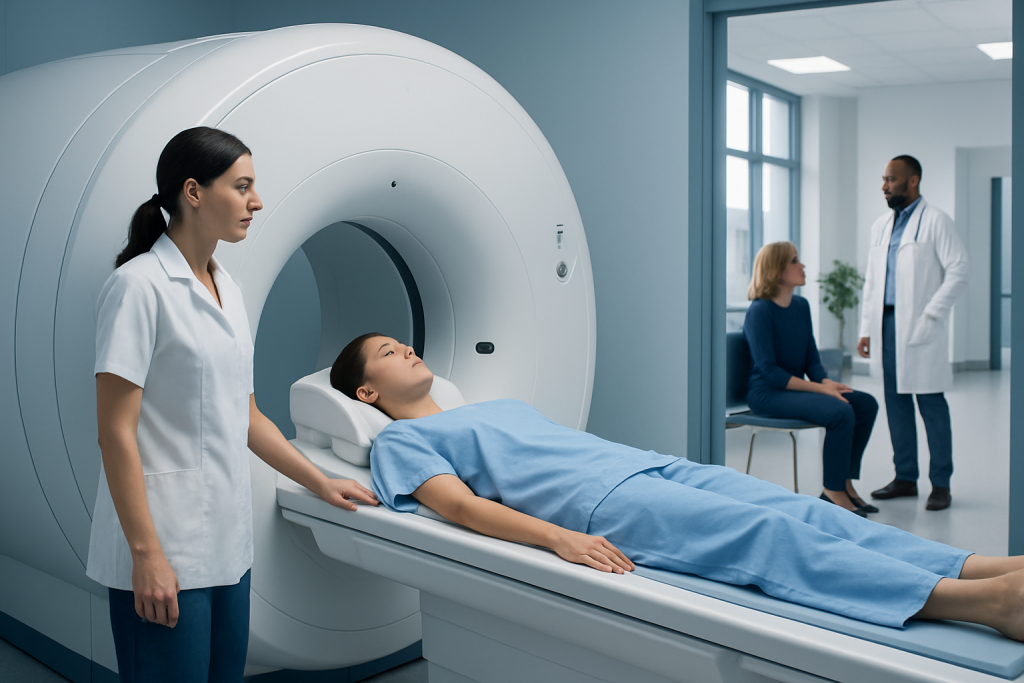
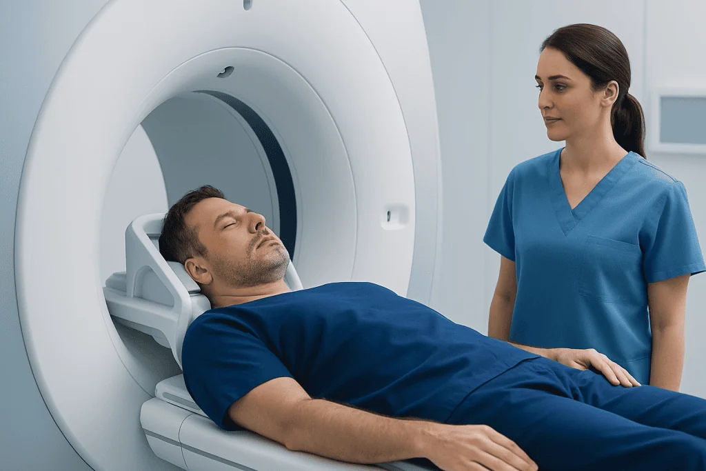
The person lies on a padded table, usually on their back, with a head coil positioned around the head. The table moves into the scanner, and staff remain in contact through an intercom. The machine makes repeated knocking or humming sounds, so earplugs or headphones are provided. It is important to lie still because movement can reduce image quality.
The examination commonly lasts between 20 and 60 minutes, depending on whether a full brain MRI is performed alongside MRA and whether contrast is used. The scan itself is painless. A call button is available so the patient can alert the radiographer if they feel uncomfortable, and people with claustrophobia may discuss support options before the appointment.
Benefits, Limitations and Possible Risks
The key benefit of MRI cerebral aneurysm assessment is detailed, noninvasive visualization of the brain and blood vessels without radiation exposure. It can help detect aneurysms, establish a baseline for monitoring, assess changes in a known aneurysm, and support treatment planning. MRI also provides information about surrounding brain structures that may be relevant to symptoms or follow-up.
No imaging test is perfect. Very small aneurysms, complex vessel anatomy, motion during scanning and artifacts from nearby metal or prior aneurysm clips can make interpretation more difficult. An MRA may occasionally suggest an abnormality that needs confirmation with CT angiography or catheter angiography. Catheter angiography is more invasive but can provide highly detailed images and may be used when treatment is being considered.
MRI does not cause pain and does not use ionizing radiation, but some people find the enclosed scanner space or noise challenging. Gadolinium contrast is generally well tolerated, although mild reactions can occur and rare serious reactions are possible. The care team weighs these considerations against the expected diagnostic benefit.
When a confirmed aneurysm needs intervention, management may include microsurgical clipping or minimally invasive endovascular techniques. A specialist can explain whether observation, brain aneurysm treatment or additional imaging is the most appropriate next step.
Results, Recovery and Next Steps
After a non-contrast scan, most people can return to usual activities immediately. If contrast was used, normal activities are also usually possible unless the patient received sedation or has been given specific instructions. People who receive sedation need someone to accompany them home and should avoid driving, alcohol and important decisions until the sedative effects have fully worn off.
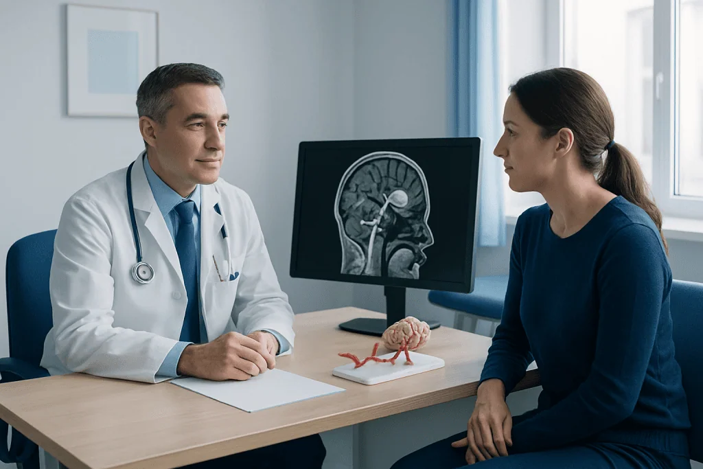
A radiologist reviews the images and sends a report to the requesting clinician. The report may describe whether an aneurysm is seen, its location and approximate measurements, whether there are features that need closer assessment, and whether other findings are present. Results are not always available immediately because careful image review is essential.
If an aneurysm is identified, the next step depends on its characteristics and the person’s overall health, family history and risk factors. Some aneurysms are monitored with repeat imaging, while others are referred to a neurovascular team. Understanding brain aneurysms can help patients prepare for a discussion about surveillance and treatment options.
At Acibadem International, multidisciplinary specialists in neurology, neuroradiology and neurosurgery at JCI-accredited hospitals assess and treat brain aneurysm concerns for international patients. Individual recommendations should always follow a personal clinical evaluation.
When to Seek Medical Care
A planned MRI appointment should not delay urgent care. A sudden, extremely severe headache that reaches maximum intensity quickly—sometimes described as the worst headache of a person’s life—can be a warning sign of bleeding around the brain. Emergency assessment is also needed for a severe headache with fainting, seizure, neck stiffness, vomiting, confusion, new weakness, facial drooping, speech difficulty, loss of balance or a change in vision.
Less urgent but prompt medical advice is appropriate for persistent new headaches, unexplained neurological symptoms, or concerns about a known aneurysm or family history. These symptoms have many possible causes, and most are not due to an aneurysm, but a clinician can determine whether imaging is needed.
People with a diagnosed aneurysm can support long-term vascular health by avoiding smoking, managing blood pressure with their healthcare team, taking prescribed medicines as directed and attending scheduled follow-up. They should not stop or start medicines, including blood pressure medicines or anticoagulants, without medical advice.
Frequently asked questions
Is MRI or MRA better for detecting a brain aneurysm?
MRI refers to the broader imaging examination of the brain, while MRA is an MRI technique designed to show blood vessels. MRA is commonly the most relevant part of an MRI cerebral aneurysm assessment, although the best test depends on the clinical situation. CT angiography or catheter angiography may be used when more detail or urgent assessment is needed.
Does an MRI cerebral aneurysm scan require contrast?
Not always. Many MRA examinations use non-contrast time-of-flight imaging to assess the arteries in the brain. Contrast may be used when it is expected to improve image quality or answer a specific clinical question.
How long does a brain aneurysm MRI take?
The scan often takes about 20 to 60 minutes, depending on the sequences required and whether contrast is used. A combined brain MRI and MRA may take longer than an MRA alone. The imaging department can provide more specific timing before the appointment.
Can MRI detect a ruptured brain aneurysm?
MRI can show signs of bleeding in some circumstances, but it is often not the first test when rupture is suspected. In an emergency, clinicians commonly use CT because it can be performed quickly and is highly useful for detecting acute bleeding. Urgent symptoms should be assessed in an emergency department rather than waiting for an outpatient MRI.
Is MRA brain aneurysm screening recommended with a family history?
It may be considered for people with a strong family history, particularly when two or more close relatives have had aneurysms or aneurysmal subarachnoid hemorrhage. The decision is individualized and should include discussion of possible benefits, limitations and follow-up implications. A clinician may also consider inherited medical conditions and other risk factors.
What happens if an MRA finds an aneurysm?
A finding does not automatically mean treatment is needed. Specialists consider the aneurysm’s size, location, shape, growth, symptoms and the person’s health before recommending surveillance or intervention. Additional imaging may be requested to clarify the result or support planning.
References
- American Heart Association
- American Stroke Association
- National Institute of Neurological Disorders and Stroke
- Radiological Society of North America
- Mayo Clinic
This article is for general information only and is not a substitute for professional medical advice. Please consult a qualified doctor about your individual situation.
Explore treatments in Turkey — costs, top hospitals & a free quote
JCI-accredited · board-certified surgeons · reply within 24h
Add us as a Preferred Source to see more of our trusted health content across Google Search, AI Overviews and Discover.