MRI Chest Wall: Preparation, Procedure and Results
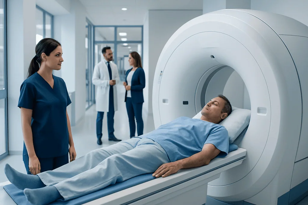
MRI chest wall uses magnetic fields and radio waves rather than ionizing radiation. It provides detailed images of chest wall soft tissues, bone marrow, muscles and areas near the lungs and pleura.
Key Takeaways
- MRI chest wall uses magnetic fields and radio waves rather than ionizing radiation.
- It provides detailed images of chest wall soft tissues, bone marrow, muscles and areas near the lungs and pleura.
- Preparation usually involves removing metal items and discussing implants, pregnancy, kidney problems and contrast allergies.
- The scan commonly takes 30 to 60 minutes, depending on the area studied and whether contrast is used.
- A chest wall MRI does not usually assess clogged coronary arteries; dedicated heart or vascular imaging may be needed.
- Urgent symptoms such as severe chest pain, breathing difficulty or coughing blood need prompt medical assessment rather than waiting for an MRI.
An MRI chest wall is a detailed, radiation-free imaging examination of the ribs, muscles, nerves, connective tissues and other structures surrounding the chest. It can help clinicians investigate a lump, pain, injury, inflammation or an abnormality seen on another imaging test.
MRI Chest Wall: What It Is and Why It Is Done
An MRI chest wall is an imaging examination that uses a strong magnetic field, radio waves and computer processing to create detailed pictures of the tissues around the chest. It does not use X-rays or other ionizing radiation. The scan can show muscles, ribs, cartilage, bone marrow, skin, fat, nerves, lymph nodes and the lining around the lungs with more soft-tissue detail than many other tests.
A clinician may request this examination when a person has a chest wall lump, localized swelling, ongoing pain, a suspected sports or traumatic injury, signs of infection or inflammation, or an area that needs further evaluation after an X-ray, ultrasound or CT scan. It may also be used to define the size and location of a known abnormality and to support treatment planning or follow-up.
The term “chest wall” refers to the structures that form and cover the rib cage, rather than the heart and lungs alone. The most useful imaging test depends on the clinical question. MRI is particularly valuable when the goal is to examine soft tissues and bone marrow in detail, while CT, ultrasound, mammography, cardiac imaging or other tests may be better for different concerns.
How an MRI Chest Wall Works and Who May Need One
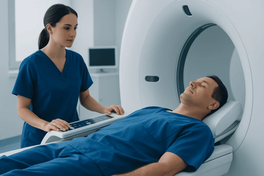
During MRI, the body is placed within a scanner containing a powerful magnet. Radiofrequency signals briefly alter the alignment of hydrogen atoms in body tissues. As these atoms return to their usual state, they release signals that a computer converts into detailed images. Different imaging sequences help distinguish fluid, fat, muscle, inflammation and other tissue characteristics.
People may be referred for chest wall MRI after a clinical examination or another scan suggests a possible problem in the chest wall. Examples include unexplained focal pain, a palpable mass, possible muscle or tendon injury, suspected infection, a rib or sternum abnormality, or concern about extension of a condition into nearby tissue. MRI findings are interpreted alongside symptoms, examination findings and previous tests.
Most people can safely have an MRI, but screening is essential. A patient should tell the imaging team about any pacemaker, implanted defibrillator, neurostimulator, cochlear implant, vascular clip, infusion pump, metal fragments, joint replacement, surgical hardware or other implanted device. Many modern implants are MRI-conditional, meaning scanning may be possible under specific safety conditions, but this must be confirmed before the appointment.
Pregnancy, reduced kidney function, a previous reaction to MRI contrast and severe claustrophobia should also be discussed in advance. These factors do not always prevent MRI, but they may affect timing, scan technique or the need for additional support.
How to Prepare for a Chest MRI

Preparation instructions vary according to the facility, the reason for the scan and whether contrast material is planned. In many cases, normal eating, drinking and prescribed medicines can continue. The imaging center should provide personalized instructions before the appointment, and these should take priority over general guidance.
Before entering the scan room, a person will be asked to remove metal-containing items. This includes jewelry, watches, hearing aids, hairpins, removable dental appliances, belts, coins, keys, bank cards and clothing with metal fasteners. Makeup, tattoos and medication patches should be mentioned, as some products can contain metallic ingredients or need special handling.
What not to do 24 hours before MRI? There is usually no universal restriction on normal activities during the 24 hours before an MRI chest wall. However, a person should not ignore specific fasting instructions, should avoid arriving with metal items or metal-containing patches, and should not stop prescribed medication unless the clinician specifically advises it. Those who may need sedation should follow instructions about food, drink, driving and arranging an escort home.
Why do you drink water before an MRI? Water is not required before every chest MRI. Some centers encourage normal hydration, especially when contrast may be given, because it supports general comfort and normal kidney function. In other situations, water intake may be limited because of fasting instructions or another health condition. The safest approach is to follow the directions supplied by the imaging team.
The MRI Chest Wall Procedure: Step by Step
On arrival, a radiology professional reviews the safety questionnaire and confirms the reason for the examination. The patient may change into a gown. If contrast is needed, a small intravenous line may be placed in the arm or hand. MRI contrast, commonly gadolinium-based, can help show blood flow, active inflammation and the characteristics of certain abnormalities.
The patient usually lies on their back on a padded table. A specialized receiving device, sometimes called a coil, may be positioned over the chest area to improve image quality. The table then moves slowly into the scanner. The body part being examined is positioned at the center of the magnet, and staff remain in contact through an intercom throughout the scan.
The scanner makes repeated loud tapping or knocking sounds while images are acquired. Earplugs or headphones are provided. Remaining still is important because movement can blur the images; the team may also ask for brief breathing instructions. A chest wall MRI commonly lasts about 30 to 60 minutes, though timing can vary with the complexity of the study and the use of contrast.
The examination is painless, but lying still in an enclosed space can feel uncomfortable for some people. Those who experience anxiety or claustrophobia should tell their clinician before the visit. Options may include reassurance, a wider-bore scanner where available, relaxation strategies or, in selected cases, prescribed sedation with appropriate monitoring and transport arrangements.
Benefits, Risks and Recovery After the Scan
A main benefit of MRI chest wall is its detailed assessment of soft tissues without radiation exposure. It can help clarify whether a finding involves muscle, fat, bone marrow, cartilage, nerves or another structure, and it can show the relationship of an abnormality to surrounding tissues. This information may guide monitoring, further testing, biopsy planning or treatment decisions.
MRI is generally considered very safe when proper screening is completed. The magnetic field can be hazardous around certain implants or loose metal objects, which is why safety questions are so important. Some people find the noise, need to remain still or enclosed scanner environment challenging. The radiology team can provide support throughout the examination.
If contrast is used, most people do not experience significant problems. Mild temporary effects, such as a cool sensation at the injection site, may occur. Serious allergic-like reactions are uncommon, but staff are trained to recognize and manage them. In people with severe kidney impairment, contrast decisions require particular care, so relevant kidney conditions and recent test results should be shared beforehand.
Recovery is usually immediate. A person can generally return to usual food, fluids and activities after a non-sedated scan. If sedation was used, the patient will need a responsible adult to accompany them home and should avoid driving, alcohol, important decisions and operating machinery for the period advised by the care team.
Understanding MRI Chest Wall Results
A radiologist, a doctor specializing in medical imaging, reviews the images and prepares a report for the clinician who requested the examination. Results may be available within several days, although timing depends on the imaging service and whether comparison with previous scans or additional specialist review is needed. The referring clinician explains what the results mean in the context of the person’s symptoms and medical history.
An MRI report may describe normal chest wall structures, inflammation, fluid, a muscle or soft-tissue injury, changes in bone marrow, infection-related features, or an area that needs further assessment. An abnormal imaging finding does not automatically indicate a serious condition. In some cases, the next step may be watchful follow-up; in others, blood tests, a different scan or tissue sampling may be appropriate.
Does chest MRI show clogged arteries? A routine MRI chest wall is not designed to diagnose clogged coronary arteries. Coronary artery disease is usually assessed with tests selected for the heart and blood vessels, such as coronary CT angiography, invasive coronary angiography, stress testing or specialized cardiac MRI in selected situations. New or concerning chest symptoms should be assessed promptly by a qualified clinician rather than interpreted through a chest wall MRI alone.
If findings suggest a condition involving more than one specialty, care may involve radiology alongside respiratory medicine, orthopedics, cardiology, oncology, surgery or other relevant teams. Acibadem International’s multidisciplinary specialists and JCI-accredited hospitals support diagnosis and treatment planning for international patients when clinically appropriate.
When to Seek Medical Care
A person should arrange medical assessment for a new or enlarging chest wall lump, persistent focal chest pain, unexplained swelling, tenderness that does not improve, or pain after an injury that limits normal movement or breathing. A clinician can determine whether MRI chest wall or another test is the most appropriate next step.
Urgent medical care is important for severe or pressure-like chest pain, sudden shortness of breath, fainting, bluish lips, coughing blood, new confusion, or chest symptoms accompanied by sweating, nausea or pain spreading to the arm, jaw, back or shoulder. These symptoms can have causes that require immediate assessment and should not wait for an outpatient imaging appointment.
People with fever, redness, warmth or rapidly increasing swelling over the chest wall should also seek timely medical advice, particularly after surgery, injury or an injection. Prompt evaluation helps clinicians identify whether infection, inflammation or another cause needs treatment.
Frequently asked questions
How long does an MRI chest wall take?
A chest wall MRI often takes about 30 to 60 minutes. The exact duration depends on the area being examined, the number of image sequences needed and whether contrast material is used. Additional time is needed for check-in, safety screening and preparation.
Do I need contrast for an MRI chest wall?
Not every chest wall MRI requires contrast. Contrast may be helpful when clinicians need more information about inflammation, infection, blood supply or the nature and extent of an abnormal area. The radiologist and referring clinician select the protocol based on the clinical question.
Can I eat before a chest MRI?
Many chest MRI examinations do not require fasting, so people can often eat and take regular medicines as usual. However, fasting may be requested when contrast, sedation or another related procedure is planned. The imaging center’s individual instructions should always be followed.
Is an MRI chest wall painful?
The MRI scan itself is painless and does not involve radiation. Some people may feel discomfort from lying still or from the position needed for imaging, especially if they already have chest pain or an injury. The staff can help with positioning and communicate with the patient during the scan.
Can I have an MRI if I have metal in my body?
It depends on the type, location and manufacturer information for the metal or implanted device. Many joint replacements and surgical implants can be scanned safely under specific conditions, while some devices require special precautions or may prevent MRI. The imaging team must review all implants and possible metal exposure before the examination.
What happens if my chest wall MRI result is abnormal?
An abnormal result is reviewed by the clinician who ordered the scan alongside symptoms, examination findings and other tests. Depending on the finding, the next step may be reassurance, follow-up imaging, referral to a specialist, laboratory testing or occasionally a biopsy. A radiology report alone does not provide a complete diagnosis.
References
- Radiological Society of North America
- American College of Radiology
- National Institute of Biomedical Imaging and Bioengineering
- U.S. Food and Drug Administration
- National Health Service
This article is for general information only and is not a substitute for professional medical advice. Please consult a qualified doctor about your individual situation.
Explore treatments in Turkey — costs, top hospitals & a free quote
JCI-accredited · board-certified surgeons · reply within 24h
Add us as a Preferred Source to see more of our trusted health content across Google Search, AI Overviews and Discover.
Check your numbers in seconds
BMI, calories, due date, blood pressure and 30+ more clinical calculators — free, instant, doctor-reviewed ranges.