MRI Em Português: Preparation, Procedure and Results
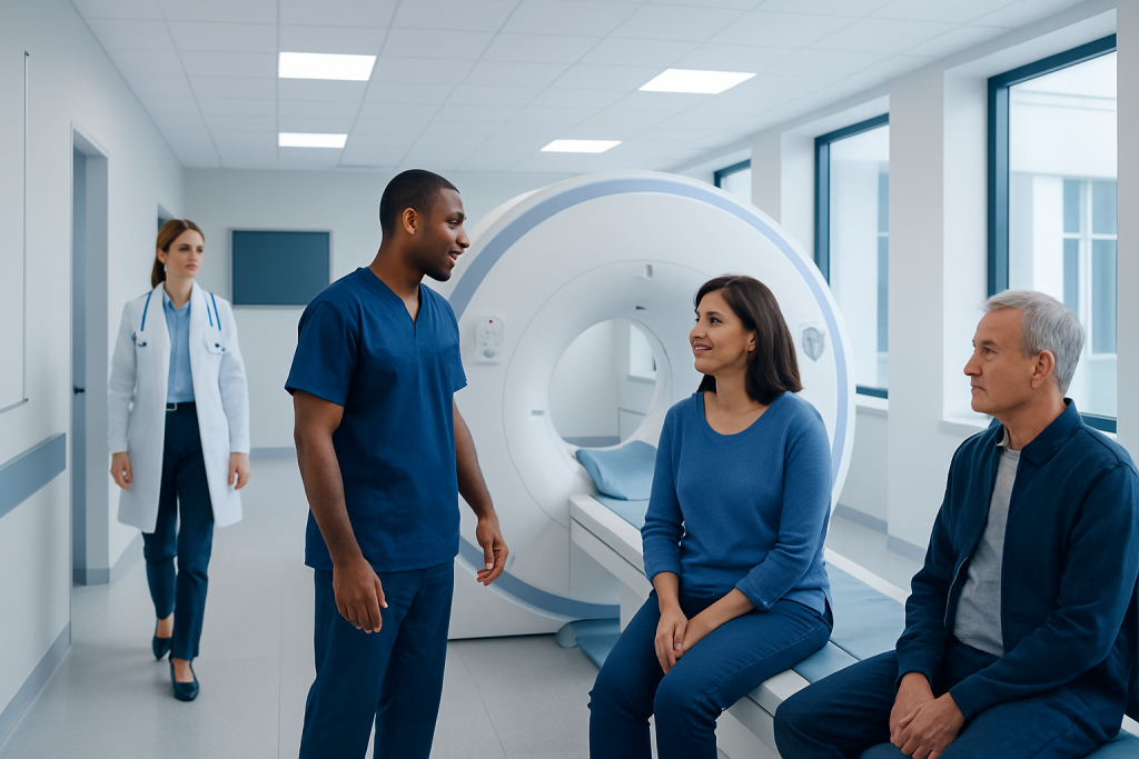
An MRI scan uses magnets and radio waves rather than X-rays or ionising radiation. Most MRI appointments take 30 to 60 minutes, although some scans take longer.
Key Takeaways
- An MRI scan uses magnets and radio waves rather than X-rays or ionising radiation.
- Most MRI appointments take 30 to 60 minutes, although some scans take longer.
- Patients should tell the imaging team about implants, metal fragments, pregnancy, kidney disease and possible contrast allergies.
- Many people can eat, drink, brush their teeth and take usual medicines before an MRI, unless the care team gives different instructions.
- Results are interpreted by a radiologist and sent to the requesting clinician, who explains what they mean in the context of symptoms and medical history.
MRI em português refers to information about magnetic resonance imaging (MRI) in Portuguese. An MRI is a non-invasive scan that uses a strong magnet and radio waves to produce detailed images of internal organs, soft tissues, joints, blood vessels and the nervous system, without using ionising radiation.
MRI em português: an overview
MRI em português means magnetic resonance imaging information for Portuguese-speaking patients. Magnetic resonance imaging, often called MRI or RM in Portuguese, is an imaging examination that creates highly detailed pictures of structures inside the body. It is commonly used to assess the brain, spine, joints, muscles, abdomen, pelvis, heart and blood vessels.
The examination uses a powerful magnetic field and radiofrequency energy to organise and measure signals from water molecules in the body. A computer converts these signals into images. Unlike X-rays and CT scans, MRI does not use ionising radiation, which can make it a useful option when detailed soft-tissue imaging is needed.
An MRI may be requested to investigate symptoms, clarify findings from another test, monitor a known condition or help plan treatment. The scan itself does not diagnose every health problem on its own; its images are interpreted alongside a person’s symptoms, examination findings and other tests.
How MRI works and who may need it
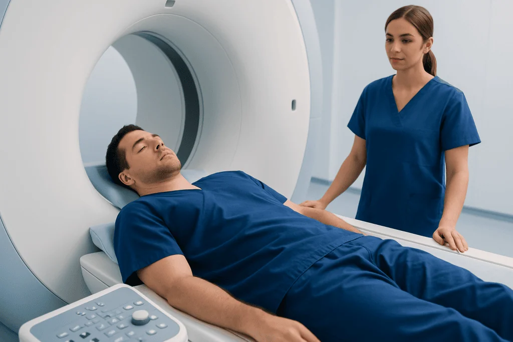
During an MRI, the patient lies still on a padded table that moves into the scanner. The scanner contains a magnet, and a special device called a coil may be placed around the body part being examined. The equipment produces loud tapping or knocking sounds while image sequences are collected.
Doctors may recommend MRI when they need clear views of soft tissues or complex anatomy. Examples include suspected brain or spinal conditions, ligament or cartilage injuries, inflammation, tumours, pelvic concerns, liver findings and some heart or vascular conditions. It can also be useful in evaluating conditions affecting the nervous system, such as multiple sclerosis.
Most people can safely have an MRI, but screening is essential. The team needs to know about pacemakers, cochlear implants, neurostimulators, aneurysm clips, medication pumps, artificial joints, stents, surgical clips, dental devices, shrapnel or previous metal injuries to the eye. Many modern implants are MRI-compatible under specific conditions, but this must be checked before scanning.
Some MRI examinations use a contrast agent, usually gadolinium-based, injected into a vein to make certain tissues or blood vessels more visible. The care team will assess whether contrast is appropriate, particularly for people with significant kidney disease, prior contrast reactions or pregnancy.
How to prepare for an MRI

Preparation depends on the body area being scanned and whether contrast or sedation is planned. The imaging centre should provide individual instructions before the appointment. In many cases, patients may eat and drink normally and continue usual prescribed medicines, but fasting may be requested for some abdominal, pelvic or contrast-enhanced examinations.
Patients should wear comfortable clothing without metal fasteners where possible. Jewellery, watches, hairpins, piercings, hearing aids, removable dental appliances, credit cards and other metallic or electronic items must be removed before entering the MRI room. Makeup, hair products and some tattoos may contain metallic pigments, so it is sensible to tell the team about them.
It is important to mention claustrophobia or anxiety in advance. The team may explain options such as music, a support person where permitted, relaxation techniques, a wider-bore scanner or prescribed sedation when clinically appropriate. Anyone receiving sedation will usually need an adult to accompany them home and should not drive afterwards.
What to do the night before an MRI? Most people do not need special preparation the night before. They should follow any fasting instructions they were given, take medicines as advised, arrange transport if sedation is planned and prepare details about implants, previous surgery, allergies and kidney problems. A good night’s rest can also make it easier to remain comfortable and still during the scan.
Can I brush my teeth before an MRI? Yes, brushing teeth is usually fine before an MRI. If the examination requires fasting, patients should avoid swallowing water or toothpaste only if their care team has specifically instructed them to take nothing by mouth. Removable dentures, retainers and dental appliances should be removed before the scan unless the imaging staff says otherwise.
What should you not do before an MRI? Patients should not enter the MRI area with metal, electronic devices or cards with magnetic strips. They should not skip important screening information about implants, possible metal fragments, pregnancy, kidney disease or previous reactions to contrast. They should also not drive themselves after sedating medication, and should follow any specific eating or drinking restrictions supplied by the imaging centre.
What happens during the MRI procedure?
At check-in, a radiographer or MRI technologist reviews the safety questionnaire and confirms the reason for the scan. The patient changes into a gown if needed and removes all relevant items. If contrast is required, an intravenous cannula may be placed in the arm or hand.
The patient lies on the scanning table, and cushions, straps or supports may be used to help keep the area still and comfortable. Earplugs or headphones are provided because the scanner is noisy. The staff remain nearby in a separate control room, can see and hear the patient, and provide a call button for communication throughout the examination.
The table moves into the scanner for image acquisition. It is important to remain as still as possible because movement can blur images and may mean that sequences need to be repeated. The patient may be asked to hold their breath briefly during scans of the chest or abdomen. If contrast is administered, a cool sensation at the injection site is possible, but many people notice little or no change.
How many hours is the MRI procedure? MRI is usually measured in minutes rather than hours. Many standard scans take about 30 to 60 minutes, while more detailed studies, multiple body areas or specialised examinations may take 60 to 90 minutes or occasionally longer. The total visit may be longer because of registration, safety screening, changing, contrast preparation and observation when sedation is used.
Benefits, limitations and possible risks
A major benefit of MRI is its detailed view of soft tissues, including the brain, spinal cord, nerves, muscles, tendons, ligaments and many internal organs. It can often provide information not visible on an X-ray or ultrasound and does so without ionising radiation. The results can help clinicians decide whether monitoring, medicines, rehabilitation, surgery or further tests may be appropriate.
MRI also has limitations. It may not be the best test for every question, and its findings can sometimes be non-specific or reveal incidental changes that are not causing symptoms. Image quality depends on remaining still, and some people find the scanner’s enclosed space uncomfortable. The requesting clinician chooses the examination based on the clinical question and individual circumstances.
Serious complications are uncommon when MRI safety procedures are followed. The strongest safety concern is an unrecognised metal or electronic implant, which is why thorough screening matters. Contrast reactions are uncommon but can occur; urgent symptoms such as trouble breathing, facial swelling, widespread hives or severe dizziness should be reported immediately to staff.
Gadolinium contrast is generally used only when it is expected to add useful information. In people with severe kidney impairment, the care team may use a different approach or avoid certain contrast agents. Pregnant patients should tell their clinician and imaging team so that the benefits and timing of MRI, with or without contrast, can be considered carefully.
After the scan and understanding MRI results
Most patients can return to normal activities straight after a routine MRI. Drinking fluids after contrast may be encouraged unless a clinician has advised fluid restriction for another health reason. If sedatives were used, temporary drowsiness and reduced coordination are expected, so driving, alcohol, operating machinery and important decisions should be avoided for the period advised by the care team.
A radiologist reviews the images and prepares a report for the doctor or specialist who requested the scan. Result timing varies with the urgency and complexity of the examination, as well as local reporting processes. The requesting clinician explains whether the findings match the person’s symptoms and what follow-up, if any, is needed.
It is helpful for patients to ask how and when results will be communicated, especially if the MRI was arranged to assess persistent or worsening symptoms. A report may use technical terms, and an imaging result should not be interpreted in isolation. The clinician can explain the significance of findings and discuss appropriate next steps.
Acibadem International’s multidisciplinary specialists and JCI-accredited hospitals support international patients who need diagnostic assessment and care planning, including MRI imaging.
When to seek medical care
An MRI appointment should be discussed with a qualified clinician when symptoms are persistent, unexplained or affecting daily life. The urgency depends on the symptom pattern and the suspected cause. Imaging is generally arranged after a medical assessment, rather than as a substitute for one.
Urgent medical assessment is important for sudden weakness or numbness on one side, difficulty speaking, sudden severe headache, new loss of vision, seizures, severe back pain with loss of bladder or bowel control, chest pain, major trauma or rapidly worsening symptoms. Emergency services should be contacted according to local guidance when these symptoms occur.
Before a scheduled MRI, patients should contact the imaging centre promptly if they discover an implant, have had recent surgery, may have metal in the eye, develop a significant illness, become pregnant or have new concerns about contrast or sedation. Sharing this information early helps the team plan the safest examination.
Frequently asked questions
Is MRI painful?
MRI is usually painless. The scanner can be loud, and lying still in one position may feel uncomfortable, but the radiographer can provide cushions, hearing protection and a way to communicate during the scan. Contrast injection may cause brief discomfort where the cannula is placed.
Can someone with braces have an MRI?
Many people with braces can have an MRI safely, but they must tell the imaging team before the examination. Braces may create image distortion, especially for scans of the head, face or neck, and the team can determine whether adjustments or a different imaging approach is needed.
Can I eat before an MRI scan?
For many MRI scans, normal eating and drinking are allowed. However, some abdominal, pelvic or contrast-enhanced examinations may require fasting for a period before the appointment. Patients should follow the specific instructions from their imaging provider.
What if I am claustrophobic during an MRI?
Claustrophobia is common and should be discussed before the appointment. The imaging team may offer reassurance, communication throughout the scan, music, positioning support, a wider scanner where available or sedation when appropriate. Planning ahead often makes the experience more manageable.
How long do MRI results take?
The timing varies according to the examination, reporting workflow and whether the result is urgent. A radiologist reviews the images and sends a report to the requesting clinician, who discusses the findings with the patient. Patients can ask the imaging centre or clinician when to expect an update.
Is MRI safe during pregnancy?
MRI without contrast may be considered during pregnancy when it is clinically necessary, particularly when the information could affect care. Gadolinium contrast is generally avoided unless there is a clear medical reason. Pregnant patients should inform their doctor and the MRI team before the scan.
References
- Radiological Society of North America
- American College of Radiology
- National Institute of Biomedical Imaging and Bioengineering
- U.S. Food and Drug Administration
- National Health Service
This article is for general information only and is not a substitute for professional medical advice. Please consult a qualified doctor about your individual situation.
Joint and spine care in Turkey — expert assessment & treatment
JCI-accredited · board-certified surgeons · reply within 24h
Add us as a Preferred Source to see more of our trusted health content across Google Search, AI Overviews and Discover.