MRI Kidney Mass: Preparation, Procedure and Results
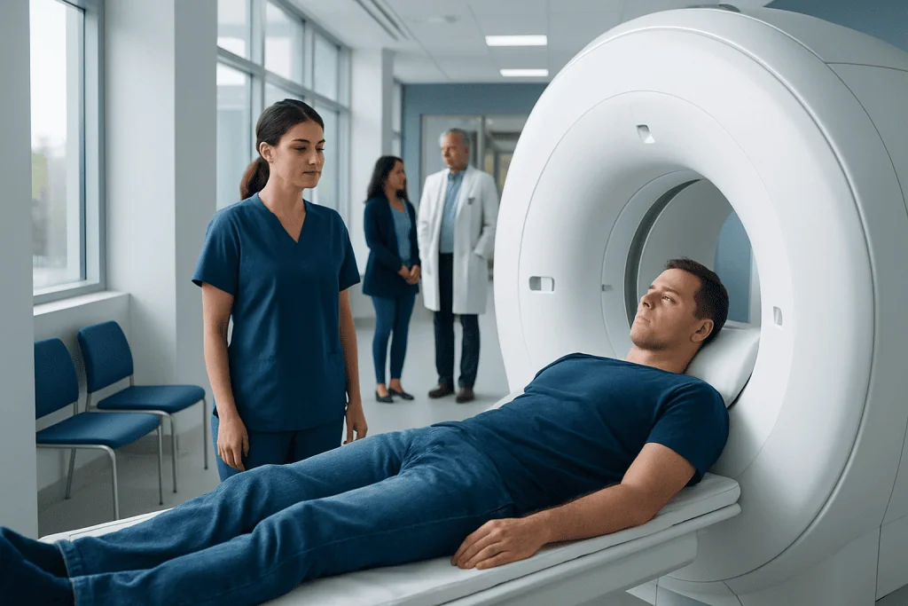
A kidney MRI produces detailed images that can help characterize a mass found on ultrasound or CT. Many kidney masses are not cancer, particularly simple fluid-filled cysts, but solid or complex lesions require expert assessment.
Key Takeaways
- A kidney MRI produces detailed images that can help characterize a mass found on ultrasound or CT.
- Many kidney masses are not cancer, particularly simple fluid-filled cysts, but solid or complex lesions require expert assessment.
- Preparation may include screening for metal implants, reviewing kidney function and following instructions about food, fluids and contrast material.
- A dedicated MRI kidney mass protocol usually includes images before and after contrast, taken in several phases.
- Most people resume usual activities immediately after the scan unless they receive sedation or have specific instructions.
- MRI findings should be interpreted alongside symptoms, blood tests and previous imaging by a urology and radiology team.
An MRI kidney mass examination is a detailed scan used to clarify whether a kidney lesion is likely to be a simple cyst, a solid growth or another finding that needs follow-up. It is painless and does not use radiation; results help the care team decide whether monitoring, further testing or treatment is appropriate.
MRI Kidney Mass: What the Scan Can Show
An MRI kidney mass scan uses a strong magnetic field and radio waves to create detailed images of the kidneys and surrounding structures. It is commonly requested after an ultrasound or CT scan identifies a lesion that cannot be fully characterized. MRI can distinguish fluid, fat, blood products and enhancing soft tissue, which helps clinicians assess the nature of a renal mass.
The examination does not use ionizing radiation. In many cases, a contrast agent containing gadolinium is given through a vein to show how a lesion receives blood over time. This pattern is valuable when distinguishing a simple cyst from a complex cyst or solid mass, although MRI alone cannot confirm every diagnosis.
A kidney mass is a broad term that includes benign cysts, noncancerous tumors, infections or inflammatory changes, and kidney cancer. The scan is therefore one part of a careful evaluation rather than a diagnosis by itself. The radiology report is usually reviewed with a urologist, who considers the lesion’s appearance, size, growth, symptoms and the person’s overall health.
How Does an MRI for a Kidney Mass Work?
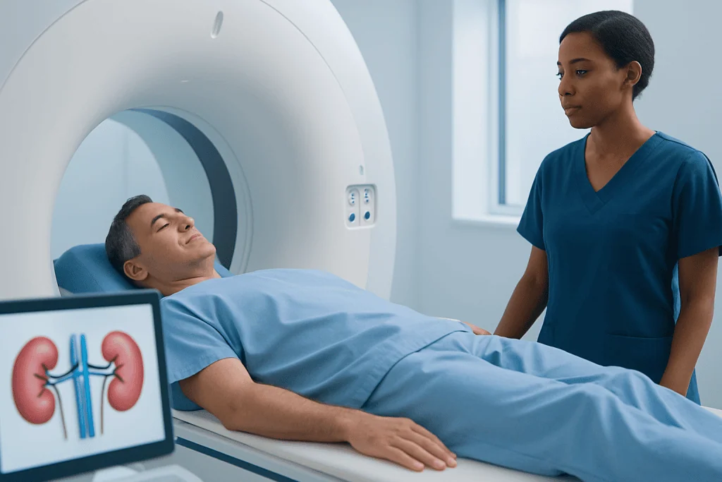
During MRI, the body lies within a scanner that temporarily aligns hydrogen particles in tissues. Radiofrequency pulses alter this alignment, and the signals released as the particles return to their usual state are processed into highly detailed images. Different MRI sequences emphasize different tissue properties, allowing radiologists to evaluate a kidney lesion from several perspectives.
A dedicated mri kidney renal mass protocol often includes images of the abdomen before contrast and repeated images after contrast is injected. The scan may also include diffusion-weighted imaging, which provides additional information about how water moves within tissue. Breath-hold instructions help reduce motion from breathing and make small lesions easier to assess.
MRI is especially useful when CT contrast is unsuitable, when a lesion remains uncertain after CT or ultrasound, or when radiation avoidance is important. However, the most appropriate test depends on the individual situation. A clinician may recommend ultrasound, CT, MRI, surveillance imaging, or another evaluation based on the initial finding and medical history.
What Percent of Kidney Masses Are Cancer?
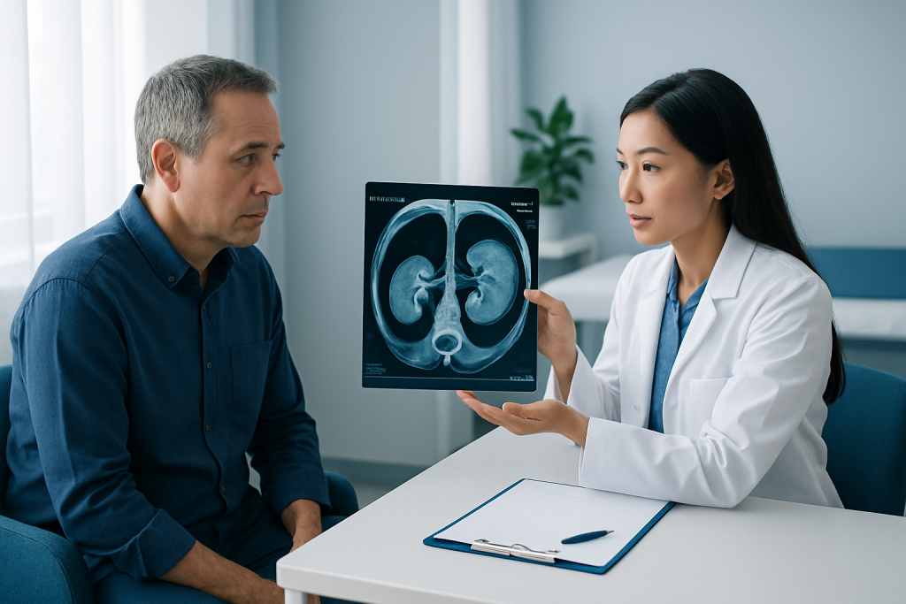
There is no single percentage that applies to every kidney mass because the likelihood of cancer depends strongly on whether the finding is solid or cystic, its size, imaging features and the person’s clinical history. Simple kidney cysts are very common and are almost always benign. In contrast, a solid enhancing kidney mass is more concerning for renal cell carcinoma, the most common type of kidney cancer in adults.
Small renal masses, generally defined as masses up to 4 cm, include both cancerous and noncancerous lesions. Published studies show that a meaningful minority of small solid masses prove benign after surgery or biopsy, while the chance of malignancy tends to increase as size increases. Imaging can estimate risk, but it may not always reliably determine whether an individual lesion is cancer.
Complex cystic lesions are commonly evaluated using imaging classifications that consider wall thickness, septa, calcification and enhancement. A urologist may recommend observation, repeat imaging, biopsy or treatment depending on the risk category. For broader information about kidney cancer evaluation, readers may find kidney cancer information helpful.
Is a 2.5 cm Kidney Mass Big?
A 2.5 cm kidney mass is considered a small renal mass. It is below the 4 cm threshold often used to describe stage T1a kidney tumors when cancer is confirmed and confined to the kidney. Small does not automatically mean harmless, but it can influence the range of management options and often allows time for thoughtful assessment.
The measurement alone does not determine whether a mass is cancerous or whether treatment is needed. Radiologists also evaluate whether it is solid or cystic, whether it enhances after contrast, whether it contains fat, where it sits within the kidney and whether there are signs of spread. Comparison with earlier scans is important because growth rate can provide useful context.
Depending on the imaging findings and the person’s health, options may include active surveillance with scheduled imaging, a biopsy, removal of the mass while preserving kidney tissue, or ablation in selected circumstances. Decisions should be made with a urologist and, when needed, radiologists and oncology specialists rather than based on size alone.
How Should I Prepare for a Kidney MRI?
MRI kidney preparation begins with a safety screening questionnaire. The imaging team needs to know about pacemakers, implanted stimulators, aneurysm clips, cochlear implants, metal fragments, joint replacements, medication pumps and prior surgery. Many modern implants can be scanned safely under specific conditions, but the MRI department must verify this before the appointment.
Instructions about eating and drinking vary by facility and whether contrast or sedation is planned. Some centers ask patients not to eat for several hours before a contrast-enhanced abdominal MRI to reduce nausea and improve image quality. The patient should follow the instructions given by the imaging center, continue regular medicines unless told otherwise, and ask in advance about diabetes medications or fasting requirements.
Before gadolinium contrast is used, the team may review kidney function with a recent blood test, especially in people with known kidney disease, diabetes, older age or other risk factors. Previous contrast reactions, pregnancy, breastfeeding and allergies should also be discussed. Gadolinium reactions are uncommon, and the team can explain whether contrast is appropriate in an individual case.
- Bring previous imaging reports and, if possible, the images themselves for comparison.
- Wear comfortable clothing without metal, or use a hospital gown.
- Remove jewelry, hearing aids, removable dental work, credit cards and other metal or electronic items.
- Tell staff about claustrophobia; some people benefit from coaching, an open MRI when available, or prescribed anxiety medication arranged in advance.
What Is the Typical MRI Protocol for Kidney Masses?
A typical mri kidney mass protocol is tailored to the question being asked, but it generally includes images through both kidneys and the upper abdomen in more than one plane. Sequences before contrast can identify fat, fluid, blood products and certain tissue characteristics. Diffusion-weighted images may add information about cellularity and lesion behavior.
If contrast is appropriate, the mri renal mass protocol commonly obtains repeated images at timed intervals after gadolinium injection. These phases assess enhancement, meaning whether a lesion takes up contrast relative to the surrounding kidney. Enhancement is an important feature because it can help differentiate a simple cyst from a solid or complex lesion.
The total examination often takes about 30 to 60 minutes, depending on the scanner, the protocol and whether contrast is needed. The patient must remain still and will hear loud tapping or knocking sounds; ear protection is provided. Staff can communicate throughout the scan, and a call button is available if the patient needs help.
There is no single mri kidney protocol for every person. A radiologist may adjust the scan to assess a previously seen lesion, evaluate possible involvement of kidney blood vessels, clarify a complex cyst, or monitor a known mass over time. This individualized approach improves the usefulness of the final report.
Step-by-Step Procedure, Recovery and Results
At check-in, the patient completes screening, changes clothing if needed and has any required blood tests reviewed. An intravenous line may be placed for contrast. The person then lies on a padded table, usually on the back, and a receiving device called a coil is positioned over the abdomen. The table moves into the scanner, though the head may remain partly outside the opening for some kidney MRI examinations.
During the scan, the technologist gives brief breathing instructions through an intercom. Contrast, if used, is injected partway through the examination. The injection may cause a brief cool sensation, but most people do not feel significant discomfort. The patient should remain still, as movement can blur images and lengthen the scan.
Recovery is usually immediate. People who do not receive sedation can typically eat, drink, drive and return to normal activities after the test. Those given a sedative need an adult to take them home and should follow the facility’s instructions about driving, work and decision-making for the rest of the day.
A radiologist reviews the images and sends a report to the referring clinician, often within several days, although timing varies. The report may describe the lesion’s size, location, enhancement pattern and whether additional follow-up is advised. If treatment is indicated, partial nephrectomy may be considered for suitable localized masses to remove the lesion while preserving as much healthy kidney tissue as possible.
Benefits, Risks and When to Seek Medical Care
The main benefit of MRI is detailed, radiation-free characterization of a kidney finding. It can reduce uncertainty after other imaging and support decisions about surveillance, biopsy or treatment. MRI is particularly valuable for assessing complex cysts and for evaluating the relationship of a mass to kidney tissue and blood vessels.
MRI is generally safe, but it is not suitable for every person or every implant. Gadolinium contrast is usually well tolerated, though allergic-like reactions can occur rarely. In people with severe kidney impairment, the team carefully considers whether contrast is necessary and which agent is safest. Claustrophobia, noise and the need to remain still can also be challenging, but staff can offer practical support.
When to seek medical care: A kidney mass found on imaging should be discussed with the clinician who ordered the scan, even when it was discovered incidentally. Prompt medical assessment is appropriate for visible blood in the urine, persistent flank or back pain, unexplained weight loss, fever with urinary symptoms, or worsening general health. These symptoms have many possible causes and do not necessarily indicate cancer, but they warrant evaluation.
People with a new imaging result should seek urgent care for severe pain, inability to pass urine, heavy bleeding in urine, fainting, or fever with chills and significant illness. Acibadem International’s multidisciplinary urology, radiology and oncology specialists at JCI-accredited hospitals can evaluate kidney masses and discuss individualized care for international patients.
Frequently asked questions
Why would a doctor order an MRI for a kidney mass?
A doctor may order MRI after ultrasound or CT detects a kidney lesion that needs closer characterization. MRI can show whether the lesion contains fluid, fat, blood products or enhancing tissue and can help guide follow-up or treatment planning.
Does an MRI kidney mass scan require contrast?
Many kidney mass MRI examinations use gadolinium contrast because enhancement is important for evaluating solid and complex cystic lesions. However, contrast is not always necessary or appropriate, particularly for people with certain medical conditions. The radiologist and referring clinician decide based on the clinical question and kidney function.
Can MRI tell if a kidney mass is cancer?
MRI can identify features that make a lesion more or less suspicious for cancer, but it cannot always provide a definitive diagnosis. Some masses may require follow-up imaging, biopsy or surgical assessment. The result should be discussed with a urologist in the context of all available information.
How long does a kidney MRI take?
A kidney MRI commonly takes around 30 to 60 minutes. The exact duration depends on whether contrast is used, how many image sequences are needed and whether repeat images are required because of motion. Patients should allow extra time for registration and safety screening.
Can I eat before an MRI of the kidneys?
Instructions vary by imaging center and by whether contrast or sedation is planned. Some facilities ask patients to avoid food for several hours before an abdominal MRI. The patient should follow the specific instructions from the imaging department and ask if they are uncertain.
What happens if the MRI shows a suspicious kidney mass?
The next step may include consultation with a urologist, review by a multidisciplinary team, surveillance imaging, biopsy or treatment. The choice depends on the size and appearance of the mass, symptoms, overall kidney function and the person's health and preferences. A suspicious result does not automatically mean immediate surgery is required.
References
- American College of Radiology
- Radiological Society of North America
- National Cancer Institute
- European Association of Urology
- National Institute of Diabetes and Digestive and Kidney Diseases
This article is for general information only and is not a substitute for professional medical advice. Please consult a qualified doctor about your individual situation.
Explore treatments in Turkey — costs, top hospitals & a free quote
JCI-accredited · board-certified surgeons · reply within 24h
Add us as a Preferred Source to see more of our trusted health content across Google Search, AI Overviews and Discover.