Nuclear Medicine Specialist: Roles, Scans and Safety

Nuclear medicine scans show how organs, bones and tissues are functioning, not only how they look. A nuclear medicine specialist selects and interprets appropriate tracer-based tests and works closely with radiologists, oncologists, cardiologists and other clinicians.
Key Takeaways
- Nuclear medicine scans show how organs, bones and tissues are functioning, not only how they look.
- A nuclear medicine specialist selects and interprets appropriate tracer-based tests and works closely with radiologists, oncologists, cardiologists and other clinicians.
- Preparation and aftercare depend on the exact scan, medication list and whether a diagnostic scan or treatment is planned.
- Most diagnostic studies involve low radiation exposure, but pregnancy, breastfeeding and kidney problems should always be discussed beforehand.
- Results are interpreted alongside symptoms, examination findings and other tests rather than in isolation.
A nuclear medicine specialist is a physician who uses small, carefully selected amounts of radioactive tracer to diagnose disease and, in some cases, provide targeted treatment. These studies help the care team see body function as well as structure, supporting more informed decisions about symptoms, diagnosis and follow-up.
What does a nuclear medicine specialist do?
A nuclear medicine specialist is a doctor with advanced training in using radioactive materials, called radiopharmaceuticals or tracers, for imaging and selected treatments. In simple terms, the nuclear medicine specialist meaning is a physician who evaluates how parts of the body are working by tracking a tiny amount of a substance designed to collect in a particular organ, tissue or type of cell.
During a nuclear medicine assessment, the specialist reviews the clinical question, chooses or approves the most suitable examination, interprets the images and communicates findings to the referring clinician. They may work alongside radiographers, technologists, medical physicists, radiologists, cardiologists, endocrinologists, surgeons and cancer specialists. The results can help confirm a diagnosis, assess disease activity, guide treatment planning or monitor response to treatment.
Nuclear medicine is different from many standard imaging tests because it often focuses on function. For example, it may show blood flow to the heart, active bone remodeling, thyroid activity, kidney drainage or the metabolism of suspicious tissue. A scan may be combined with CT or MRI in hybrid imaging, such as SPECT/CT or PET/CT, to show both functional and anatomical detail.
How nuclear medicine procedures work
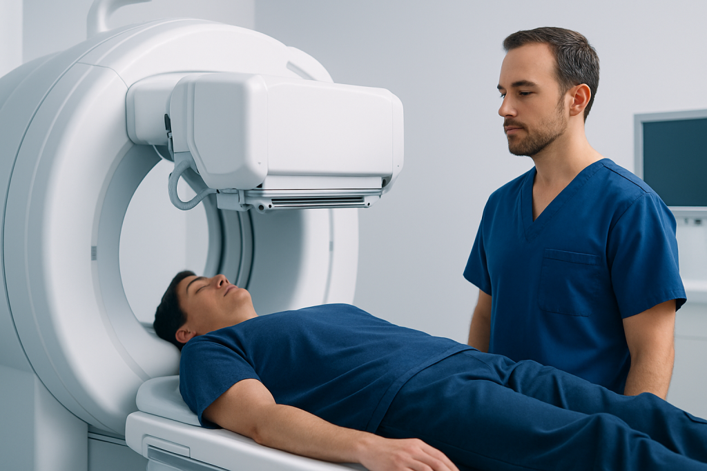
Most diagnostic nuclear medicine procedures begin with a tracer administered by injection, swallowed as a capsule or liquid, or breathed in as a gas. The tracer travels through the body and concentrates in the tissue being studied. A special camera detects the small amount of energy released by the tracer and creates images for the nuclear medicine specialist to assess.
The type of examination depends on the medical question. A bone scan may identify areas of increased bone activity; a myocardial perfusion scan assesses blood supply to heart muscle; thyroid studies assess thyroid function; and PET imaging can help evaluate certain cancers, inflammation or brain disorders. A tracer is not the same as contrast dye used for CT or MRI, although some studies may involve both.
Professional nuclear medicine procedure guidelines are designed to keep radiation exposure as low as reasonably achievable while obtaining useful images. The amount of tracer is individualized according to the test and may take body size, kidney function and clinical need into account. Patients should rely on instructions from their imaging team rather than generic online nuclear medicine procedures PDF documents, since protocols can differ between examinations and hospitals.
Who may need a nuclear medicine assessment?

A doctor may refer someone to a nuclear medicine specialist when symptoms, laboratory results or other scans leave an important question unanswered. The examination is chosen to answer a specific clinical question, such as whether chest symptoms may relate to reduced blood flow to the heart, whether bone pain is associated with an active bone process, or whether a thyroid gland is overactive.
In cancer care, PET/CT and other studies can help characterize a suspected abnormality, determine the extent of some cancers, assess whether treatment is working or investigate possible recurrence. Nuclear medicine imaging may also be used in cardiology, endocrinology, kidney medicine, neurology, orthopedics and infection assessment. It does not replace a complete clinical evaluation, biopsy when needed, or other imaging tests.
Pregnant people, those who may be pregnant, and people who are breastfeeding should tell the referring doctor and nuclear medicine department before an appointment. The team can determine whether the scan should be delayed, modified or performed with particular precautions. Patients should also report allergies, reduced kidney function, recent imaging tests and all medicines, vitamins and supplements they use.
What happens during a nuclear medicine scan?
Before the appointment, the department provides scan-specific preparation instructions. At check-in, a technologist or nurse confirms identity, medical history, medications and pregnancy or breastfeeding status. The patient can ask why the examination is being performed, which tracer will be used, how long the visit may take and what instructions apply afterward.
The tracer is then given in the planned way. Imaging may begin immediately, or there may be a waiting period while the tracer reaches the target tissue. During imaging, the patient is usually asked to lie still on a padded table while a gamma camera, SPECT/CT scanner or PET/CT scanner collects images. The equipment does not normally cause pain, though remaining still or lying flat can be uncomfortable for some people.
Some tests have additional steps. A cardiac stress study may involve monitored exercise or medication that simulates the effect of exercise. A renal scan may include drinking fluids or receiving a medicine that helps assess urine drainage. A nuclear medicine specialist or trained team member supervises the procedure and can respond if a patient feels unwell.
- Appointment length can range from under an hour to several hours, depending on the study and tracer uptake time.
- Patients should bring a current medication list and relevant previous imaging information if requested.
- It is important to follow the department’s instructions even when they differ from general online advice.
What not to do before a nuclear bone scan?
Before a nuclear bone scan, patients should not stop prescribed medicines, fast, or avoid fluids unless the imaging department specifically instructs them to do so. Many bone scans require no special dietary preparation, but individual instructions can vary if the scan is combined with another examination or if medical conditions require adjustments.
Patients should not forget to tell the team about pregnancy, possible pregnancy or breastfeeding. They should also report recent barium studies, contrast examinations, surgeries, fractures, infections and any known cancer history, since these details can affect image interpretation. Wearing comfortable clothing and avoiding unnecessary metal objects may make the visit easier, although the department will explain whether clothing must be changed.
It is also wise not to rely on advice intended for a different nuclear medicine test. For instance, fasting may be important before some PET scans but is not routinely required for every bone scan. The safest preparation is to contact the nuclear medicine department if instructions are unclear or if a scheduled medication might interfere with the study.
Benefits, recovery and what to avoid after a scan
The main benefit of nuclear medicine is that it can reveal changes in tissue function that may not be visible on structural imaging alone. This may help clinicians diagnose conditions earlier, distinguish active from inactive disease, select further testing or monitor treatment. However, an abnormal result is not always a diagnosis by itself, and a normal result does not rule out every possible cause of symptoms.
Recovery after a diagnostic scan is usually straightforward. Most people return to normal daily activities the same day unless they received sedation, underwent a stress test, or were given separate instructions. The tracer naturally loses radioactivity and is cleared from the body over time, commonly through urine and, for some tracers, stool.
What should someone avoid after a nuclear medicine scan? The answer depends on the tracer and the examination. Many patients are advised to drink extra fluids if medically appropriate, urinate regularly and wash hands carefully after using the toilet. For a short period, the department may recommend limiting prolonged close contact with pregnant people, infants or young children. Any restrictions should come directly from the nuclear medicine team, particularly after therapeutic radioactive treatments, which have different aftercare needs from diagnostic imaging.
Risks from diagnostic nuclear medicine scans are generally low. They include exposure to a small amount of ionizing radiation, brief discomfort or bruising at an injection site, and rare allergic-type reactions. The team weighs these considerations against the expected clinical value of the study and uses established safety procedures.
What diseases can nuclear medicine detect?
Nuclear medicine can help detect or assess a wide range of conditions, depending on the tracer and scan type. Examples include coronary artery disease and impaired heart blood flow, overactive or underactive thyroid conditions, bone infection, fractures that may be difficult to see on early X-rays, kidney drainage problems, certain lung circulation problems, dementia patterns and some inflammatory disorders.
It also has an important role in cancer care. PET/CT and other nuclear medicine studies may help evaluate cancers such as lymphoma, lung cancer, colorectal cancer, breast cancer, prostate cancer and neuroendocrine tumors, among others. These tests can help show whether tissue is metabolically active or has features that warrant further investigation, but results may still require correlation with pathology, blood tests and other imaging.
A bone scan can show increased bone turnover from many causes, including arthritis, healing injuries, infection and cancer spread to bone. Therefore, a scan result must be interpreted in the context of the person’s medical history and other findings. A nuclear medicine specialist provides the imaging interpretation, while the wider care team explains what the result means for the individual patient.
When to seek medical care and how results are used
Medical care should be sought promptly for severe or worsening chest pain, sudden shortness of breath, fainting, new weakness, confusion, severe persistent headache, coughing up blood, or other sudden concerning symptoms. These symptoms need urgent assessment and should not wait for an outpatient imaging appointment. After a scan, patients should contact the imaging department or a doctor if they develop concerning symptoms such as significant swelling, breathing difficulty, widespread rash or a reaction that feels severe.
For non-urgent concerns, patients should arrange a medical review when unexplained pain, weight change, fatigue, persistent cough, bone pain, changes in thyroid-related symptoms or other ongoing symptoms do not improve. A clinician can decide whether nuclear medicine imaging is useful or whether another test is more appropriate.
The nuclear medicine report is generally sent to the referring clinician, who discusses the findings in relation to the patient’s symptoms and next steps. At Acibadem International, multidisciplinary specialists in JCI-accredited hospitals can coordinate nuclear medicine assessment and treatment planning for international patients when indicated. Patients should ask their doctor when results are expected and whether further testing, follow-up or referral is needed.
How much money does a nuclear medicine doctor make?
A nuclear medicine doctor’s income varies substantially by country, healthcare system, years of experience, subspecialty training, workplace setting and employment arrangement. It may also differ between academic hospitals, public health services, private practices and cancer centers. For these reasons, there is no single accurate salary figure that applies internationally.
Patients do not need to consider a specialist’s income when deciding whether a scan is appropriate. More useful questions include whether the examination addresses a clear medical question, what preparation is required, how results will affect care and whether an alternative test may be suitable. The referring clinician and nuclear medicine team can explain the purpose of the recommended procedure.
Frequently asked questions
Is a nuclear medicine specialist the same as a radiologist?
Both specialties use medical imaging, but their training and roles may differ by country and institution. Nuclear medicine specialists focus on tracer-based diagnostic imaging and targeted radionuclide therapies, while radiologists interpret a broad range of imaging tests such as X-rays, CT, MRI and ultrasound. In many hospitals, the specialties work closely together, especially for PET/CT and SPECT/CT imaging.
Is radiation from a nuclear medicine scan safe?
Diagnostic nuclear medicine studies use carefully selected, small amounts of radioactive tracer. The team follows radiation safety principles and considers whether the expected information from the test justifies the exposure. Patients who are pregnant, could be pregnant or are breastfeeding should always notify the department before the scan.
Can a person drive after a nuclear medicine scan?
Most people can drive and return to ordinary activities after a routine diagnostic scan. Driving may not be appropriate if sedation was used, if the person feels unwell after a stress test, or if the care team gives different instructions. Patients should confirm plans for travel home when booking the appointment.
How long does it take to receive nuclear medicine results?
Timing varies by examination, the need for image processing and whether comparison with prior studies is required. The nuclear medicine specialist reviews the images and prepares a report for the referring clinician. Patients can ask the clinic when their result is likely to be discussed.
Do all nuclear medicine scans require an injection?
No. Many studies use an injection, but some tracers are swallowed or inhaled. The method depends on the organ being studied and the purpose of the test. The imaging team explains the planned tracer and administration method before the procedure.
Can nuclear medicine diagnose cancer on its own?
Nuclear medicine can provide valuable evidence about the location, extent or activity of some cancers, but it does not always establish a diagnosis by itself. Inflammatory conditions, infection and normal healing can sometimes create scan findings that need further evaluation. Doctors may combine the scan with clinical examination, laboratory testing, CT or MRI, and biopsy when appropriate.
References
- International Atomic Energy Agency
- Society of Nuclear Medicine and Molecular Imaging
- European Association of Nuclear Medicine
- Radiological Society of North America
- National Cancer Institute
This article is for general information only and is not a substitute for professional medical advice. Please consult a qualified doctor about your individual situation.
Medical weight-loss options in Turkey — costs & eligibility
JCI-accredited · board-certified surgeons · reply within 24h
Add us as a Preferred Source to see more of our trusted health content across Google Search, AI Overviews and Discover.