

Obstructive Shock — Explained by Medical Evidence, Not Myths

Obstructive shock happens when blood flow is mechanically blocked, often around the heart, lungs, or major blood vessels. Common causes include pulmonary embolism, cardiac tamponade, and tension pneumothorax.
Key Takeaways
- Obstructive shock happens when blood flow is mechanically blocked, often around the heart, lungs, or major blood vessels.
- Common causes include pulmonary embolism, cardiac tamponade, and tension pneumothorax.
- Symptoms can include low blood pressure, rapid breathing, chest pain, confusion, and bluish skin or lips.
- Diagnosis usually relies on urgent clinical assessment, imaging, and bedside ultrasound.
- Treatment focuses on restoring blood flow quickly while supporting breathing, circulation, and oxygen delivery.
- Anyone with possible shock symptoms needs emergency medical care right away.
Obstructive shock is a medical emergency in which a physical blockage prevents blood from circulating effectively, so the body's organs do not get enough oxygen. It is different from other forms of shock because the main problem is not blood loss or infection, but an obstruction that must be identified and treated quickly.
Overview
Obstructive shock is a severe form of circulatory failure caused by a physical blockage that prevents the heart from filling properly or stops blood from moving forward through the lungs or major vessels. In simple terms, the body may still have blood volume, but the blood cannot circulate where it needs to go. This leads to dangerously low oxygen delivery to the brain, kidneys, heart, and other organs.
Medical teams consider obstructive shock a time-sensitive emergency because the problem usually does not improve unless the obstruction is relieved. Unlike some common explanations online, obstructive shock is not a vague state of “poor circulation.” It has specific mechanical causes, such as a blood clot in the lungs, pressure around the heart, or trapped air in the chest.
It belongs to the broader group of shock states, but it differs from hypovolemic shock, where blood volume is lost, and septic shock, where infection causes widespread vessel changes. In obstructive shock, the central issue is blockage. Identifying the exact cause is critical because treatment for one cause may not help another.
How obstructive shock affects the body
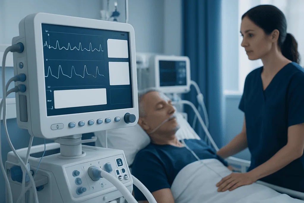
For organs to work normally, oxygen-rich blood must return to the heart, pass through the lungs, and then be pumped out to the body. In obstructive shock, one of these steps is interrupted. The result is a fall in cardiac output, meaning the heart cannot send enough blood forward even if it is beating quickly.
This can happen in several ways. A large clot in the lung arteries can sharply increase pressure in the right side of the heart, making it difficult to move blood through the lungs. Fluid or blood collecting around the heart can squeeze it so it cannot fill well between beats. Air trapped under pressure in the chest can shift structures and reduce blood returning to the heart.
As circulation worsens, the body tries to compensate by increasing heart rate and narrowing some blood vessels. These short-term responses may temporarily support blood pressure, but they cannot solve the mechanical problem. Without urgent treatment, tissue oxygen levels fall, organ function declines, and the condition can become fatal.
Symptoms and warning signs

Symptoms of obstructive shock can develop suddenly and may become severe within minutes or hours. A person may appear very unwell, weak, pale, sweaty, or confused. Low blood pressure is common, but not every patient has clearly low readings at the very beginning.
Common symptoms and signs include:
- Shortness of breath or rapid breathing
- Chest pain or chest pressure
- Fast heart rate
- Dizziness, fainting, or near-fainting
- Cold, clammy skin
- Blue or gray lips, fingernails, or skin
- Confusion, agitation, or reduced alertness
- Very low urine output
Some clues may point toward the cause. Sudden breathlessness and chest pain can suggest a large pulmonary embolism. Neck vein swelling, muffled heart sounds, or severe low blood pressure may raise concern for cardiac tamponade. Sharp breathing difficulty with one-sided chest symptoms may suggest tension pneumothorax.
Because these symptoms overlap with heart attack, severe infection, or other emergencies, it is not safe to diagnose obstructive shock at home. Any suspected shock symptoms should be treated as an emergency.
Main causes and risk factors
The best-known causes of obstructive shock are pulmonary embolism, cardiac tamponade, and tension pneumothorax. Less commonly, severe narrowing or obstruction of major heart valves, a mass pressing on large blood vessels, or complications involving the aorta can interfere with circulation enough to produce shock.
Pulmonary embolism occurs when a blood clot, usually from a deep vein in the leg or pelvis, travels to the lungs and blocks pulmonary arteries. When the clot is large, pressure rises abruptly inside the lung circulation and the right side of the heart may fail. This can lead to severe low blood pressure and signs of shock. Related conditions such as deep vein thrombosis may increase this risk.
Cardiac tamponade happens when fluid, blood, or sometimes pus builds up in the sac around the heart. The pressure around the heart becomes high enough to prevent normal filling. It may develop after trauma, certain cancers, infections, kidney failure, or as a complication of heart procedures.
Tension pneumothorax develops when air enters the space around a lung and becomes trapped under pressure. This pressure can collapse the lung and push on the heart and major veins, reducing blood return to the heart. It can occur after chest injury, lung disease, mechanical ventilation, or occasionally without an obvious cause.
Risk factors vary by cause, but may include recent surgery, prolonged immobility, active cancer, pregnancy, smoking, lung disease, major trauma, central venous procedures, and known clotting disorders.
How doctors diagnose obstructive shock
Diagnosis begins with rapid assessment of airway, breathing, circulation, mental status, oxygen level, and blood pressure. Doctors look for signs that suggest a mechanical obstruction rather than dehydration or infection alone. The pattern of symptoms, examination findings, and bedside monitoring often guides the first steps.
Bedside ultrasound is especially valuable because it can quickly show fluid around the heart, strain on the right side of the heart, or a collapsed lung. In emergency and intensive care settings, ultrasound can help doctors make decisions within minutes. Electrocardiography, chest X-ray, blood tests, arterial blood gas testing, and continuous heart monitoring are also commonly used.
When the person is stable enough, more detailed imaging may be needed. A CT pulmonary angiogram is often used when a large pulmonary embolism is suspected. Echocardiography can identify tamponade physiology and assess heart function. If pneumothorax is likely, doctors may treat it immediately rather than wait for extensive imaging.
Because the condition can worsen rapidly, diagnosis and treatment often happen at the same time. For example, a patient with suspected tamponade may need urgent drainage, while someone with severe clot-related obstruction may need embolism treatment without delay.
Treatment options
Treatment of obstructive shock has two goals: support vital functions and remove the obstruction as quickly as possible. Supportive care may include oxygen, intravenous fluids in selected cases, medications to maintain blood pressure, close monitoring, and help with breathing. The exact approach depends on the cause and the person’s overall condition.
For pulmonary embolism causing shock, treatment may involve blood-thinning medicine, clot-dissolving therapy, catheter-based procedures, or surgery in carefully selected patients. The decision depends on severity, bleeding risk, and how unstable the patient is. In some centers, advanced options are coordinated by multidisciplinary teams experienced in cardiology care.
For cardiac tamponade, urgent drainage of fluid from around the heart is often necessary. This procedure, called pericardiocentesis, relieves pressure so the heart can fill and pump more effectively. If there is trauma or ongoing bleeding, surgery may be needed.
For tension pneumothorax, immediate decompression of the chest is required, followed by placement of a chest drain to allow the trapped air to escape. If the obstruction is due to a structural heart or vessel problem, treatment may involve emergency procedures, surgery, or specialized cardiovascular surgery.
After the immediate crisis is controlled, doctors continue evaluating the underlying cause, prevent complications, and plan follow-up care. Recovery depends on how quickly circulation was restored, the cause of the obstruction, and whether other organs were affected.
Prevention and self-care
Obstructive shock itself is not usually something a person can prevent directly at home, but some underlying causes can be reduced in risk. Preventing blood clots is one important example. After surgery or long periods of limited movement, people may be advised to walk early, use compression devices, stay hydrated, or take prescribed preventive medication.
Managing known heart and lung conditions also matters. People with cancer, clotting disorders, severe lung disease, or a history of thromboembolism should follow their treatment plan carefully and attend regular medical follow-up. Chest injuries and invasive procedures should be monitored for warning signs such as worsening shortness of breath, chest pain, or lightheadedness.
Self-care is not a substitute for emergency treatment when shock is suspected. A person should not attempt to drive themselves if they feel faint, severely short of breath, or confused. Calling emergency services is the safest step.
Near the end of recovery, some patients need rehabilitation, repeated imaging, blood-thinning follow-up, or specialist review. For international patients, Acibadem International’s multidisciplinary specialists and JCI-accredited hospitals diagnose and treat complex emergency and cardiovascular conditions in a coordinated setting.
When to seek medical care
Possible obstructive shock requires immediate emergency medical care. A person should seek urgent help right away for severe shortness of breath, chest pain, fainting, blue lips, sudden confusion, or signs of very low blood pressure such as extreme weakness and cold, clammy skin.
Emergency evaluation is also important after chest trauma, recent major surgery, or known clotting risk if new breathing difficulty or collapse develops. People with suspected pulmonary embolism, tamponade, or pneumothorax should not wait to see whether symptoms pass on their own.
Even if the cause turns out to be different, symptoms of shock should always be assessed promptly by qualified clinicians. Early treatment greatly improves the chance of stabilizing circulation and protecting organs.
Frequently asked questions
What is obstructive shock in simple terms?
Obstructive shock is a dangerous condition where something physically blocks normal blood flow through the heart, lungs, or major blood vessels. Because blood cannot circulate properly, the body's organs do not receive enough oxygen and urgent treatment is needed.
Is obstructive shock the same as a heart attack?
No. A heart attack is caused by blocked blood flow to part of the heart muscle, while obstructive shock describes a broader emergency where circulation is blocked in a way that prevents effective pumping or filling of the heart. However, both can cause chest pain and collapse, so emergency evaluation is important.
What are the most common causes of obstructive shock?
The most common causes are pulmonary embolism, cardiac tamponade, and tension pneumothorax. Each one interferes with blood flow in a different way, which is why doctors work quickly to identify the exact cause.
Can obstructive shock be treated?
Yes, but it requires urgent hospital care. Treatment focuses on supporting breathing and blood pressure while removing the blockage, such as draining fluid around the heart, decompressing the chest, or treating a major blood clot.
How do doctors tell obstructive shock apart from other types of shock?
Doctors use symptoms, physical examination, blood pressure and oxygen monitoring, blood tests, and imaging such as bedside ultrasound, echocardiography, chest X-ray, or CT scans. The overall pattern helps them determine whether a mechanical blockage is present.
Can someone recover fully from obstructive shock?
Many people can recover, especially when the cause is recognized and treated quickly. Recovery depends on the underlying cause, how long organs were under stress, and whether there were complications such as heart strain or kidney injury.
References
- World Health Organization
- American Heart Association
- National Heart, Lung, and Blood Institute
- Society of Critical Care Medicine
- European Society of Cardiology
This article is for general information only and is not a substitute for professional medical advice. Please consult a qualified doctor about your individual situation.
Explore treatments in Turkey — costs, top hospitals & a free quote
JCI-accredited · board-certified surgeons · reply within 24h
Check your numbers in seconds
BMI, calories, due date, blood pressure and 30+ more clinical calculators — free, instant, doctor-reviewed ranges.

More from the Health Library



Related Specialists

Dr. Ayhan Ulusoy
Orthopedic Surgery & Traumatology
Assoc. Prof. Dr. Selim Aydin
Cardiovascular Surgery
Dr. Recep Murat Mert
Emergency Service
Dr. Serkan Ünsal
Intensive Care