Pelvic Phleboliths: A Complete Medical Overview
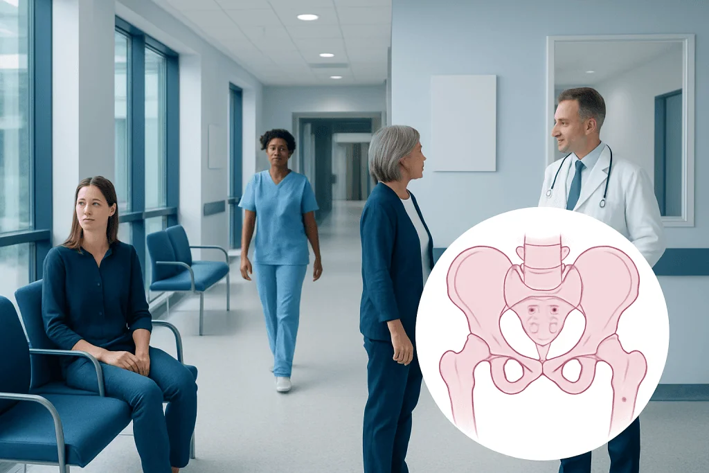
Pelvic phleboliths are usually harmless calcified deposits within pelvic veins. Most people have no symptoms, and the finding does not need treatment by itself.
Key Takeaways
- Pelvic phleboliths are usually harmless calcified deposits within pelvic veins.
- Most people have no symptoms, and the finding does not need treatment by itself.
- They can sometimes be confused with ureter stones on imaging, so careful diagnosis matters.
- Evaluation focuses on ruling out other causes of pelvic, urinary, or abdominal pain.
- Medical care is important if pain is severe, persistent, or associated with urinary symptoms, fever, or blood in the urine.
Pelvic phleboliths are small calcium deposits that form inside veins in the pelvis. They are usually benign, often cause no symptoms, and are commonly discovered incidentally during imaging done for another reason.
Overview: What pelvic phleboliths are
Pelvic phleboliths are small, round calcifications that develop inside veins in the pelvis. In simple terms, they are tiny hardened deposits that can form when a small blood clot in a vein gradually becomes calcified over time. On imaging tests such as X-rays or CT scans, they often appear as smooth, circular spots and are usually an incidental finding rather than a sign of serious disease.
For many people, the main concern is not the phlebolith itself but what it might be mistaken for. Pelvic phleboliths can resemble urinary tract stones, especially stones in the lower ureter, because both can appear as small calcified structures in the pelvis. This is why doctors interpret them in context, using symptoms, physical examination, and sometimes additional imaging.
Phleboliths are common in adults and tend to be seen more often with increasing age. Their presence alone usually does not mean there is active vein disease, infection, or cancer. In most cases, no treatment is needed for the phlebolith itself, but the reason imaging was performed in the first place may still need proper assessment.
Symptoms and what a person may notice
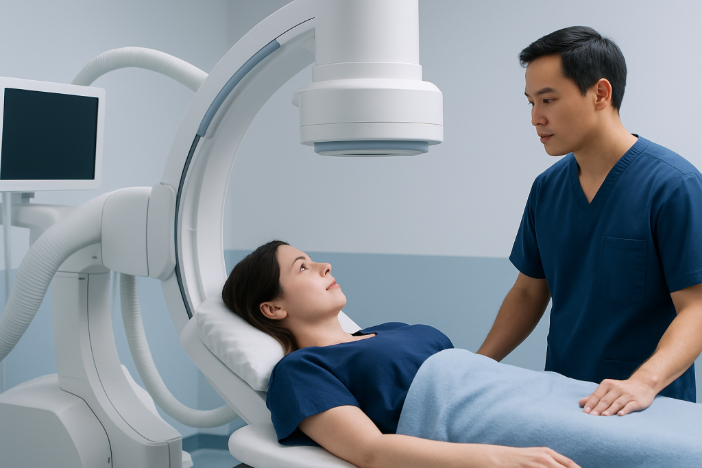
Most pelvic phleboliths do not cause symptoms. Many people learn they have them only after having an imaging test for abdominal pain, pelvic discomfort, back pain, urinary symptoms, or a routine health evaluation. Because they are usually silent, they are often considered a normal incidental radiologic finding in adults.
When symptoms are present, they are often related to another condition rather than to the phlebolith itself. For example, pelvic pain may stem from urinary stones, gynecologic conditions, bowel disorders, or musculoskeletal causes. Doctors therefore avoid assuming that a calcified spot on a scan is the explanation for symptoms without considering the full clinical picture.
If a person has signs that suggest a urinary stone rather than a phlebolith, the pattern may include sudden side or groin pain, nausea, urinary urgency, painful urination, or blood in the urine. In that setting, the main goal of evaluation is to distinguish a benign pelvic phlebolith from kidney stones or other causes of obstruction.
Why pelvic phleboliths form and who is more likely to have them

Pelvic phleboliths form when a small clot or area of slow blood flow within a vein becomes organized and gradually calcifies. They are most often linked to normal age-related changes in the veins rather than to a dangerous process. Veins in the pelvis can be prone to sluggish circulation, which helps explain why this is a common location for phleboliths.
Several factors may make phleboliths more likely. These include increasing age, varicose-type venous changes, prolonged sitting or standing, and conditions associated with venous stasis. Some people with pelvic venous congestion or other chronic vein changes may have them more often, although the calcifications themselves do not necessarily cause symptoms.
Doctors also consider the broader context of pelvic health. In some patients, pelvic imaging is done to investigate chronic pelvic pain or venous disorders, and this can reveal both phleboliths and underlying conditions such as varicose veins. Even then, the mere presence of pelvic phleboliths does not automatically mean treatment is needed.
How doctors tell phleboliths from other pelvic calcifications
The most important part of diagnosis is distinguishing pelvic phleboliths from other calcified structures, especially ureter stones. A doctor usually starts with the person’s symptoms, medical history, and physical examination. If symptoms are absent and the finding is classic on imaging, no further workup may be needed.
On X-ray or CT, phleboliths often appear round and may show a characteristic central lucency, sometimes described as a small lighter center. Their location also matters: they are usually seen along venous pathways in the pelvis. Ureter stones may look denser, follow the expected course of the ureter, and be associated with swelling of the urinary tract on ultrasound or CT.
Imaging tools can help when the diagnosis is uncertain. A doctor may recommend MRI or more commonly CT or ultrasound depending on the suspected cause of symptoms. If urinary obstruction is a concern, evaluation may involve urologic assessment and, when clinically appropriate, procedures such as ureteroscopy for confirmed stones rather than for phleboliths.
Other pelvic calcifications can include lymph node calcifications, calcified fibroids, vascular calcifications, or less commonly masses with calcified components. This is why image interpretation by an experienced radiologist is important, particularly when pelvic pain, urinary symptoms, or atypical imaging features are present.
Treatment options and when no treatment is needed
Pelvic phleboliths themselves usually do not require treatment. If they are discovered incidentally and the person has no concerning symptoms, doctors often simply explain the finding and provide reassurance. This approach helps avoid unnecessary tests or procedures for a benign condition.
Treatment is directed only if there is an underlying problem causing symptoms. For instance, if imaging and symptoms point to a ureter stone, urinary tract infection, pelvic vein disorder, or another pelvic condition, care is tailored to that diagnosis. The management plan may range from observation and pain relief to specialty procedures depending on the cause.
In selected cases, further evaluation by vascular, urology, gynecology, or radiology specialists may be helpful. If a pelvic venous condition is suspected, doctors may consider vein-focused assessment and, in some situations, minimally invasive embolization procedures for the venous disorder, not for the phlebolith itself.
Near the end of a diagnostic pathway, patients who need coordinated assessment may benefit from centers with multiple specialties. Acibadem International’s multidisciplinary specialists and JCI-accredited hospitals evaluate pelvic imaging findings and treat related urinary, vascular, and pelvic conditions for international patients.
Self-care, monitoring, and prevention
Because pelvic phleboliths are usually harmless and often longstanding, there is no specific home treatment to remove them. For most people, the practical approach is to understand the diagnosis, keep copies of imaging reports when possible, and discuss any new symptoms with a doctor rather than assuming they are related to the phleboliths.
There is also no guaranteed way to prevent phleboliths. Still, general vein and overall health habits may support good circulation and lower the risk of venous stasis. These include staying physically active, avoiding very long periods of immobility when possible, maintaining hydration, and following medical advice for conditions that affect circulation.
If a person has recurrent pelvic discomfort, urinary symptoms, or a history of stones, tracking symptom timing can be useful. Helpful details include where the pain starts, whether it radiates to the groin, whether urination changes, and whether there is fever or visible blood in the urine. This information can help a clinician decide whether the issue is likely unrelated to phleboliths and whether additional testing is needed.
- Stay active with regular movement during the day.
- Seek evaluation for new urinary or pelvic symptoms.
- Follow treatment plans for known vein or urinary conditions.
- Avoid self-diagnosing from imaging reports alone.
When to seek medical care
Medical attention is advisable when pelvic phleboliths are reported on imaging and the person also has symptoms that could suggest another condition. These include persistent or severe pelvic pain, flank pain, painful urination, urinary frequency, blood in the urine, fever, vomiting, or trouble passing urine. In these situations, the key issue is to rule out urinary obstruction, infection, or another cause that may need prompt treatment.
A person should also speak with a doctor if pain is recurring, if the imaging findings were described as uncertain, or if there is a history of kidney stones, pelvic vein disease, or gynecologic problems. Pregnant individuals and people with complex medical histories should be assessed professionally rather than relying on incidental imaging findings alone.
Urgent care may be needed for severe pain, inability to urinate, high fever, fainting, or symptoms of significant infection. Even though pelvic phleboliths are usually benign, new or intense symptoms should not be assumed to come from them without a clinical evaluation.
Frequently asked questions
Are pelvic phleboliths dangerous?
Pelvic phleboliths are usually not dangerous. They are commonly benign calcified deposits in pelvic veins and often do not cause symptoms or require treatment.
Can pelvic phleboliths cause pain?
Most pelvic phleboliths do not cause pain. If someone has pelvic, abdominal, or flank pain, doctors usually look for another cause first, such as a urinary stone, infection, or a gynecologic or bowel condition.
How are pelvic phleboliths different from kidney stones?
Pelvic phleboliths are calcifications inside veins, while kidney stones form in the urinary tract. They can look similar on imaging, especially in the pelvis, but doctors use the location, shape, symptoms, and sometimes additional scans to tell them apart.
Do pelvic phleboliths need to be removed?
In most cases, no. Because they are usually harmless incidental findings, removal is not needed, and treatment is aimed only at any separate condition that may be causing symptoms.
Can pelvic phleboliths go away on their own?
They generally do not disappear, because they are calcified deposits. However, they often remain stable and do not cause problems, so their presence alone is usually not a reason for concern.
What tests are used to diagnose pelvic phleboliths?
Pelvic phleboliths are most often seen on X-rays or CT scans. Ultrasound, MRI, and urine testing may also be used when doctors need to rule out urinary stones or other causes of symptoms.
References
- National Institute of Diabetes and Digestive and Kidney Diseases
- American College of Radiology
- Radiological Society of North America
- National Health Service
- Merck Manual Consumer Version
This article is for general information only and is not a substitute for professional medical advice. Please consult a qualified doctor about your individual situation.
Explore treatments in Turkey — costs, top hospitals & a free quote
JCI-accredited · board-certified surgeons · reply within 24h
Add us as a Preferred Source to see more of our trusted health content across Google Search, AI Overviews and Discover.