Screening Plate: How It Works, Results and What to Expect

A screening mammogram uses low-dose X-rays to create images, sometimes called plates, of each breast. Compression is an important part of standard mammography because it improves image quality and reduces radiation exposure.
Key Takeaways
- A screening mammogram uses low-dose X-rays to create images, sometimes called plates, of each breast.
- Compression is an important part of standard mammography because it improves image quality and reduces radiation exposure.
- A screening result may be normal, may recommend routine follow-up, or may request additional imaging for clarification.
- Being called back after a screening mammogram is common and does not by itself mean cancer is present.
- New breast symptoms should be assessed by a clinician even if a recent screening mammogram was normal.
A screening plate usually refers to an X-ray image obtained during a screening mammogram. These images allow trained radiologists to look for breast changes that may be too small to feel, while most people with an abnormal screening result do not have breast cancer.
What Is a Screening Plate?
A screening plate is a term sometimes used for one of the images produced during a screening mammogram. Mammography uses low-dose X-rays to examine breast tissue for changes that may not cause symptoms or be felt during a breast examination. Today, the images are usually digital rather than physical film plates, but the older wording is still sometimes used.
For most screening mammograms, images are taken from more than one angle for each breast. This helps the radiologist view tissue that overlaps differently in each projection and compare both breasts. The purpose is to identify findings such as a new mass, an area of distortion, or small calcium deposits that may need a closer look.
A screening plate is not a diagnosis. It is one part of a preventive health assessment, and its meaning depends on the full set of images, prior mammograms when available, personal history, and the radiologist’s interpretation.
How Does Mammogram Screening Work?
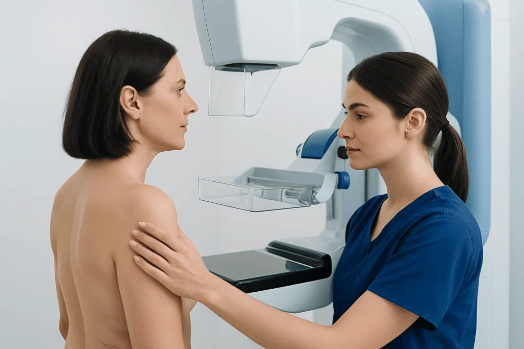
Mammogram screening is designed for people who do not have a new breast symptom. During the examination, a radiographer positions one breast at a time on the mammography unit. A clear plastic paddle briefly and firmly compresses the breast while an X-ray image is taken.
Compression spreads breast tissue into a thinner layer. This reduces motion, limits overlapping tissue, and can make subtle changes easier to detect. It also allows the machine to use less radiation than would otherwise be needed. Although it can feel uncomfortable, the pressure lasts only for the few seconds needed for each image.
Some centers use digital breast tomosynthesis, often called 3D mammography. It collects multiple low-dose images from different angles and reconstructs thin image slices. This can help the radiologist assess areas where normal tissue overlaps, but the best screening approach depends on the person, local practice, and available technology.
The term screening portion may be used when discussing the preventive mammogram images separately from any extra diagnostic views. If an image needs clarification, additional views or ultrasound may be arranged rather than trying to interpret a single image in isolation.
How Is a Screening Test Done?

Before the appointment, the person may be asked to avoid deodorant, powder, lotion, or perfume on the chest and underarm area because some products can leave particles that resemble tiny calcium deposits on an X-ray. They should tell the imaging team about breast implants, previous breast surgery, pregnancy or possible pregnancy, breastfeeding, and any symptoms or prior abnormal imaging.
At the appointment, the person changes from the waist up and stands beside the mammography machine. The radiographer positions the breast carefully, applies compression, and takes standard images. The process is repeated on the other side. Positioning can be adjusted to ensure that as much breast tissue as possible is included.
The examination itself commonly takes around 15 to 30 minutes, though timing varies. The X-ray exposure for each image is very brief. Most people can leave immediately after the study and return to normal daily activities without a recovery period.
Screening for breast changes does not replace breast awareness. People should become familiar with what is normal for their own breasts and report a new lump, skin change, nipple change, or persistent focal pain. Advice sometimes described as examining the breasts each month should not be viewed as a substitute for scheduled screening or clinical assessment of symptoms.
Who Is a Candidate for Screening Mammography?
Screening recommendations differ among countries and professional organizations because they balance the likely benefits of earlier detection against the possibility of false-positive results and additional testing. Eligibility is usually based on age and average or increased breast cancer risk. A clinician can help determine when to start, how often to screen, and which imaging method is most appropriate.
People at increased risk may need an individualized plan. Factors that can influence this include a strong family history of breast or ovarian cancer, known inherited genetic variants, previous chest radiation at a young age, certain high-risk breast biopsy findings, or a personal history of breast cancer. In some situations, breast MRI may be recommended in addition to mammography.
Mammography is generally safe, and the radiation dose is low. However, pregnancy, possible pregnancy, or breastfeeding should always be discussed with the care team so that imaging can be planned appropriately. People with implants can still have mammograms, but additional positioning and views may be needed.
A person with a new symptom is not having a routine screening situation. They may require diagnostic mammography, ultrasound, or another targeted evaluation. This distinction matters because diagnostic imaging is tailored to the particular concern rather than following only standard screening views.
Can You Show Me a Video of the Process of a Mammogram?
A written health guide cannot replace an in-person demonstration or provide a procedure video, but many accredited breast imaging departments and recognized public health organizations offer educational mammogram videos. These typically show the changing area, the standing position at the machine, breast placement, short periods of compression, and the image-taking process.
When choosing a video, it is helpful to use material from a hospital, national cancer organization, radiology society, or public health authority. Videos can prepare a person for the practical steps, but the radiographer may position each person differently to obtain safe, high-quality images.
Anyone worried about pain, mobility, implants, a prior difficult mammogram, or a disability should contact the imaging center before the appointment. The team can discuss reasonable adjustments, including additional time, positioning support, or communication needs.
Is It Possible to Have a Mammogram Without Compression?
Standard mammography requires some degree of breast compression. It is a core part of the examination because it improves visibility of breast tissue, decreases blurring from movement, and helps keep the radiation dose as low as reasonably achievable. A mammogram performed without adequate compression may be less reliable and may need to be repeated.
The radiographer aims to use the minimum compression needed to obtain diagnostic-quality images. The pressure should be brief, and the person can communicate at any point if it is too painful or if they need a pause. Scheduling after menstrual-related breast tenderness has eased may help some people feel more comfortable.
For certain clinical questions, other tests such as breast ultrasound or MRI may be useful. However, they are not direct replacements for mammography in every person or screening setting. The imaging team selects tests based on breast density, symptoms, risk factors, previous imaging, and the question being investigated.
People who experience significant discomfort should not simply avoid screening. Discussing previous pain with the radiographer and referring clinician can help identify practical options for making the visit more manageable.
Understanding Screening Results and Follow-Up
Screening results meaning can be difficult to interpret without context. A report may indicate that no suspicious abnormality was seen and advise returning at the usual screening interval. It may also identify a finding that requires additional images, ultrasound, or comparison with older mammograms before the radiologist can make a clearer assessment.
A callback is not the same as a cancer diagnosis. Dense breast tissue, overlapping tissue, harmless cysts, stable benign changes, or an image that needs a different angle can all lead to further assessment. If an area remains concerning after diagnostic imaging, a needle biopsy may be recommended to examine cells or tissue directly.
Radiology reports often use a structured assessment system to communicate whether routine screening, short-interval follow-up, further imaging, or biopsy is advised. The referring clinician or breast imaging team should explain what the category means for the individual and what the next step is.
Bringing prior mammograms, or ensuring the new facility can obtain them, can be very helpful. Comparison over time enables radiologists to see whether a finding is new, stable, or changing. The question “what are we checking for in plate 1” is best answered as part of the entire image set: each view contributes different information rather than serving as a standalone test.
When to Worry About Mammogram Results and Seek Medical Care
It is understandable to feel anxious while waiting for mammogram results, especially if additional testing is requested. The appropriate response is to complete the recommended follow-up promptly and ask the care team to explain the reason for the next test. Most callbacks are resolved without a cancer diagnosis, but timely clarification is important.
A person should contact a doctor without waiting for their next routine screening appointment if they notice a new breast or underarm lump, persistent one-sided breast change, skin dimpling or thickening, nipple inversion that is new, bloody or spontaneous nipple discharge, or a new persistent area of focal pain. These signs can have non-cancer causes, but they deserve assessment.
Urgent medical attention is appropriate for severe breast pain with fever, marked redness, swelling, or feeling unwell, particularly during breastfeeding, as infection may need treatment. Mammograms do not diagnose every breast condition, and a normal recent screening examination should not prevent someone from seeking care for a new symptom.
Acibadem International’s multidisciplinary specialists and JCI-accredited hospitals support international patients who need breast imaging evaluation and follow-up planning. A qualified clinician can review individual risk factors, symptoms, and imaging results to recommend appropriate care.
Frequently asked questions
What does a screening plate mean in a mammogram?
A screening plate generally means one mammogram image used to examine breast tissue. Modern mammograms are usually stored as digital images, although the term plate may still be used informally. Radiologists interpret all images together rather than relying on one view alone.
How long does a screening mammogram take?
The appointment commonly takes about 15 to 30 minutes, depending on positioning, the need for additional views, and the imaging center's workflow. The actual X-ray exposure lasts only seconds for each image. Most people return to normal activities immediately afterward.
Does mammogram compression damage breast tissue?
Mammogram compression is designed to be brief and should not damage breast tissue. It may cause temporary discomfort or tenderness, particularly in people with sensitive breasts. Tell the radiographer if the pressure is painful so they can pause or adjust positioning when possible.
Why am I being called back after a screening mammogram?
A callback means the radiologist needs more information, such as additional mammogram views, an ultrasound, or comparison with previous images. This is common and may result from overlapping tissue, a benign-looking change, or an image that was not fully clear. A callback alone does not mean that cancer has been found.
Can I have a mammogram if I have breast implants?
Yes, people with breast implants can usually have mammograms. The imaging team should be told in advance because special positioning and additional views may be needed to visualize as much breast tissue as possible. The team can also discuss any implant-related concerns before the examination.
Should I wait for my next mammogram if I find a lump?
No. A new lump or other breast symptom should be discussed with a doctor promptly, even if a routine screening mammogram is scheduled soon or was recently normal. The clinician may arrange diagnostic imaging tailored to the symptom.
References
- American Cancer Society
- U.S. Food and Drug Administration
- Radiological Society of North America
- American College of Radiology
- World Health Organization
This article is for general information only and is not a substitute for professional medical advice. Please consult a qualified doctor about your individual situation.
Oncology care in Turkey — second opinion and treatment plan
JCI-accredited · board-certified surgeons · reply within 24h
Add us as a Preferred Source to see more of our trusted health content across Google Search, AI Overviews and Discover.