Spleen MRI: Preparation, Procedure and Results
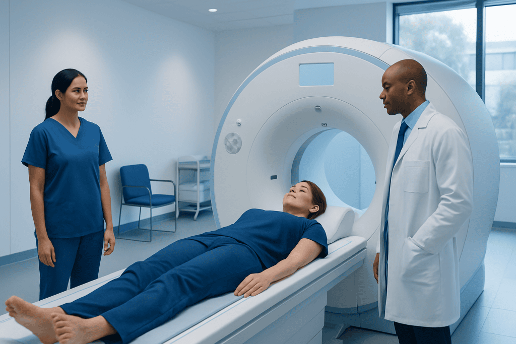
A spleen MRI produces detailed images of splenic tissue and nearby abdominal organs without X-ray radiation. Preparation may include screening for metal implants, discussing kidney health and arranging contrast when needed.
Key Takeaways
- A spleen MRI produces detailed images of splenic tissue and nearby abdominal organs without X-ray radiation.
- Preparation may include screening for metal implants, discussing kidney health and arranging contrast when needed.
- Most spleen lesions found incidentally are benign, but imaging features and personal medical history guide assessment.
- The examination is painless, although remaining still in a confined scanner and holding the breath briefly can be challenging for some people.
- Urgent medical assessment is needed for severe left upper abdominal pain, especially after an injury, fainting or signs of internal bleeding.
A spleen MRI is a detailed, noninvasive imaging examination that uses a powerful magnet and radio waves to evaluate the spleen. It may help clarify an unexpected finding on ultrasound or CT, investigate symptoms, or assess known conditions without using ionizing radiation.
Overview: What Is a Spleen MRI?
A spleen MRI, also called a spleen MRI scan, is an imaging test that creates detailed pictures of the spleen, a small organ in the upper left side of the abdomen. The examination uses a strong magnetic field and radio waves rather than X-rays. It can help clinicians examine the spleen’s size, structure, blood supply and any areas that look different from normal tissue.
Doctors may request this scan after an ultrasound or CT has shown an unclear spot, called a lesion. It may also be used to assess an enlarged spleen, possible infection, inflammation, blood-related conditions, trauma, or the extent of certain cancers. MRI is particularly useful for characterizing tissue and can sometimes reduce the need for further invasive testing.
A radiologist interprets the images in the context of the person’s symptoms, previous scans and medical history. A spleen MRI result alone does not always establish a diagnosis; it is one part of a wider clinical assessment.
How a Spleen MRI Works and Who May Need One
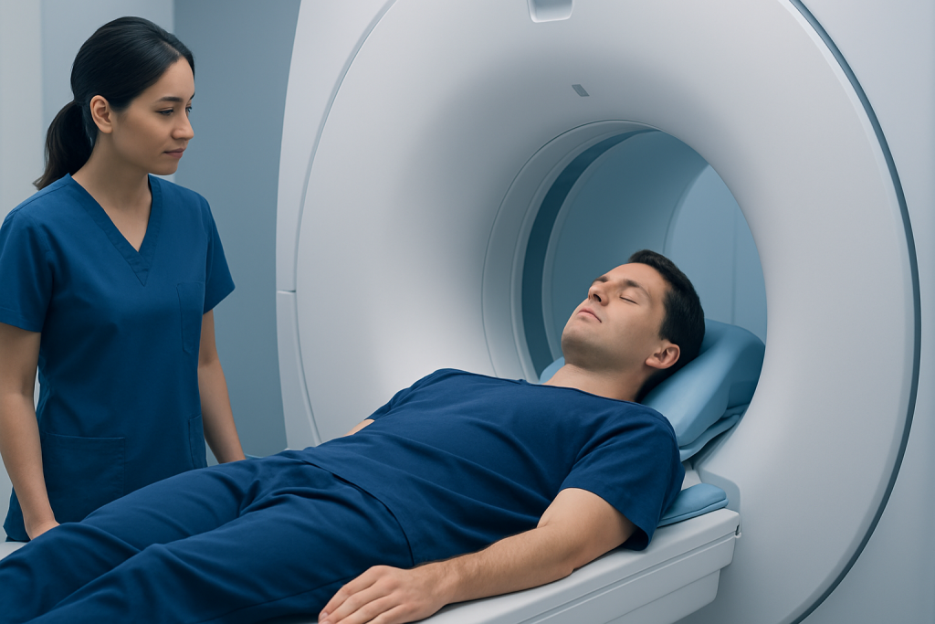
MRI equipment aligns hydrogen particles in the body using a magnetic field, then uses radiofrequency pulses to obtain signals from tissues. A computer converts these signals into highly detailed images. Different imaging sequences, often referred to as a spleen MRI protocol, allow the radiology team to compare tissue characteristics, fluid content and blood flow.
Some examinations include an intravenous contrast agent containing gadolinium. Contrast can make blood vessels and differences between normal and abnormal tissue easier to see. Whether it is required depends on the reason for the scan and findings on earlier imaging. The care team reviews kidney function and any previous contrast reactions before recommending it.
A spleen MRI may be appropriate for adults or children when detailed assessment is needed and MRI is safe for them. It is not suitable for everyone. Certain implanted devices, metal fragments, some surgical clips and older pacemakers may prevent MRI or require specialist safety review. Pregnancy, severe claustrophobia and difficulty lying flat should also be discussed before the appointment.
Preparing for a Spleen MRI

The imaging department provides individualized instructions, so these should always take priority. People are usually asked to complete an MRI safety questionnaire and inform staff about any pacemaker, implant, hearing device, neurostimulator, metal injury, surgery, pregnancy, kidney disease or allergy history. They should also bring previous imaging reports or discs if requested.
For some abdominal MRI examinations, fasting for several hours beforehand may improve image quality by reducing movement in the stomach and bowel. Other scans do not require fasting. Patients should ask whether they may eat, drink and take their usual medicines before the examination rather than making changes independently.
What not to do 24 hours before MRI? Most people do not need major restrictions 24 hours before a spleen MRI. They should not stop prescribed medication unless a clinician specifically tells them to do so, and they should avoid arriving with metal items such as jewelry, watches, hairpins, belts, bank cards or electronic devices. If fasting instructions were given, they should avoid food and drinks during the stated period; alcohol or recreational substances should be avoided if they could affect comfort, safety or the ability to remain still.
People who are concerned about enclosed spaces can contact the imaging team ahead of time. In some circumstances, a clinician may discuss relaxation approaches or medicine for anxiety. If sedating medicine is used, the patient will need someone else to take them home and should follow the team’s instructions regarding food, drink and driving.
Step-by-Step: What Happens During the Procedure?
On arrival, a radiographer checks the safety questionnaire, explains the process and asks the patient to change into a gown if necessary. All metal and electronic belongings are stored safely outside the scan room. If contrast is planned, staff may place a small cannula in a vein, usually in the arm or hand.
The patient lies on a movable table, commonly on their back, with a receiver coil positioned around the abdomen. The table moves gradually into the scanner. The machine makes repetitive tapping, knocking and humming sounds, so earplugs or headphones are provided. The radiographer watches and speaks with the patient from an adjacent room, and the patient can use an alarm device if they need assistance.
During a spleen MRI scan, the radiographer may ask for short breath-holds, often lasting only several seconds, to reduce motion on the images. Remaining as still as possible is important. If contrast is injected, some people notice a cool sensation in the arm; this usually passes quickly. The scan commonly takes around 30 to 60 minutes, though timing varies with the planned sequences and whether contrast is used.
What if I have to pee during an MRI? The patient should tell the radiographer as soon as possible using the call button or intercom. In many cases, staff can pause the scan safely and help the patient use the toilet before continuing. It is better to communicate discomfort than try to endure it, as movement can affect image quality.
Benefits, Risks and Recovery Timeline
The main benefit of MRI is its ability to provide detailed soft-tissue images without ionizing radiation. For splenic findings, it may help distinguish common benign conditions from findings that need closer follow-up. It can also provide useful information about adjacent structures, such as the liver, pancreas, lymph nodes and abdominal blood vessels.
The scan itself is painless. The most common challenges are noise, the need to lie still and a feeling of confinement. Serious problems are uncommon, but MRI safety screening is essential because the magnetic field can move, heat or interfere with certain metal objects and implanted devices. Contrast reactions are uncommon and are usually mild, though the imaging team is prepared to manage them.
Most people can return to normal activities immediately after a non-sedated scan. If contrast was used, normal hydration is generally encouraged unless a clinician has advised fluid restriction. Anyone who received sedation should not drive, drink alcohol, operate machinery or make important decisions until the effects have fully worn off, according to the care team’s guidance.
There is usually no physical recovery period after a spleen MRI. The radiologist reviews the images and sends a report to the referring clinician, who explains the results and any next steps. This may include comparison with prior scans, laboratory tests, follow-up imaging or referral to an appropriate specialist.
Understanding Spleen MRI Results
A report may describe the spleen as normal in size and appearance, enlarged, injured, congested or containing one or more lesions. Lesions may be cysts, vascular growths such as hemangiomas, areas related to infection or inflammation, changes following injury, or less commonly tumors. The report may also mention findings outside the spleen that are relevant to the clinical question.
What percentage of spleen lesions are cancerous? There is no single reliable percentage that applies to every person or every type of splenic lesion. Many lesions discovered incidentally in people without a known cancer are benign, while the likelihood of malignancy is higher when there is a history of cancer, concerning symptoms, multiple abnormalities or suspicious imaging features. A radiologist considers lesion appearance, contrast behavior, size, growth over time and the individual’s clinical history rather than relying on a percentage alone.
Not every unclear finding needs immediate treatment. Depending on the result, clinicians may recommend no action, repeat imaging after a defined interval, blood tests, consultation with hematology, gastroenterology or oncology, or occasionally a biopsy or surgery. Decisions should be individualized because biopsy of the spleen requires careful planning due to bleeding risk.
At Acibadem International, multidisciplinary specialists in JCI-accredited hospitals can coordinate imaging review and further assessment for international patients when a spleen finding needs evaluation.
When to Seek Medical Care
What are the first signs of a bad spleen? The spleen can be affected by many different conditions, so there is no single early warning sign. Possible symptoms include discomfort or fullness beneath the left ribs, feeling full quickly after eating, unexplained fatigue, fever, repeated infections, easy bruising or symptoms related to anemia. These symptoms are nonspecific and can have many causes, but they should be discussed with a qualified clinician if they persist or worsen.
Urgent medical assessment is important after a blow to the upper left abdomen, lower left chest or left shoulder, particularly if pain is increasing. Emergency care is also needed for severe abdominal pain, dizziness, fainting, confusion, marked weakness, rapid heartbeat, pale or clammy skin, or trouble breathing, as these can be signs of significant internal bleeding or another serious condition.
People should contact their doctor promptly if they have a known enlarged spleen and develop new pain, fever or increasing abdominal fullness. Those with an abnormal MRI result should arrange follow-up with the clinician who ordered the scan, even when they feel well, so the finding can be interpreted appropriately and a clear care plan can be made.
Frequently asked questions
Is a spleen MRI painful?
A spleen MRI is not painful. The person needs to lie still on a padded table and may be asked to hold their breath briefly. Some people find the scanner noise or enclosed space uncomfortable, but the radiographer remains in contact throughout the examination.
Do I need contrast for a spleen MRI?
Not every spleen MRI requires contrast. Gadolinium contrast may be used when the clinician needs more information about blood flow or the characteristics of a splenic lesion. The team will review kidney health, pregnancy status and prior contrast reactions before using it.
How long does a spleen MRI take?
Many examinations take approximately 30 to 60 minutes. The exact duration depends on the imaging protocol, whether contrast is used and whether additional abdominal images are required. Allow extra time for registration, screening and preparation.
Can I eat before a spleen MRI?
The answer depends on the type of examination and the imaging department’s instructions. Some abdominal MRI studies require fasting for several hours, while others do not. Patients should follow the instructions provided by their care team and continue prescribed medicines unless told otherwise.
Can an MRI tell whether a spleen lesion is benign?
MRI can often provide strong clues about whether a lesion has benign or concerning features, especially when contrast-enhanced images are available. However, no imaging test is perfect, and some findings require follow-up scans, blood tests or specialist review. The result should be interpreted alongside the patient’s history and other investigations.
Can I have an MRI if I have a pacemaker or metal implant?
Some modern implanted devices are MRI-conditional and can be scanned under specific safety procedures, while others may not be suitable. Patients should never assume an implant is safe for MRI. They should provide the device information to the imaging department well before the appointment so the team can assess it.
References
- American College of Radiology
- Radiological Society of North America
- National Institute of Diabetes and Digestive and Kidney Diseases
- Merck Manual Professional Edition
- National Cancer Institute
This article is for general information only and is not a substitute for professional medical advice. Please consult a qualified doctor about your individual situation.
Explore treatments in Turkey — costs, top hospitals & a free quote
JCI-accredited · board-certified surgeons · reply within 24h
Add us as a Preferred Source to see more of our trusted health content across Google Search, AI Overviews and Discover.

More from the Health Library



Related Specialists

Prof. Dr. Gamze Erfan
Dermatology
Dr. Yalçın Culha
Inpatient Clinic Physicians Clinical Service
Prof. Dr. İbrahim Bildirici
Gynecology & Obstetrics
Dr. Mahmut Baktir
Pediatrics