Vur Ultrasound: Preparation, Procedure and Results

VUR ultrasound is painless, radiation-free and commonly used to monitor kidney and bladder health. A renal and bladder ultrasound may show kidney swelling, scarring concerns, size differences or bladder-emptying issues, but it cannot reliably grade reflux.
Key Takeaways
- VUR ultrasound is painless, radiation-free and commonly used to monitor kidney and bladder health.
- A renal and bladder ultrasound may show kidney swelling, scarring concerns, size differences or bladder-emptying issues, but it cannot reliably grade reflux.
- A voiding cystourethrogram (VCUG) is commonly used when clinicians need to confirm or grade VUR.
- Children with recurrent urinary tract infections, prenatal kidney dilation or known VUR may need ultrasound follow-up.
- Most children with VUR do well with monitoring, infection prevention and, when appropriate, medication or surgery.
A VUR ultrasound is a noninvasive imaging test that uses sound waves to examine the kidneys, ureters and bladder in people with vesicoureteral reflux (VUR). It does not usually show reflux happening directly, but it can identify urinary tract changes that help clinicians assess kidney health and plan follow-up.
What Is a VUR Ultrasound?
A VUR ultrasound, also called a renal and bladder ultrasound, is an imaging examination used to assess the urinary system in a person with vesicoureteral reflux (VUR). VUR occurs when urine flows backward from the bladder toward one or both ureters and kidneys. The ultrasound uses high-frequency sound waves, not radiation, to create images of the kidneys and bladder.
The scan does not usually capture the backward flow of urine itself. Instead, it looks for effects that may be associated with reflux, such as widening of the kidney’s drainage system, changes in kidney size, structural differences, bladder abnormalities or urine remaining after the bladder is emptied. It is often one part of a broader evaluation of vesicoureteral reflux.
Ultrasound is particularly useful because it is comfortable and safe for people of all ages, including infants and children. A clinician may request it after a urinary tract infection, following an abnormal prenatal scan, when VUR is suspected, or during ongoing monitoring of known reflux.
How It Works, Who May Need It and What It Can Show
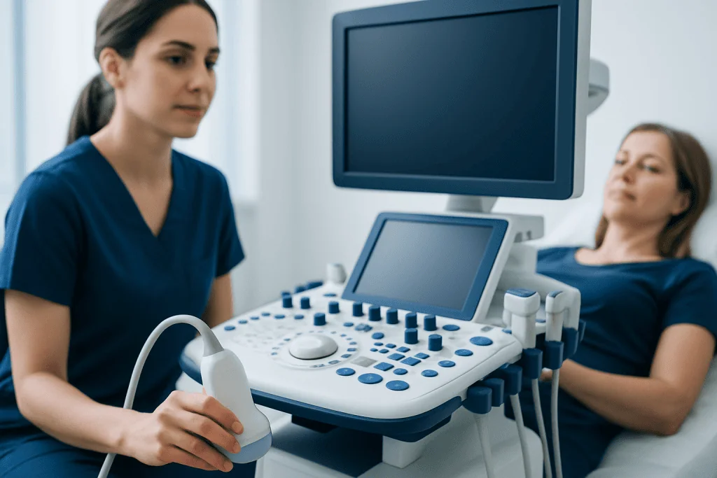
During an ultrasound, a sonographer moves a small handheld device called a transducer over the abdomen and lower back. The transducer sends sound waves into the body and receives returning echoes, which a computer turns into real-time images. A clear gel is placed on the skin to help transmit the sound waves.
A VUR ultrasound may be appropriate for infants, children or adults who have recurrent urinary tract infections, unexplained fever with suspected urinary infection, known VUR, kidney dilation seen before birth, urinary symptoms, or a family history of urinary tract abnormalities. It may also be used after treatment to monitor kidney growth and drainage.
The examination can assess kidney size, shape and position; dilation of the renal pelvis or ureters; possible stones or other structural findings; bladder wall appearance; and, in some cases, the amount of urine left after voiding. A normal ultrasound is reassuring, but it does not rule out VUR. If reflux must be confirmed or graded, a clinician may recommend a voiding cystourethrogram (VCUG) or another specialized imaging study.
- Benefit: no radiation, injections or sedation are usually needed.
- Limitation: it cannot reliably determine whether urine flows backward during urination.
- Clinical role: it provides a baseline and helps track possible urinary tract changes over time.
How Should I Prepare for a Urinary Bladder Ultrasound?

Preparation for a urinary bladder ultrasound is usually simple. The imaging center will provide instructions based on the person’s age, symptoms and whether the kidneys, bladder or both are being examined. In many cases, the person is asked to arrive with a comfortably full bladder because this improves the view of the bladder and nearby structures.
Adults and older children may be asked to drink water before the appointment and avoid urinating until after the first images are taken. The amount and timing vary by age and facility, so it is best to follow the specific instructions provided. Infants and young children are not usually expected to hold urine for long periods; caregivers may simply be asked to offer fluids before the visit when practical.
People should generally continue usual medicines unless their clinician advises otherwise. Comfortable, two-piece clothing can make the scan easier, and a parent or caregiver may remain with a child during the test. Inform the imaging team about recent urinary symptoms, prior surgery, current infection or difficulty emptying the bladder.
VUR Ultrasound Procedure: Step by Step
A VUR ultrasound is performed in an imaging department, clinic or hospital and commonly takes about 20 to 40 minutes, although timing can vary. The person lies on an examination bed, usually first on the back and sometimes on either side or the stomach. For babies and younger children, gentle positioning and reassurance are typically all that is required.
The sonographer applies warm or room-temperature gel to the abdomen, side and lower back, then moves the transducer over the area. Images are taken of each kidney and the bladder. If a full bladder assessment is needed, the person may be asked to urinate partway through the appointment so the bladder can be scanned again afterward.
The scan is painless. Mild pressure from the transducer may feel uncomfortable if the bladder is very full or the abdomen is tender, but it should not cause pain. There are no needles, contrast dye or recovery room for a standard ultrasound. A radiologist reviews the images and sends a report to the requesting clinician, who explains what the findings mean in the context of symptoms and other test results.
Results, Recovery, Benefits and Possible Risks
After a VUR ultrasound, normal activities, eating and drinking can resume immediately. The gel is wiped away, and there is no radiation exposure or medication effect to recover from. Results may be available the same day in some settings, but the formal report often takes longer depending on local processes.
Possible findings include normal kidneys and bladder; hydronephrosis, meaning dilation of the kidney’s urine-collecting area; differences in kidney size; bladder distension; or residual urine after voiding. These findings do not automatically mean that reflux is severe. They help the clinical team decide whether observation, repeat imaging, urine testing, a VCUG or referral to pediatric urology or nephrology is appropriate.
The main advantages of ultrasound are safety, convenience and the ability to monitor changes over time without radiation. Risks are minimal and generally limited to temporary discomfort from a full bladder or light probe pressure. Ultrasound findings should always be interpreted alongside the person’s infection history, urine tests, blood pressure, kidney function when relevant and other imaging.
How Serious Is Vesicoureteral Reflux?
Vesicoureteral reflux ranges from mild to more significant. Many children with low-grade VUR have no lasting kidney problems and may outgrow reflux as the urinary tract matures. The concern is not simply reflux itself, but the possibility that bacteria can travel upward during a urinary tract infection and contribute to kidney infection or, less commonly, kidney scarring.
The likelihood of complications depends on factors such as the grade of reflux, age, whether infections recur, the presence of kidney abnormalities, bladder and bowel habits, and whether treatment plans are followed. Prompt assessment of fever or possible urinary infection is important in infants and young children, as symptoms may be nonspecific.
Clinicians individualize care. This may include observation with periodic ultrasounds, urine testing when symptoms occur, strategies to prevent constipation and support regular bladder emptying, preventive antibiotics for selected patients, or surgery when reflux persists with breakthrough infections or kidney concerns. VUR treatment options are chosen according to the individual’s anatomy, infection history and kidney findings.
Is VUR More Common in Boys or Girls?
VUR is diagnosed more often in girls overall, especially after urinary tract infections, because urinary infections are more common in girls after infancy. However, among newborns and young infants identified because of prenatal urinary tract dilation or early urinary problems, VUR may be recognized relatively often in boys as well.
Sex is only one part of the picture. Family history, urinary tract anatomy, prenatal ultrasound findings, bowel and bladder function, and previous infections may all influence whether testing is considered. Siblings of a child with VUR may have a higher likelihood of reflux, although routine testing is not necessary for every sibling.
Regardless of sex, a child with fever without an obvious cause, painful urination, foul-smelling urine, new wetting after toilet training, abdominal or back pain, or recurrent urinary infections should be assessed by a qualified clinician. Testing decisions are based on age, symptoms and medical history rather than sex alone.
Is VUR Surgery Successful? When to Seek Medical Care
VUR surgery is generally considered when reflux is persistent or high grade, when urinary infections continue despite preventive measures, or when there are concerns about kidney damage. Procedures may include endoscopic injection of a bulking material near the ureter opening or ureteral reimplantation surgery. The best approach depends on the reflux grade, urinary tract anatomy, age and prior treatment history.
Both surgical approaches can be effective, but no procedure is appropriate for every person and outcomes vary. Endoscopic treatment is less invasive and may be suitable for selected children, while reimplantation has a longer recovery but may be recommended for more complex cases. A pediatric urologist can explain expected benefits, limitations, follow-up testing and alternatives. For children requiring intervention, pediatric urology care can coordinate imaging, infection prevention and surgical planning.
Medical care should be sought promptly for fever in an infant or young child, fever with urinary symptoms, pain in the side or back, vomiting with suspected urinary infection, blood in the urine, reduced urine output, marked sleepiness, or a child who appears significantly unwell. Acibadem International’s multidisciplinary specialists and JCI-accredited hospitals provide diagnostic assessment and treatment planning for international patients with urinary tract conditions.
Frequently asked questions
Can an ultrasound diagnose VUR?
An ultrasound cannot reliably diagnose or grade VUR because it does not usually show urine flowing backward from the bladder to the kidneys. It can identify changes that may be linked to reflux, such as kidney dilation or structural differences. A VCUG is commonly used when confirmation or grading of VUR is needed.
Does a VUR ultrasound use radiation?
No. Ultrasound uses sound waves to create images and does not expose the person to ionizing radiation. This makes it useful for children who may need repeat imaging during follow-up.
Does a urinary bladder ultrasound hurt?
The test is usually painless. A person may feel pressure from the probe or discomfort from holding a full bladder, but there are no needles or contrast injections in a standard ultrasound.
Why is the bladder scanned before and after urination?
Scanning before urination shows the bladder when it is full and can help assess its shape and surrounding structures. A scan after urination can estimate whether urine remains in the bladder. Residual urine may be relevant when evaluating urinary symptoms or recurrent infections.
Will a child with VUR always need surgery?
No. Many children, especially those with lower-grade reflux, are monitored and may improve as they grow. Surgery is usually considered only when there are persistent concerns such as recurrent infections, higher-grade reflux or evidence of kidney impact.
How often is ultrasound repeated for VUR?
The schedule depends on the child’s age, reflux severity, kidney findings and history of infections. Some children need periodic imaging, while others need less frequent follow-up. The treating clinician will recommend an individualized monitoring plan.
References
- American Academy of Pediatrics
- National Institute of Diabetes and Digestive and Kidney Diseases
- European Association of Urology
- RadiologyInfo.org
- National Kidney Foundation
This article is for general information only and is not a substitute for professional medical advice. Please consult a qualified doctor about your individual situation.
Ultrasound in Turkey — costs, top hospitals & a free quote
JCI-accredited · board-certified surgeons · reply within 24h
Add us as a Preferred Source to see more of our trusted health content across Google Search, AI Overviews and Discover.

Check-up & Preventive Medicine
Comprehensive health screenings designed for early detection and prevention.
3 specialists in this unit