What Can a Chest Xray Show: Preparation, Procedure and Results

Chest X-rays use a small amount of ionizing radiation to produce images of chest structures. They may help identify pneumonia, fluid around the lungs, collapsed lung, fractures, and some heart or lung changes.
Key Takeaways
- Chest X-rays use a small amount of ionizing radiation to produce images of chest structures.
- They may help identify pneumonia, fluid around the lungs, collapsed lung, fractures, and some heart or lung changes.
- A normal chest X-ray does not rule out all causes of cough, chest pain, or shortness of breath.
- Most people need little preparation, and the examination itself usually takes only a few minutes.
- Results should be interpreted alongside symptoms, examination findings, and, when needed, further tests.
A chest X-ray is a quick imaging test that creates pictures of the lungs, heart, ribs, and surrounding chest structures. It can help clinicians identify patterns that may suggest infection, fluid buildup, lung disease, injury, or changes in heart size, although it cannot diagnose every chest condition on its own.
Overview: What Can a Chest Xray Show?
What can a chest xray show? A chest X-ray can show broad changes in the lungs, airways, heart silhouette, ribs, diaphragm, and tissues within the chest. It is commonly used to look for signs of infection, fluid, injury, or chronic lung and heart-related changes, helping a clinician decide whether further assessment is needed.
Also called a chest radiograph, this test uses a controlled, low dose of X-ray radiation to make a two-dimensional image. Air-filled lungs appear relatively dark, while denser structures such as bones, the heart, and fluid appear lighter. Radiologists are trained to recognize patterns within these shades and compare them with prior images when available.
A chest X-ray is often an early investigation rather than a final answer. Symptoms, medical history, examination findings, blood tests, and more detailed imaging may all be important when a result is unclear or when symptoms persist despite a normal image.
How a Chest X-Ray Works and Who May Need One
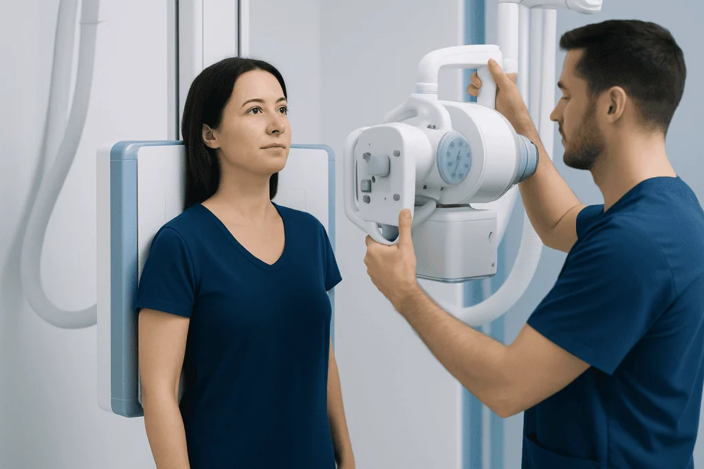
During the test, an X-ray machine sends a brief beam of radiation through the chest to a detector. Different tissues absorb different amounts of radiation, creating the image. Most examinations include a front view and a side view because two perspectives can help localize a finding more accurately.
A clinician may request a chest X-ray for cough that does not improve, fever with respiratory symptoms, shortness of breath, chest discomfort, coughing up blood, suspected chest injury, or unexplained changes seen during a physical examination. It may also be used to monitor selected known lung or heart conditions, assess the position of medical devices, or provide information before certain procedures.
Chest X-rays can be performed for adults and children when clinically appropriate. Pregnancy should always be mentioned before imaging, even if it is only possible. The imaging team can consider whether the test should be postponed, modified, or replaced, while recognizing that an urgently needed X-ray can still be performed with appropriate precautions.
What Illnesses Can a Chest X-Ray Show?

A chest X-ray can reveal findings that are consistent with several illnesses, but the image alone does not always establish a diagnosis. For example, areas of increased whiteness in a lung may occur with pneumonia, but they can also be caused by fluid, partial lung collapse, inflammation, or other processes. The report is therefore interpreted in the clinical context.
Conditions and abnormalities a chest X-ray may help identify include:
- Infections such as pneumonia and, in some circumstances, tuberculosis-related changes.
- Fluid around the lungs, called pleural effusion, or fluid-related congestion linked to heart failure.
- A collapsed lung, also known as pneumothorax, and areas of partial lung collapse.
- Chronic changes associated with conditions such as chronic obstructive pulmonary disease (COPD), although additional breathing tests are often needed.
- Changes in heart size or lung blood vessels that can support assessment of some heart conditions.
- Rib fractures and certain other bony chest injuries.
- Some lung masses or nodules, which require further evaluation and are not always visible on a standard X-ray.
When an X-ray raises concern for a lung nodule, complex infection, blood clot, or another issue not clearly defined by radiography, a clinician may recommend more detailed imaging. Computed tomography (CT) scanning provides cross-sectional images and can show many chest structures in greater detail.
Do I Need to Do Anything to Prepare for a Chest X-Ray?
Most people do not need special preparation for a chest X-ray. There is usually no need to fast, change regular meals, or stop medications unless a clinician gives specific instructions for another test planned on the same day.
Before the examination, the patient may be asked to change into a gown and remove necklaces, metal clothing fasteners, body piercings, hearing aids, or other objects that could obscure the chest image. It is helpful to bring previous imaging reports or ensure the imaging center can access earlier studies, as comparison can help identify whether a change is new or longstanding.
The patient should tell the radiographer about possible pregnancy, recent surgery, mobility limitations, or difficulty standing. Images can often be obtained while seated or lying down if needed. Clear communication allows the team to make the examination as safe and comfortable as possible.
Chest X-Ray Procedure, Benefits, Risks, and Recovery
For a typical standing examination, the patient stands against an image detector, usually facing it for the front view. The radiographer positions the shoulders and arms, then asks the patient to take a deep breath and hold it briefly while the image is captured. A side-view image may then be taken with the arms raised or placed out of the way.
The actual image capture takes only seconds, and the whole appointment is generally brief. The examination is painless. Remaining still and taking a full breath are important because movement or shallow breathing can reduce image quality and sometimes require a repeat image.
The main benefit is that chest X-ray imaging is fast, widely used, and useful for evaluating many urgent and non-urgent chest concerns. It uses a relatively low radiation dose, but no radiation exposure is completely without risk. The clinical team requests the test when its expected information is considered more valuable than this small exposure. There is no physical recovery period: most people can return to usual activities immediately after the test.
A radiologist reviews the images and sends a report to the requesting clinician. In urgent situations, results may be reviewed promptly; in other settings, reporting times vary. The clinician who requested the X-ray can explain what the findings mean for the individual patient.
When Should I Worry About Chest X-Ray Results?
An abnormal chest X-ray result should be taken seriously, but it does not automatically mean a severe illness is present. Terms such as “opacity,” “infiltrate,” “shadow,” “nodule,” or “enlarged cardiac silhouette” describe an imaging appearance. They often need comparison with previous images, symptom review, and sometimes follow-up testing before their cause is known.
It is important to discuss results promptly with the clinician who ordered the test, particularly if the report recommends repeat imaging, CT scanning, laboratory testing, or referral to a specialist. Follow-up is especially important if a possible mass, unexplained fluid, persistent lung collapse, or significant infection is reported.
Urgent medical assessment is appropriate for chest symptoms that are severe, worsening, or accompanied by warning signs. These include significant difficulty breathing, new or persistent chest pressure, blue or gray lips, fainting, confusion, coughing up more than small streaks of blood, or a high fever with marked weakness. Emergency services should be contacted for symptoms that may indicate an emergency.
What Will Not Show Up on a Chest X-Ray?
A chest X-ray has important limitations. It may miss small lung nodules, early or mild pneumonia, small blood clots in the lungs, subtle rib injuries, and conditions that do not cause obvious structural changes. A normal image also cannot reliably exclude asthma, early COPD, many airway disorders, or all forms of heart disease.
Chest X-rays do not directly show detailed blood vessels, coronary artery blockages, heart rhythm problems, or the function of the heart. Depending on the concern, a clinician may use tests such as electrocardiography, echocardiography, lung function testing, blood tests, or CT imaging. Magnetic resonance imaging (MRI) may be useful for selected heart, soft-tissue, spine, or chest-wall concerns, although it is not usually the first test for common lung symptoms.
Because the image is two-dimensional, structures can overlap and hide abnormalities. This is one reason a clinician may recommend follow-up even after a normal X-ray when symptoms are persistent, unexplained, or concerning.
Next Steps and When to Seek Medical Care
After a chest X-ray, the next step depends on the reason for testing and the report. A result consistent with a straightforward infection may lead to treatment and, in selected cases, a follow-up image to confirm that the change has cleared. Other results may need further imaging, respiratory testing, cardiac evaluation, or consultation with a relevant specialist.
People should arrange a medical review for a cough lasting more than a few weeks, recurring fever, unexplained weight loss, breathlessness that affects daily activities, chest pain, or coughing up blood. New chest symptoms should not be self-diagnosed from an imaging report alone. A qualified clinician can connect the report to the person’s overall health and recommend appropriate care.
For international patients who need coordinated assessment, Acibadem International’s multidisciplinary specialists and JCI-accredited hospitals diagnose and treat chest and respiratory conditions. Further evaluation may include bronchoscopy when clinicians need to examine the airways or collect samples for specific indications.
Frequently asked questions
How long does a chest X-ray take?
The image capture itself usually takes only a few seconds. Allowing time for registration, changing clothes, positioning, and any additional views, the appointment is often completed within a short visit.
Can a chest X-ray show lung cancer?
A chest X-ray can sometimes show a lung mass or other abnormality that requires further investigation. However, it can miss small or early cancers and cannot confirm cancer by itself. CT imaging and, when appropriate, tissue testing may be needed.
Can pneumonia be missed on a chest X-ray?
Yes. Early, mild, or dehydrated-stage pneumonia may not always be visible, and some X-ray findings can be difficult to distinguish from other conditions. If symptoms strongly suggest pneumonia, a clinician may recommend follow-up assessment or additional testing.
Is a chest X-ray painful?
No, a chest X-ray is not painful. The patient may need to hold a position and take a deep breath briefly, which can be uncomfortable for someone with chest pain or severe breathlessness, but the radiographer can provide support.
Can I eat and drink before a chest X-ray?
Yes, most chest X-rays do not require fasting, so normal eating and drinking are usually fine. If another procedure is scheduled at the same time, different preparation instructions may apply.
How soon will chest X-ray results be available?
Timing depends on the care setting and how urgently the test is needed. Images may be reviewed quickly in emergency care, while formal reports for non-urgent tests may take longer. The requesting clinician can explain how and when results will be communicated.
References
- Radiological Society of North America
- American College of Radiology
- National Heart, Lung, and Blood Institute
- National Health Service
- Mayo Clinic
This article is for general information only and is not a substitute for professional medical advice. Please consult a qualified doctor about your individual situation.
Explore treatments in Turkey — costs, top hospitals & a free quote
JCI-accredited · board-certified surgeons · reply within 24h
Add us as a Preferred Source to see more of our trusted health content across Google Search, AI Overviews and Discover.

More from the Health Library



Related Specialists

Dr. Cemalettin Sakal
Emergency Service
Dr. Süreyya Menteş
Gynecology & Obstetrics
Dt. Tuğçe Atçakan
Dentistry
Dr. Mehriban Allahverdiyeva
Inpatient Clinic Physicians Clinical Service