Atypical Mole Nevus: What Patients Need to Know

An atypical mole nevus is usually benign, but it can look similar to melanoma. People with several atypical moles may have a higher risk of developing melanoma over time.
Key Takeaways
- An atypical mole nevus is usually benign, but it can look similar to melanoma.
- People with several atypical moles may have a higher risk of developing melanoma over time.
- Warning signs include changes in size, shape, color, border, or new symptoms such as itching or bleeding.
- Dermatologists diagnose suspicious moles through skin examination and, when needed, biopsy.
- Sun protection and routine skin self-checks help reduce risk and support earlier detection.
An atypical mole nevus is a mole with irregular features that can look different from common moles. Most are not cancer, but they can resemble melanoma and may indicate a higher overall risk of skin cancer, so regular skin checks are important.
What is an atypical mole nevus?
An atypical mole nevus, also called a dysplastic nevus, is a mole that has some unusual features when compared with an ordinary mole. It may be larger, have less even color, or show a border that looks less smooth or more blurred. In many cases, it is still a benign growth of pigment-producing skin cells.
What matters most for patients is that an atypical mole nevus can sometimes look similar to early melanoma. For that reason, it should not be judged by appearance alone at home. A clinician, usually a dermatologist, assesses the mole in the context of the person’s skin type, family history, sun exposure, and whether the mole has changed over time.
Having one atypical mole does not mean a person has skin cancer. However, people who have many atypical moles may have a higher lifetime risk of melanoma than those without them. This is why follow-up, skin awareness, and prevention are central parts of care rather than reasons for panic.
How atypical moles look and feel
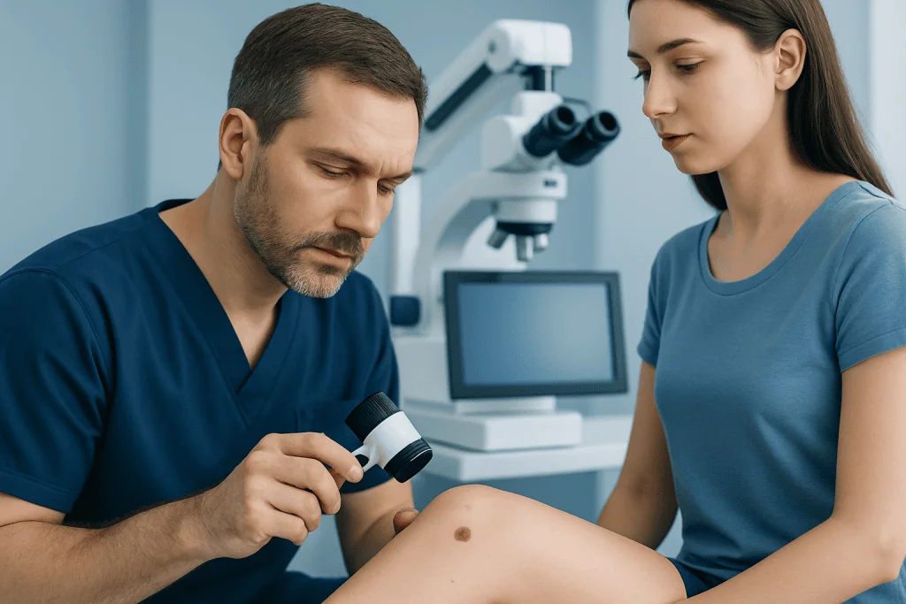
Atypical moles often stand out from common moles because they do not follow the usual pattern of being small, round, evenly colored, and sharply bordered. They may be flat or slightly raised and can appear tan, brown, pink, or a mix of shades. Some have a darker center and lighter edges.
Doctors often use the ABCDE guide when evaluating pigmented lesions. This does not diagnose a mole by itself, but it helps identify features that deserve attention.
- Asymmetry: one half does not match the other
- Border irregularity: edges look ragged, blurred, or uneven
- Color variation: more than one color or uneven pigment
- Diameter: often larger than common moles, though size alone is not enough
- Evolving: any change in shape, size, color, or symptoms
Not every atypical mole causes symptoms. When symptoms do occur, they may include itching, tenderness, crusting, or occasional bleeding. These changes do not always mean cancer, but they should prompt a medical review because melanoma can present in similar ways. For related warning signs, patients may also read about skin cancer.
Why atypical moles develop and who is at higher risk
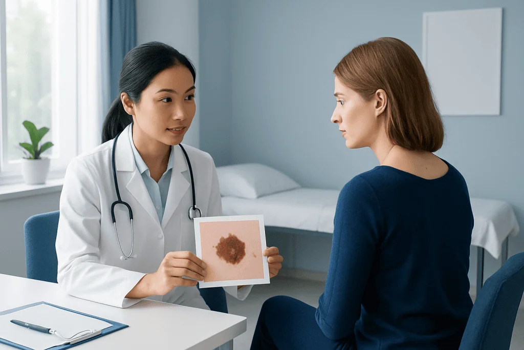
Atypical moles develop because of a combination of inherited traits and environmental exposure. Genetics plays a major role in how many moles a person has and how their skin responds to sunlight. Some families tend to have more atypical moles, and a family history of melanoma is especially relevant.
Ultraviolet radiation from the sun or tanning beds can contribute to mole changes and skin cell damage. This is why sun protection is recommended even for people who already have stable moles. Repeated sunburns, especially in childhood or adolescence, can increase future skin cancer risk.
Risk tends to be higher in people with fair skin, light eyes, freckles, many moles, a personal history of skin cancer, or a close relative with melanoma. Still, atypical moles can occur in people of any skin tone. Individual risk is best assessed by a doctor rather than by counting risk factors alone.
It is also helpful to understand that an atypical mole is not the same thing as melanoma, and most never become cancerous. The larger concern is that they can act as markers of increased overall risk, which is why skin surveillance is often advised.
How doctors diagnose an atypical mole nevus
Diagnosis begins with a careful skin examination. The doctor asks when the mole first appeared, whether it has changed, and whether there is a personal or family history of atypical moles or melanoma. They may compare the mole with others on the body and look for what dermatologists sometimes call the “ugly duckling” sign, meaning a spot that looks different from a person’s usual pattern of moles.
Dermatologists often use dermoscopy, a handheld device that magnifies the skin and helps reveal pigment structures not visible to the naked eye. This can improve the assessment of whether a mole appears benign, atypical, or suspicious enough to sample. In some clinics, total-body photography or digital mole mapping may also be used for people with many moles.
If the mole has concerning features, a biopsy may be recommended. During a biopsy, the doctor removes part or all of the lesion and sends it to a laboratory for microscopic examination. This is the only way to confirm whether the lesion is an atypical nevus, another benign lesion, or melanoma. Patients who need tissue evaluation may undergo a skin biopsy as part of diagnosis.
Treatment and follow-up options
Treatment depends on what the mole looks like, whether it is changing, and what the biopsy shows if one is done. Many atypical moles do not require immediate removal if they appear stable and not strongly suspicious. In these cases, the dermatologist may recommend observation with regular checkups and photographs to monitor for change.
If a mole is clearly suspicious or the biopsy shows significant atypia, complete removal may be advised. The goal is to ensure accurate diagnosis and reduce the chance that an early melanoma could be missed. When removal is needed, the doctor will explain the method, healing process, and whether further treatment is necessary.
Some patients benefit from a structured dermatology follow-up plan, especially if they have numerous atypical moles or a family history of melanoma. This may include periodic examinations and education on self-monitoring. If cancer is detected, care may involve dermatology evaluation and treatment and, when appropriate, skin cancer treatment.
Near the end of the care pathway, some international patients seek assessment in centers with coordinated specialties. Acibadem International’s multidisciplinary specialists and JCI-accredited hospitals diagnose and treat skin lesions, including suspicious moles, with individualized planning.
Prevention and self-care
Prevention focuses on reducing ultraviolet exposure and noticing changes early. No self-care method can guarantee that a mole will stay benign, but healthy sun habits can lower cumulative skin damage. This is especially important for people with many moles, a history of sunburn, or a family history of melanoma.
Practical steps include using a broad-spectrum sunscreen, wearing protective clothing and a wide-brimmed hat, seeking shade when sunlight is strongest, and avoiding tanning beds. Sunscreen should be applied carefully and reapplied as directed on the product label, especially after swimming or sweating.
Monthly skin self-checks can help people become familiar with their usual moles. A mirror or help from a partner can make it easier to inspect the scalp, back, and other hard-to-see areas. It can be useful to take dated photographs so that subtle changes are easier to recognize over time.
Patients should avoid trying to remove or treat moles at home. Picking, scratching, or using unproven products can delay diagnosis, cause irritation, and make medical assessment more difficult. If there is concern about a spot, the safest next step is a professional skin examination.
When to seek medical care
A short delay is usually not harmful for a stable mole that has looked the same for years, but a changing lesion should not be ignored. Medical review is recommended if a mole becomes noticeably larger, more irregular, darker, more varied in color, or different from surrounding moles. New symptoms such as itching, pain, crusting, or bleeding are also reasons to arrange an appointment.
People with many atypical moles, a personal history of melanoma, or a close family history of melanoma should ask a doctor how often they need skin checks. In these higher-risk groups, regular surveillance can be more important than waiting for symptoms to appear. A clinician can help decide whether observation is enough or whether a lesion should be sampled.
Urgent evaluation is especially important if a new dark spot appears in adulthood and changes quickly, or if a mole looks very different from the rest. While many skin lesions turn out to be benign, it is safer to have uncertain lesions assessed early. Patients who are worried about a suspicious lesion may also wish to learn more about melanoma and how it is distinguished from atypical moles.
Frequently asked questions
Is an atypical mole nevus cancer?
No. An atypical mole nevus is usually benign, but it has features that can resemble melanoma. Because appearance alone cannot always tell the difference, a doctor may recommend monitoring or biopsy if the mole looks suspicious.
Can an atypical mole turn into melanoma?
Some atypical moles may develop cancerous changes, but most do not become melanoma. More importantly, having atypical moles can be a marker of higher overall melanoma risk, which is why regular skin checks are often advised.
What does an atypical mole usually look like?
It may be larger than a common mole and have uneven color, irregular borders, or an asymmetric shape. Some are flat, while others are slightly raised. Any mole that changes over time deserves medical attention.
Should every atypical mole be removed?
Not always. Many atypical moles can be safely observed if they are stable and do not have strongly suspicious features. Removal is more likely when a mole changes, looks concerning on examination, or has significant atypia on biopsy.
How often should someone check their moles?
A monthly self-check is a practical habit for many people, especially those with many moles. The right schedule for professional skin exams depends on personal risk factors such as family history, past skin cancer, and the number of atypical moles.
Are atypical moles caused only by sun exposure?
No. Genetics also plays an important role, and some people are naturally more likely to develop atypical moles. Sun exposure and tanning beds can still contribute to skin damage and may increase the risk of concerning changes.
References
- American Academy of Dermatology
- National Cancer Institute
- American Cancer Society
- Mayo Clinic
- World Health Organization
This article is for general information only and is not a substitute for professional medical advice. Please consult a qualified doctor about your individual situation.
Oncology care in Turkey — second opinion and treatment plan
JCI-accredited · board-certified surgeons · reply within 24h
Add us as a Preferred Source to see more of our trusted health content across Google Search, AI Overviews and Discover.