Oral Tumors Pictures: A Complete Medical Overview

Oral tumors can be benign or malignant, and they may appear as lumps, ulcers, patches, or thickened areas in the mouth. Pictures are useful for awareness, but they cannot confirm a diagnosis because many harmless and serious conditions can look similar.
Key Takeaways
- Oral tumors can be benign or malignant, and they may appear as lumps, ulcers, patches, or thickened areas in the mouth.
- Pictures are useful for awareness, but they cannot confirm a diagnosis because many harmless and serious conditions can look similar.
- Warning signs include a sore that does not heal, unexplained bleeding, pain, numbness, difficulty swallowing, or a persistent lump.
- Risk factors include tobacco use, alcohol, human papillomavirus infection, sun exposure to the lips, and long-term irritation.
- Diagnosis usually involves a clinical examination, imaging when needed, and a biopsy to identify the exact type of lesion.
- Early evaluation improves treatment planning and may allow simpler, less invasive care.
Oral tumors pictures may help a person recognize concerning changes such as persistent sores, lumps, red or white patches, or growths on the lips, tongue, gums, or roof of the mouth. However, images alone cannot diagnose the cause, so any lesion that lasts more than two weeks should be assessed by a qualified doctor or dentist.
Overview: What oral tumors pictures can and cannot show
Oral tumors pictures usually show visible changes inside or around the mouth, such as a raised lump, a crater-like ulcer, a smooth swelling, or a red, white, or mixed red-and-white patch. These changes may affect the lips, gums, tongue, floor of the mouth, inner cheeks, hard palate, or the area behind the last molars. Some lesions look small and harmless at first, while others are larger, irregular, or ulcerated.
It is important to understand that a picture cannot tell whether a lesion is benign, precancerous, inflammatory, infectious, or cancerous. Many conditions can resemble one another in photographs. For example, a traumatic ulcer from biting the cheek, a canker sore, a mucous cyst, and an early oral cancer can each appear as a sore or bump, even though they need very different management.
A more helpful way to use oral tumors pictures is as a guide for self-awareness. They can help a person notice patterns that should not be ignored, especially a lesion that persists, enlarges, bleeds, or feels firm. If a visible change in the mouth lasts longer than two weeks, a professional examination is recommended rather than relying on online image comparison.
How oral tumors may look in the mouth
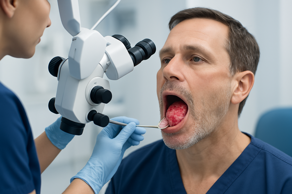
There is no single appearance that defines an oral tumor. Some tumors form a painless lump under the lining of the mouth. Others look like a persistent ulcer with a hard edge, a cauliflower-like growth, or a flat patch with an abnormal color. Early lesions may be subtle and easy to miss, especially if they are on the side of the tongue or under the tongue.
Benign tumors often grow slowly and may be smooth, mobile, and well-defined, although not always. Malignant tumors may be firm, irregular, fixed to deeper tissue, or prone to bleeding, but these features are not universal. A suspicious lesion may also cause nearby teeth to feel loose, change the fit of dentures, or create discomfort when chewing or speaking.
Common visual patterns include:
- White patches that do not wipe off
- Red patches or red-and-white mixed patches
- A sore that does not heal
- A thickened or roughened area
- A lump on the tongue, gums, lip, or palate
- An ulcer with a firm base
- Swelling under the tongue or inside the cheek
Because appearance varies so much, doctors do not diagnose by sight alone. They consider the lesion’s location, texture, duration, growth pattern, symptoms, and a person’s medical history before deciding whether testing is needed.
Symptoms that may suggest a concerning oral lesion

Some oral tumors cause no symptoms at first and are found during a routine dental examination. When symptoms do occur, they may include a persistent sore, a new lump, unexplained mouth pain, tenderness, numbness, or a burning sensation. A lesion that keeps returning in the same place also deserves attention.
As a lesion becomes larger or more invasive, a person may notice bleeding, bad breath that does not improve, pain with chewing, difficulty swallowing, a feeling that something is stuck in the throat, ear pain on one side, or trouble moving the tongue or jaw. Changes in speech or voice can also occur if the tumor affects nearby structures.
Not every persistent mouth symptom means cancer. Conditions such as fungal infection, chronic irritation, autoimmune inflammation, and benign salivary gland growths can produce similar complaints. Even so, symptoms that last beyond two weeks should not be dismissed, particularly in adults with risk factors.
Some oral lesions are related to broader conditions that may involve other tissues as well. In selected cases, doctors may evaluate for associated head and neck disease, including oral cancer or nearby tongue cancer when the location and examination findings raise concern.
Causes and risk factors
Oral tumors develop for different reasons depending on the type of lesion. Benign tumors may arise from salivary tissue, connective tissue, blood vessels, nerves, or the mucosal lining. Malignant tumors most often begin in the thin surface lining of the mouth, but they can also arise from salivary glands, lymphoid tissue, or, more rarely, bone and soft tissues.
Several factors increase the risk of oral cancer and certain precancerous changes. The best established risk factors include tobacco use in any form, regular heavy alcohol use, and infection with high-risk types of human papillomavirus in specific areas of the mouth and throat. Prolonged sun exposure can contribute to lip cancer, especially on the lower lip.
Other possible contributors include increasing age, poor oral hygiene, chronic irritation from rough teeth or ill-fitting dental appliances, immune suppression, previous head and neck cancer, and diets low in fruits and vegetables. Chronic irritation alone does not necessarily cause cancer, but it can make lesions more noticeable or complicate healing.
Importantly, oral tumors can also occur in people without obvious risk factors. That is one reason self-examination and regular dental visits remain valuable. A new or changing lesion should be assessed on its own merits, even in a person who does not smoke or drink alcohol.
How doctors diagnose oral tumors
Diagnosis begins with a careful history and examination. A doctor or dentist asks how long the lesion has been present, whether it has changed in size or appearance, and whether there are symptoms such as pain, bleeding, numbness, swallowing difficulty, or weight loss. During the examination, the clinician checks the entire mouth, tongue, throat entrance, jaw function, and lymph nodes in the neck.
If a lesion appears suspicious or does not resolve, the next step is often a biopsy. This is the most reliable way to determine whether a lesion is benign, precancerous, or malignant. A pathologist examines the tissue under a microscope and identifies the exact diagnosis, which then guides treatment decisions.
Imaging may be recommended when there is concern about the size, depth, or spread of a lesion. Depending on the case, this may include ultrasound, CT, MRI, or PET-based imaging. If the lesion seems to arise from a salivary gland or deep tissue space, imaging helps show its relationship to nearby nerves, bone, and muscles.
In some situations, specialist evaluation is useful, especially if surgery may be needed. This can include assessment by experts in ear, nose, and throat care or oral and maxillofacial surgery for lesions involving the tongue, jaw, palate, salivary glands, or complex areas of the mouth.
Treatment options and what they depend on
Treatment depends entirely on the diagnosis. Some benign lesions can simply be monitored if they are small and not causing symptoms. Others are removed because they interfere with eating, speaking, denture fit, comfort, or because their exact nature is uncertain. If chronic irritation is contributing, correcting the source may be part of care.
When a lesion is cancerous, treatment planning is based on the tumor type, size, location, depth, lymph node involvement, and the person’s overall health. Surgery is commonly used for many oral cancers and for a range of benign tumors. In certain cases, treatment may also include radiation therapy, medical oncology treatment, or reconstruction to restore speech, swallowing, and appearance as much as possible.
For advanced or complex disease, multidisciplinary care is often helpful. This may involve specialists in oncology along with surgeons, radiologists, pathologists, dentists, speech therapists, and nutrition professionals. The goal is not only tumor control, but also preserving quality of life and everyday function.
Near the end of the care pathway, patients may benefit from rehabilitation and close follow-up to monitor healing and detect recurrence or new lesions early. Acibadem International’s multidisciplinary specialists and JCI-accredited hospitals diagnose and treat oral tumors for international patients when specialist evaluation is needed.
Prevention, self-checks, and everyday mouth care
Not all oral tumors can be prevented, but several practical steps may reduce risk and help with earlier detection. Avoiding tobacco, limiting alcohol, protecting the lips from sun exposure, and maintaining good oral hygiene are among the most important measures. Regular dental examinations are valuable because some lesions are painless and can be found before they cause obvious symptoms.
A simple self-check can be done in good light using a mirror. A person can look at the lips, gums, roof of the mouth, inner cheeks, top and underside of the tongue, and floor of the mouth. Any new patch, swelling, ulcer, or color change that does not improve should be noted, especially if it is getting larger or feels firm.
It can help to document a lesion by noting the date it first appeared and whether there are symptoms such as pain, bleeding, or trouble eating. Photos taken over time may be useful to show a doctor how the area has changed, but they should not delay an appointment if the lesion is persistent.
People who wear dentures or have broken teeth should also address friction or trauma promptly. Although many irritative lesions are benign, a sore spot that continues after the source of rubbing is corrected should still be rechecked by a professional.
When to seek medical care
Medical or dental care should be sought if a mouth sore, patch, or lump lasts longer than two weeks. An earlier appointment is appropriate if the lesion is growing quickly, bleeds easily, is very painful, causes numbness, or makes it hard to chew, swallow, or speak. A neck lump together with a mouth lesion also deserves prompt assessment.
Urgent evaluation is especially important for people with significant risk factors such as tobacco use, heavy alcohol use, previous oral cancer, or immune suppression. Even in lower-risk individuals, it is safest to have persistent changes examined rather than waiting for them to disappear on their own.
In children and younger adults, many oral growths are benign, but persistent or unexplained lesions still need professional review. The same is true for anyone who notices a nonhealing area after dental treatment or trauma. A timely exam can often provide reassurance, and if treatment is needed, it allows care to begin sooner.
Frequently asked questions
Can oral tumors pictures tell if a lesion is cancer?
No. Pictures can show that a lesion looks unusual, but they cannot confirm whether it is benign, precancerous, infectious, or malignant. A clinical examination and, when needed, a biopsy are required for an accurate diagnosis.
What does an oral tumor usually look like?
An oral tumor may look like a lump, a nonhealing ulcer, a white patch, a red patch, or a thickened area of tissue. Some are painless and subtle, while others may bleed, feel firm, or interfere with speaking and eating.
Are all mouth lumps dangerous?
No. Many mouth lumps are caused by irritation, cysts, infections, or other benign conditions. However, any lump that persists, enlarges, or is associated with other symptoms should be examined by a doctor or dentist.
How long should someone wait before getting a mouth sore checked?
A general rule is to seek evaluation if a sore, patch, or lump lasts more than two weeks. Earlier assessment is sensible if there is severe pain, bleeding, numbness, rapid growth, or trouble swallowing or speaking.
Who is at higher risk for oral cancer?
People who use tobacco, drink alcohol heavily, have high-risk HPV infection, or have significant sun exposure to the lips are at increased risk. Still, oral cancer can also occur in people without these risk factors, so persistent lesions should not be ignored.
How are oral tumors treated?
Treatment depends on the exact diagnosis. Benign lesions may be observed or removed, while cancerous tumors may need surgery, radiation therapy, medical oncology treatment, or a combination of approaches.
This article is for general information only and is not a substitute for professional medical advice. Please consult a qualified doctor about your individual situation.
Explore treatments in Turkey — costs, top hospitals & a free quote
JCI-accredited · board-certified surgeons · reply within 24h
Add us as a Preferred Source to see more of our trusted health content across Google Search, AI Overviews and Discover.

More from the Health Library

Lacosamide: A Complete Medical Overview

Yellow Spot in Skin: What Patients Need to Know

Bondage: An Evidence-Based Guide for Patients

Water Poisoning: A Complete Medical Overview

Dapagliflozin: What Patients Need to Know