Herniated Umbilical Hernia: A Complete Medical Overview

A herniated umbilical hernia causes a bulge near the belly button when tissue pushes through a weak area in the abdominal wall. In adults, umbilical hernias often do not go away on their own and may slowly enlarge over time.
Key Takeaways
- A herniated umbilical hernia causes a bulge near the belly button when tissue pushes through a weak area in the abdominal wall.
- In adults, umbilical hernias often do not go away on their own and may slowly enlarge over time.
- Sudden pain, redness, vomiting, or a bulge that cannot be pushed back may suggest incarceration or strangulation and needs urgent medical care.
- Diagnosis is usually based on physical examination, with ultrasound or CT used when the findings are unclear or complications are suspected.
- Treatment depends on symptoms, hernia size, and risk of complications, but surgery is the only definitive repair.
A herniated umbilical hernia is a condition in which fat or part of the intestine pushes through a weak spot near the belly button, creating a visible or palpable bulge. Many cases are uncomplicated, but some can become painful or trapped and should be assessed by a doctor promptly.
Overview
A herniated umbilical hernia is a protrusion of tissue through a weak point in the abdominal wall at or near the belly button. The bulge may contain fatty tissue or part of the intestine. In many people, it is soft and more noticeable when standing, coughing, straining, or lifting.
The term often refers to an umbilical hernia that has become more prominent or symptomatic. In everyday use, people may say “herniated umbilical hernia” to describe a belly button hernia that is bulging outward, causing discomfort, or raising concern about complications. While the wording is repetitive medically, the underlying issue is clear: tissue is pushing through where the abdominal wall is weaker.
Umbilical hernias occur in both children and adults, but the clinical approach differs. In infants, many small umbilical hernias close naturally as the abdominal muscles strengthen. In adults, however, these hernias are less likely to resolve without treatment and may gradually enlarge. Adult cases are also more likely to require planned surgical repair, especially if symptoms develop.
Most umbilical hernias are not emergencies. Still, some can become incarcerated, meaning the bulging tissue gets trapped, or strangulated, meaning its blood supply becomes impaired. Recognizing the difference between a reducible, uncomplicated hernia and one that needs urgent attention is an important part of safe care.
Symptoms and How It May Feel
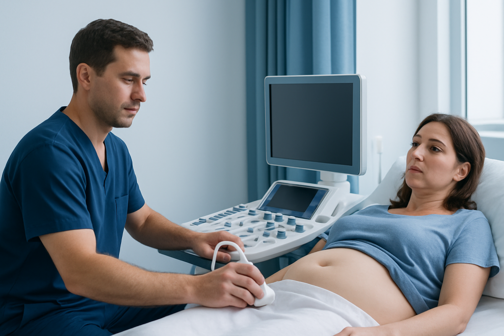
The most common sign of a herniated umbilical hernia is a bulge or swelling at the navel. It may be small at first and become more obvious during activities that increase abdominal pressure, such as coughing, laughing, bending, or lifting. Some people notice that the bulge flattens when lying down.
Symptoms vary. Some people have no pain and discover the hernia incidentally, while others describe pressure, tenderness, a pulling sensation, or mild aching around the belly button. Discomfort may worsen after meals, prolonged standing, exercise, or heavy physical work.
Warning symptoms suggest possible complications and should not be ignored. These include a bulge that becomes firm or cannot be gently pushed back in, increasing pain, skin redness or warmth over the area, nausea, vomiting, abdominal swelling, or difficulty passing stool or gas. These features can indicate an incarcerated or strangulated hernia and need prompt medical evaluation.
Because symptoms can overlap with other abdominal wall conditions, a doctor may also consider alternatives such as inguinal hernia or other causes of a painful abdominal lump. A careful examination helps clarify what is present and whether it is likely to need observation or repair.
Causes and Risk Factors

An umbilical hernia develops when there is a weakness in the abdominal wall near the umbilical ring, combined with pressure from inside the abdomen. In adults, this weakness may have been present for years and become more noticeable later in life as abdominal pressure rises or tissues lose strength.
Several factors increase the chance of developing a herniated umbilical hernia. These include obesity, pregnancy, repeated heavy lifting, chronic coughing, constipation with straining, fluid buildup in the abdomen, and prior abdominal surgery. Conditions that raise abdominal pressure over time can enlarge an existing weak spot and make a hernia more likely.
Age can also play a role, because connective tissues may become less resilient over time. Some hernias appear after significant weight changes or after repeated physical strain. In people with multiple hernias or a known connective tissue problem, doctors may assess the abdominal wall more broadly.
It is helpful to distinguish an umbilical hernia from other ventral abdominal wall hernias. A doctor may compare it with hiatal hernia only to explain that not all hernias are visible externally; a hiatal hernia occurs inside the body, whereas an umbilical hernia forms a bulge at the navel. This difference matters because symptoms, examination findings, and treatment plans are not the same.
Diagnosis and Assessment
Diagnosis usually begins with a medical history and physical examination. The doctor will ask when the bulge first appeared, whether it changes in size, and whether there is pain, nausea, vomiting, or bowel symptoms. Examination is often performed while the person is standing and again while lying down, sometimes with gentle coughing or straining to make the hernia more apparent.
A key part of the assessment is whether the hernia is reducible. A reducible hernia can be gently returned into the abdomen, while an incarcerated hernia cannot. The doctor also looks for signs of skin changes, tenderness, and bowel involvement, which can help identify whether urgent treatment is needed.
Imaging is not always necessary for straightforward cases, but it can be useful when the diagnosis is uncertain, the hernia is small, the person has a larger body habitus, or complications are suspected. Ultrasound may show the abdominal wall defect and what is protruding through it. CT imaging can provide more detail if there is concern about obstruction, incarceration, or another abdominal condition.
Preoperative assessment may include review of medications, smoking status, weight, and conditions such as diabetes or chronic lung disease. This broader evaluation helps guide timing of surgery and lowers the risk of recurrence or wound-related problems after repair.
Treatment Options
Treatment depends on age, symptoms, hernia size, and the risk of complications. In infants and young children, small umbilical hernias are often monitored because many close naturally. In adults, watchful waiting may be reasonable for some very small, asymptomatic hernias, but many eventually need repair if they enlarge, become uncomfortable, or interfere with daily life.
Surgery is the only definitive treatment for an adult umbilical hernia. The goal is to return the protruding tissue to the abdomen and close the weakness in the abdominal wall. Depending on the size of the defect and individual factors, repair may be done with sutures alone or reinforced with mesh to reduce the chance of recurrence. Some procedures are performed through a traditional open approach, while others are suitable for minimally invasive repair, such as laparoscopic surgery.
Urgent surgery may be needed if the hernia is incarcerated or strangulated. In those situations, the surgeon evaluates whether the trapped tissue is healthy after blood flow is restored. Emergency treatment focuses on protecting the intestine and repairing the abdominal wall safely.
For people preparing for repair, doctors may recommend measures such as weight management, smoking cessation, control of chronic cough or constipation, and review of blood-thinning medication use. When abdominal wall reconstruction is complex or recurrent hernias are involved, patients may benefit from assessment by teams experienced in general surgery and abdominal wall hernia repair. In selected cases where imaging is needed before treatment, MRI or other scans may help clarify anatomy, although MRI is not routinely required for most straightforward umbilical hernias.
Prevention and Self-care
Not every umbilical hernia can be prevented, but reducing excess pressure on the abdominal wall can help lower risk and may slow progression of a known hernia. Practical measures include reaching and maintaining a healthy weight, managing chronic cough, avoiding unnecessary straining during bowel movements, and using proper lifting technique.
For people who already have a diagnosed umbilical hernia, self-care is supportive rather than curative. Activities that trigger discomfort may need to be modified, especially heavy lifting or intense abdominal strain. If constipation is present, improving hydration, fiber intake, and bowel habits may reduce repeated pressure at the hernia site.
Abdominal binders or support garments may provide temporary comfort in some cases, but they do not close the defect or replace medical care. Folk remedies such as taping coins over the navel are not recommended and can irritate the skin without treating the underlying problem.
After surgical repair, following postoperative instructions carefully is important. This often includes wound care, gradual return to activity, avoiding heavy lifting for a period advised by the surgeon, and attending follow-up appointments to check healing and watch for recurrence.
When to Seek Medical Care
Medical review is appropriate for any new or persistent bulge near the belly button, especially in adults. Even if the hernia is not painful, a doctor can confirm the diagnosis, explain whether observation is reasonable, and discuss whether repair may be advisable later.
Prompt medical attention is recommended if the bulge becomes more painful, increases in size, or no longer goes back in when lying down or with gentle pressure. Nausea, vomiting, abdominal distension, fever, skin redness, or sudden severe tenderness over the hernia are warning signs that should be assessed urgently because they may indicate incarceration or strangulation.
People who are pregnant, have significant obesity, liver disease with abdominal fluid, or a history of prior hernia repair may benefit from earlier evaluation because management decisions can be more individualized. Children with pain, vomiting, or a discolored, firm umbilical bulge should also be seen promptly.
Near the end of the care pathway, some patients seek coordinated evaluation from centers experienced in abdominal wall disorders. Acibadem International’s multidisciplinary specialists at JCI-accredited hospitals diagnose and treat hernia conditions for international patients, including planned and urgent surgical care when appropriate.
Frequently asked questions
Is a herniated umbilical hernia the same as an umbilical hernia?
In practice, people often use the phrase to mean an umbilical hernia that is visibly bulging or causing symptoms. Medically, an umbilical hernia already refers to tissue pushing through a weakness near the belly button, so “herniated” does not describe a separate disease.
Can an umbilical hernia heal on its own?
In infants and some young children, small umbilical hernias may close as the abdominal wall matures. In adults, spontaneous closure is uncommon, and the hernia may remain the same size or gradually enlarge over time.
When is an umbilical hernia an emergency?
It may be an emergency if the bulge becomes trapped, very painful, red, firm, or impossible to push back in. Vomiting, abdominal swelling, or signs of bowel obstruction also require urgent medical attention because they may suggest incarceration or strangulation.
Does every adult umbilical hernia need surgery?
Not always immediately. A doctor may recommend observation for a small, symptom-free hernia in selected adults, but surgery is the only definitive repair and is commonly advised if symptoms, enlargement, or complication risk are present.
What does umbilical hernia surgery involve?
Surgery generally returns the protruding tissue to the abdomen and closes the weakness in the abdominal wall. Depending on the hernia, the repair may use stitches alone or mesh reinforcement, and it may be done through an open or minimally invasive approach.
Can exercise make a herniated umbilical hernia worse?
Activities that sharply increase abdominal pressure, such as heavy lifting or intense straining, can make symptoms more noticeable and may enlarge the bulge over time. Gentle activity is often still possible, but the safest plan depends on the hernia size, symptoms, and a doctor’s advice.
This article is for general information only and is not a substitute for professional medical advice. Please consult a qualified doctor about your individual situation.
Persistent digestive symptoms? Get evaluated in Turkey
JCI-accredited · board-certified surgeons · reply within 24h
Add us as a Preferred Source to see more of our trusted health content across Google Search, AI Overviews and Discover.

More from the Health Library

Dong Quai — Explained by Medical Evidence, Not Myths

5 Htp — Explained by Medical Evidence, Not Myths

Personality Histrionic — Explained by Medical Evidence, Not Myths

Blood Brain Barrier: A Complete Medical Overview

Knot on Top of Foot — Explained by Medical Evidence, Not Myths