Phleboliths in the Pelvis: A Complete Medical Overview

Phleboliths in the pelvis are usually benign and do not need treatment on their own. They are often found incidentally during imaging for pelvic pain, urinary symptoms, or abdominal complaints.
Key Takeaways
- Phleboliths in the pelvis are usually benign and do not need treatment on their own.
- They are often found incidentally during imaging for pelvic pain, urinary symptoms, or abdominal complaints.
- A main diagnostic challenge is telling pelvic phleboliths apart from ureteral stones.
- Symptoms such as severe pain, blood in urine, fever, or new swelling usually point to another condition rather than the phlebolith itself.
- Evaluation may include imaging and a medical review to rule out urinary, vascular, or pelvic disorders.
Phleboliths in the pelvis are small calcium deposits that form inside veins, most often after an old blood clot in a superficial pelvic vein has healed. They are very common, usually cause no symptoms, and are often discovered by chance on an X-ray or CT scan done for another reason.
Overview: what phleboliths in the pelvis mean
Phleboliths in the pelvis are tiny round calcium deposits inside pelvic veins. In most people, they represent an old, healed process in which a small clot formed in a vein and later calcified. They are commonly seen in adults on routine imaging and, by themselves, are generally not dangerous.
Many people first hear the term after an X-ray, CT scan, or other imaging test reports “pelvic phleboliths.” This can sound concerning, but in most cases it is simply an incidental finding. The important question is not whether the calcifications are present, but whether a person’s symptoms might actually be due to another cause.
Pelvic phleboliths matter clinically mainly because they can resemble stones in the urinary tract, especially a stone in the lower ureter. Radiologists and doctors use the shape, location, and surrounding features on imaging to tell these apart. This distinction helps avoid unnecessary worry and ensures that painful conditions such as kidney or ureter stones are not missed.
Are pelvic phleboliths symptoms or a harmless finding?
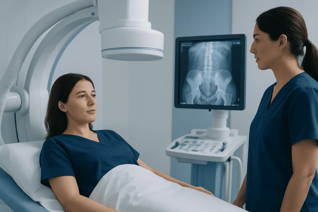
Most pelvic phleboliths do not cause symptoms. They are usually a harmless finding that appears on imaging done for another reason, such as lower abdominal pain, back pain, urinary symptoms, or a routine health evaluation. Many people live with them for years without knowing they are there.
When symptoms are present, doctors usually look for another explanation rather than assuming the phlebolith is the cause. For example, cramping pain, burning with urination, blood in the urine, fever, or nausea may be more suggestive of a urinary stone, infection, or another pelvic condition. Chronic pelvic pain may also come from gynecologic, gastrointestinal, musculoskeletal, or vascular causes.
Rarely, phleboliths may be associated with underlying venous abnormalities, such as varicose veins or a venous malformation. In that setting, the broader vascular problem may cause discomfort, fullness, or swelling, while the calcified phlebolith itself is more of a marker than the direct source of symptoms. If pelvic discomfort is ongoing, a clinician may also consider related conditions such as pelvic congestion syndrome depending on the person’s history and imaging findings.
Why phleboliths form: causes and risk factors

Phleboliths are thought to form when slow blood flow in a vein contributes to a small clot, which later hardens and calcifies over time. This process is more likely in veins where blood can pool or move sluggishly. In the pelvis, this can happen because of the anatomy of the venous system and long-term pressure changes in the area.
They are seen more often with increasing age and may be more common in people who have venous insufficiency, varicose veins, or pelvic vein enlargement. Some individuals with chronic constipation, a sedentary lifestyle, or prior pelvic surgery may also have factors that affect pelvic blood flow, though the presence of phleboliths alone does not prove disease.
In some cases, multiple pelvic phleboliths are found in people with venous malformations. These are abnormal clusters of veins that can develop in soft tissues and sometimes become painful or swollen. If imaging suggests an underlying vein problem, a doctor may recommend further assessment and, when appropriate, treatment options such as embolization or other vascular procedures.
How doctors tell phleboliths from kidney or ureter stones
The most important practical issue with phleboliths in the pelvis is distinguishing them from stones in the ureter, the tube that carries urine from the kidney to the bladder. On imaging, both can appear as small bright spots of calcium in the lower pelvis. However, their significance is very different: a ureter stone may explain sudden severe pain, while a phlebolith often does not require treatment.
Radiologists look for several clues. Phleboliths are often round, may have a central lucency or lighter center, and are usually located beside rather than within the ureter. A ureter stone is more likely to sit directly along the course of the ureter and may be associated with upstream urinary tract dilation. CT scanning is especially helpful when symptoms make the distinction important.
If a person has flank pain, nausea, urinary urgency, painful urination, or blood in the urine, a urinary stone may need prompt evaluation. In such cases, the doctor may investigate for kidney stones and use urine tests, ultrasound, or CT imaging to confirm the cause. The scan report may mention both a possible stone and incidental pelvic phleboliths, which is one reason clinical interpretation matters.
Diagnosis and medical evaluation
Pelvic phleboliths are most often diagnosed on imaging. Plain X-rays can show them as small round calcifications in the pelvis. CT scans provide more detail and are commonly used when a person has abdominal or urinary symptoms. Ultrasound is less likely to identify phleboliths directly, but it may help assess nearby organs or blood flow when another problem is suspected.
Diagnosis is not only about identifying the calcification. The doctor also considers where it is located, whether it follows the expected path of the ureter, and whether there are signs of obstruction, inflammation, or a vein abnormality. A person’s symptoms, physical examination, and medical history guide how much further testing is needed.
Additional evaluation may include urine tests, kidney function tests, pelvic ultrasound, or vascular imaging when indicated. If pelvic venous disease is suspected, a specialist in vascular or interventional medicine may review the case. In selected patients with persistent symptoms related to abnormal pelvic veins, procedures such as venography can help define the anatomy and support treatment planning.
Treatment options and when no treatment is needed
Most phleboliths in the pelvis do not need treatment. If they are found incidentally and the person has no symptoms that suggest another illness, reassurance is usually all that is required. Follow-up is generally not necessary unless the doctor is monitoring a related condition.
Treatment is considered only when there is an underlying problem. For example, if imaging and symptoms point to a ureter stone, urinary infection, or another pelvic disorder, treatment is directed at that cause. If phleboliths are part of a venous malformation or pelvic venous insufficiency, management may involve symptom control, specialist assessment, and occasionally minimally invasive therapy.
Interventional treatment can be helpful in carefully selected patients with symptomatic venous disorders. Depending on the diagnosis, options may include sclerotherapy or embolization to treat abnormal or enlarged veins. Near the end of the care pathway, some international patients choose evaluation at centers such as Acibadem International, where multidisciplinary specialists in JCI-accredited hospitals assess vascular and pelvic conditions using coordinated imaging and treatment planning.
Self-care, prevention, and daily concerns
There is no specific way to prevent every phlebolith, and most do not require lifestyle changes once discovered. Still, habits that support healthy circulation may be reasonable for overall vascular health. These include regular physical activity, avoiding prolonged sitting when possible, staying hydrated, and discussing persistent constipation or pelvic pressure with a doctor.
People who have known varicose veins, venous insufficiency, or symptoms of pelvic venous congestion may benefit from individualized advice. Depending on the situation, a clinician may suggest exercise, weight management, bowel habit support, or measures that reduce prolonged strain. These steps are supportive rather than curative for phleboliths themselves.
It is also helpful to keep medical records of prior imaging. If a future scan again shows pelvic calcifications, comparison with older images can confirm that the findings are stable and longstanding. This may reduce repeat testing and help clinicians focus on the true cause if new symptoms develop.
When to seek medical care
A person should seek medical care if pelvic or flank pain is severe, sudden, or keeps returning, especially if it is accompanied by nausea, vomiting, fever, chills, blood in the urine, or trouble passing urine. These features may suggest a urinary stone, infection, or another condition that needs prompt treatment rather than an incidental phlebolith.
Medical review is also important for new pelvic swelling, unexplained leg swelling, persistent pelvic pressure, or pain that affects daily life. Although phleboliths are usually harmless, these symptoms may point to venous disease, gynecologic problems, bowel conditions, or musculoskeletal causes that deserve a proper diagnosis.
For people who have already been told they have pelvic phleboliths, the finding itself is usually reassuring. Still, any new or changing symptoms should be discussed with a qualified doctor so that other causes can be ruled out safely and accurately.
Frequently asked questions
Are phleboliths in the pelvis dangerous?
In most cases, no. Pelvic phleboliths are usually benign calcifications inside veins and are commonly found by chance on imaging. They generally do not become cancerous or require treatment unless another underlying condition is present.
Can phleboliths in the pelvis cause pain?
Most do not cause pain directly. If a person has pelvic, abdominal, or urinary symptoms, doctors usually look for another cause such as a ureter stone, infection, pelvic venous disorder, or a gynecologic or bowel condition.
How are phleboliths different from kidney stones?
Phleboliths are calcifications in veins, while kidney or ureter stones form in the urinary tract. On scans, they can look similar, especially in the pelvis, so radiologists use location and imaging features to tell them apart. Symptoms like blood in the urine or classic colicky flank pain are more suggestive of a urinary stone.
Do pelvic phleboliths need to be removed?
Usually not. If they are incidental and there is no related venous disorder, removal is not necessary. Treatment focuses on any underlying condition that may be causing symptoms.
Can phleboliths go away on their own?
Phleboliths are calcified deposits, so they often remain visible over time. They may stay stable for years and usually do not cause problems. Their persistence on imaging is not usually a sign of worsening disease.
Should someone with pelvic phleboliths see a specialist?
Not always. Many people only need an explanation from the doctor who ordered the scan. A specialist may be helpful if symptoms persist, if there is uncertainty about whether the finding is a urinary stone, or if imaging suggests a venous malformation or pelvic venous disease.
References
- American College of Radiology
- National Institute of Diabetes and Digestive and Kidney Diseases
- Radiological Society of North America
- Society of Interventional Radiology
- National Library of Medicine
This article is for general information only and is not a substitute for professional medical advice. Please consult a qualified doctor about your individual situation.
Explore treatments in Turkey — costs, top hospitals & a free quote
JCI-accredited · board-certified surgeons · reply within 24h
Add us as a Preferred Source to see more of our trusted health content across Google Search, AI Overviews and Discover.

More from the Health Library

Ors for Electrolyte Imbalance: What Patients Need to Know

Forward Head Posture: A Complete Medical Overview

What is doa? A Doctor-Reviewed Answer

Flagyl and — Explained by Medical Evidence, Not Myths

Magnesium Oxide: What Patients Need to Know