Picture of Spine: An Evidence-Based Guide for Patients

A "picture of spine" may mean an anatomy diagram or a medical image such as an X-ray, CT scan, or MRI. The spine has three main sections—cervical, thoracic, and lumbar—plus the sacrum and coccyx.
Key Takeaways
- A "picture of spine" may mean an anatomy diagram or a medical image such as an X-ray, CT scan, or MRI.
- The spine has three main sections—cervical, thoracic, and lumbar—plus the sacrum and coccyx.
- X-rays show bone alignment well, while MRI is better for discs, nerves, and the spinal cord.
- Not every episode of back or neck pain needs imaging; the choice depends on symptoms and examination findings.
- Urgent medical care is important for severe weakness, bowel or bladder changes, major trauma, or fever with back pain.
A picture of spine usually refers to an image or diagram that shows the vertebrae, discs, spinal cord, and the spine’s natural curves. Different types of spine pictures serve different purposes: diagrams help explain anatomy, while X-rays, CT scans, and MRIs help doctors evaluate pain, injury, posture, or nerve-related symptoms.
What a Picture of Spine Shows
A picture of spine can mean two different things. It may be a simple medical illustration used to explain anatomy, or it may be a diagnostic image taken with X-ray, CT, or MRI. For patients, the most helpful starting point is knowing that each type of picture answers a different question.
An anatomy picture of the spine usually labels the vertebrae, discs, spinal cord, nerves, and the normal front-to-back curves of the neck, upper back, and lower back. These diagrams are useful for understanding where symptoms begin and how posture, aging, injury, or wear-and-tear may affect the spine over time.
Medical imaging goes further by helping doctors look for problems such as fractures, misalignment, arthritis, disc changes, narrowing around nerves, or signs of infection or tumors. A single image does not always explain pain by itself, so healthcare professionals interpret spine pictures together with symptoms, physical examination, and medical history.
Basic Spine Anatomy in Simple Terms
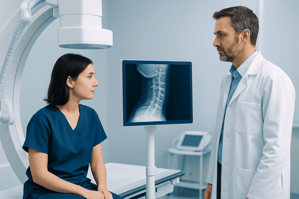
The spine is the body’s central support column. It helps a person stand upright, bend, twist, and protect the spinal cord. A standard picture of spine often divides it into regions: the cervical spine in the neck, the thoracic spine in the upper and mid-back, and the lumbar spine in the lower back. Beneath these are the sacrum and coccyx.
The spine is made of stacked bones called vertebrae. Between most vertebrae are discs, which act like cushions and help absorb shock. Small joints connect the bones at the back, and ligaments and muscles help keep the spine stable while allowing movement.
The spinal cord passes through a canal in the center of the vertebrae. Nerves branch off from the cord and travel to the shoulders, arms, chest, abdomen, legs, and feet. When a picture of spine is reviewed, doctors often look not only at the bones but also at the spaces where nerves exit, because narrowing in these areas can contribute to pain, numbness, or weakness.
- Cervical spine: supports the head and allows neck motion
- Thoracic spine: attaches to the ribs and supports the upper body
- Lumbar spine: bears much of the body’s weight
- Sacrum and coccyx: form the base of the spine and connect to the pelvis
Common Types of Spine Pictures

X-ray is often the simplest spine picture. It is commonly used to check bone alignment, fractures, scoliosis, and some signs of arthritis or degeneration. X-rays are quick and widely available, but they do not show discs, nerves, or the spinal cord in as much detail as MRI.
CT scans provide more detailed cross-sectional images of bone and can be especially helpful after trauma or when complex bony anatomy needs a closer look. In some situations, CT may be used when MRI is not suitable. MRI is usually the most informative test for soft tissues, including discs, ligaments, spinal cord, and nerve roots.
Sometimes, a doctor may begin with conservative care before ordering imaging, especially if symptoms are short-lived and there are no warning signs. Imaging becomes more important when there has been injury, persistent symptoms, progressive neurologic changes, or concern for conditions such as infection, severe nerve compression, or a structural disorder like scoliosis.
How Doctors Read a Spine Image
When specialists look at a picture of spine, they assess overall alignment first. They check whether the spine’s natural curves are preserved and whether the vertebrae are positioned normally. They also look for fractures, slippage of one vertebra over another, and changes related to aging, such as bone spurs or narrowing of disc spaces.
On MRI or CT, attention often turns to the discs, spinal canal, and nerve openings. Doctors evaluate whether a disc is bulging or herniated, whether the canal is narrowed, and whether there is pressure on a nerve root or the spinal cord. These findings may relate to symptoms such as arm pain from the neck or leg pain from the lower back.
It is important to know that many imaging findings are common even in people without pain. Mild disc bulges or age-related wear do not always need treatment. The most useful interpretation connects the image with where symptoms are felt, how long they have lasted, and whether movement, coughing, walking, or certain positions make them better or worse.
What Problems a Picture of Spine May Help Detect
A spine image may help identify several categories of problems. These include injury, degenerative changes, deformity, inflammation, infection, and, less commonly, tumors. The goal is not simply to find an abnormality but to determine whether that finding explains the person’s symptoms and whether it changes treatment decisions.
Common examples include fractures after falls or accidents, disc herniation causing nerve irritation, and age-related changes such as spinal stenosis or osteoarthritis. In some people, a picture of spine may show a curvature problem like kyphosis or a side-to-side curve such as scoliosis. In others, imaging helps investigate long-standing neck pain, lower back pain, or symptoms linked to a herniated disc.
Imaging may also be used before planning procedures or surgery. For example, MRI can help show whether symptoms are likely related to nerve compression and whether options such as rehabilitation, injections, or spine surgery should be discussed. In complex cases, assessment by specialists in orthopedics, neurosurgery, physical medicine, or pain management may be appropriate.
When Imaging Is Needed—and When It May Not Be
Many episodes of back or neck pain improve with time, activity modification, and supportive care. In these cases, an immediate picture of spine is not always necessary. Clinical guidelines often recommend imaging only when symptoms persist, do not respond to initial treatment, or suggest a specific underlying problem.
Imaging is more likely to be recommended after significant trauma, in older adults with possible fracture risk, or when there are neurologic symptoms such as weakness, radiating pain, numbness, balance changes, or loss of coordination. Doctors may also order imaging if symptoms raise concern for infection, inflammatory disease, or cancer-related spread to the spine.
Some patients worry when imaging is not ordered right away. In many situations, this is because early imaging does not improve outcomes and may reveal age-related findings that are not actually causing symptoms. A careful history and physical examination are often the best first steps before deciding whether a spine MRI or another test is needed.
Self-Care, Treatment, and Recovery
Treatment depends on what the picture of spine shows and how severe the symptoms are. Many spine conditions are managed without surgery. Common approaches include guided exercise, posture and movement advice, physical therapy, activity modification, heat or cold packs, and medicines recommended by a doctor for short-term symptom relief.
If there is nerve irritation, a structured rehabilitation plan may help improve flexibility, core support, and overall function. In some cases, doctors may suggest targeted procedures or further evaluation with physical therapy and rehabilitation to support recovery and reduce the chance of recurring symptoms. For deformities or ongoing pain, treatment is individualized according to age, imaging findings, and daily limitations.
Surgery is usually considered when there is spinal instability, a significant fracture, progressive neurologic loss, severe nerve compression, or symptoms that do not improve with appropriate non-surgical care. Decisions are made carefully and typically rely on both imaging and clinical examination. Near the end of the care pathway, patients seeking specialist evaluation may also consider centers such as Acibadem International, where multidisciplinary specialists in JCI-accredited hospitals diagnose and treat spine conditions for international patients.
When to Seek Medical Care
Medical care should be sought promptly for back or neck pain after a fall, car accident, sports injury, or other significant trauma. Urgent attention is also important for new weakness in an arm or leg, difficulty walking, loss of bowel or bladder control, numbness around the groin area, or severe pain that is sudden and intense.
A doctor should also evaluate spine symptoms that last more than a few weeks, keep returning, disturb sleep, or interfere with work, mobility, or daily activities. Fever, unexplained weight loss, a history of cancer, or immune system problems together with back pain are additional reasons not to delay assessment.
For mild pain without warning signs, simple measures such as staying gently active, avoiding prolonged bed rest, and using clinician-approved supportive care may help while monitoring symptoms. If there is uncertainty about whether a picture of spine is needed, a qualified healthcare professional can decide which exam or imaging test is most appropriate.
Frequently asked questions
What is the best picture of spine for back pain?
There is no single best test for every person. X-rays are useful for alignment and fractures, while MRI is better for discs, nerves, and the spinal cord. The right choice depends on symptoms, examination findings, and whether there are warning signs.
Can an X-ray show a slipped disc?
An X-ray does not show a disc directly in the same detail as MRI. It may show indirect signs such as narrowed disc spaces or alignment changes, but MRI is usually preferred when a disc problem is suspected. A doctor decides based on symptoms and physical examination.
What does a normal spine picture look like?
A normal spine image generally shows good alignment, preserved vertebral height, and natural curves in the neck, upper back, and lower back. On MRI, the spinal cord and nerve spaces also appear without significant compression. Normal appearance can vary somewhat by age and body structure.
Why might a doctor not order imaging right away?
Many cases of neck or back pain improve without immediate scans. Early imaging may not change treatment and can reveal common age-related findings that are not causing symptoms. Doctors usually reserve imaging for persistent pain, trauma, or neurologic warning signs.
Is MRI better than CT for the spine?
MRI is usually better for soft tissues such as discs, nerves, ligaments, and the spinal cord. CT is often better for fine detail in bone and may be especially useful after injury. Each test has a specific role, and sometimes both are helpful.
Can a spine picture explain all back pain?
Not always. Some people have clear findings on imaging but little pain, while others have significant symptoms with only subtle changes. Spine pictures are most useful when interpreted together with a person’s history, symptoms, and examination.
This article is for general information only and is not a substitute for professional medical advice. Please consult a qualified doctor about your individual situation.
Joint and spine care in Turkey — expert assessment & treatment
JCI-accredited · board-certified surgeons · reply within 24h
Add us as a Preferred Source to see more of our trusted health content across Google Search, AI Overviews and Discover.

More from the Health Library

Dextrose: An Evidence-Based Guide for Patients

Magnesium for ADHD: A Complete Medical Overview

Cerebral Hypoxia: An Evidence-Based Guide for Patients

Low Carb Vegetables: An Evidence-Based Guide for Patients

Verbal Abuse: A Complete Medical Overview

Rezurock: A Complete Medical Overview
Physical Medicine & Rehabilitation Specialists at Acibadem

Fzt. Aylin Kurt
Physical Medicine & Rehabilitation
Dr. Aynur Göksel
Physical Medicine & Rehabilitation
Dr. Ayşe Esra Sırmagül
Physical Medicine & Rehabilitation
Prof. Dr. Aziz Kaya Alturfan
Orthopedic Surgery & Traumatology