Black Spot on Inside Cheek of Mouth: A Complete Medical Overview

Many dark spots inside the cheek are benign, including small bruises, pigmentation, or deposits from dental work. A spot that lasts more than two weeks, grows, changes color, or bleeds should be checked by a dentist or doctor.
Key Takeaways
- Many dark spots inside the cheek are benign, including small bruises, pigmentation, or deposits from dental work.
- A spot that lasts more than two weeks, grows, changes color, or bleeds should be checked by a dentist or doctor.
- Diagnosis may include an oral examination, review of dental history, and sometimes a biopsy.
- Treatment depends on the cause and may range from simple observation to removal or treatment of an underlying condition.
- Avoiding tobacco, reducing cheek biting, and keeping regular dental visits can support early detection and prevention.
A black spot on inside cheek of mouth can have many causes, from harmless pigmentation and minor injury to medication-related changes or, less commonly, a condition that needs prompt evaluation. The most important next step is to notice how long the spot has been present, whether it is changing, and whether it comes with pain, bleeding, swelling, or other symptoms.
What a black spot inside the cheek can mean
A black spot on inside cheek of mouth is not a diagnosis by itself. It is a visible sign that can come from several different causes, including a small blood blister after accidental biting, natural melanin pigmentation, a stain from dental materials, or a less common growth that needs closer attention.
In many people, especially when the spot is small, flat, and unchanged over time, the cause is benign. Still, the inside of the mouth is a sensitive area where color changes can sometimes signal a problem that should not be ignored. A spot that is new, persistent, enlarging, irregular, painful, or associated with bleeding deserves a professional assessment.
Rather than focusing only on the color, clinicians usually look at the full picture: the size, shape, texture, exact location, whether the area is raised or flat, and how long it has been present. A patient’s age, tobacco exposure, medications, dental work, and history of trauma also help guide the evaluation.
Common benign causes of a dark spot in the mouth
One of the most common explanations is minor trauma. Accidentally biting the cheek, irritation from sharp teeth, braces, or hot foods can cause a small bruise or blood-filled blister. These spots may look red, purple, brown, or nearly black and often improve as the tissue heals.
Natural oral pigmentation is another frequent cause. Some people normally have darker patches inside the mouth because of melanin, the same pigment that affects skin color. These areas are often symmetrical, painless, and stable over time. A smoker may also develop pigment changes, especially with long-term tobacco exposure.
Dental materials can sometimes create a localized dark mark. An amalgam tattoo happens when tiny particles from a dental filling become embedded in the soft tissue, producing a blue-gray or black spot. It is usually harmless and may sit close to a tooth that has been restored.
Less commonly, dark spots may be linked to medication effects or systemic conditions that increase pigmentation. For example, some medicines can alter mucosal color, and certain hormone or metabolic disorders may also contribute. In those cases, the mouth finding is only one part of a broader clinical picture.
Possible symptoms and warning signs

A harmless pigmented area is often flat, painless, and discovered by chance. However, symptoms matter. A dark spot that is tender, swollen, ulcerated, raised, crusted, or repeatedly irritated may suggest trauma, infection, inflammation, or another active process that should be examined.
Changes over time are especially important. Clinicians become more concerned when a spot gets larger, develops uneven borders, shows multiple shades of black, brown, blue, red, or white, or starts to bleed without clear injury. Numbness, difficulty chewing, or a lump in the neck are also signs that call for timely assessment.
Sometimes the dark area is not a true pigment spot at all but part of another mouth condition. For example, an ulcer, vascular lesion, or growth may have a dark surface because of dried blood or tissue breakdown. If there are other mouth sores or persistent changes, a dentist or oral specialist may also consider related oral conditions such as mouth sores.
- Often reassuring features: small size, no pain, no growth, clear history of cheek biting or known dental material nearby
- Features that need review: lasting more than two weeks, bleeding, irregular shape, color change, thickening, ulceration, or repeated recurrence
Less common but important causes doctors consider
Although uncommon, a persistent black or dark brown mouth lesion can sometimes represent a melanocytic lesion, such as an oral melanotic macule, nevus, or very rarely oral melanoma. Oral melanoma is rare but serious, which is why unexplained pigmented lesions are evaluated carefully rather than dismissed based on appearance alone.
Blood vessel-related lesions and collections of blood can also appear dark. A venous lake, varix, or resolving hematoma may look blue-black. These can be benign, but they may need examination to distinguish them from pigmented lesions.
Another consideration is a growth involving the lining of the mouth. While many oral growths are noncancerous, a lesion that hardens, ulcerates, or persists may need a biopsy to rule out cancer or precancerous change. If the area is suspicious, the clinician may also evaluate it in the broader context of oral cancer.
Because the causes overlap in appearance, self-diagnosis is limited. Photos and online descriptions can be helpful for awareness, but they cannot replace an in-person examination when a lesion is new or changing.
How diagnosis is made
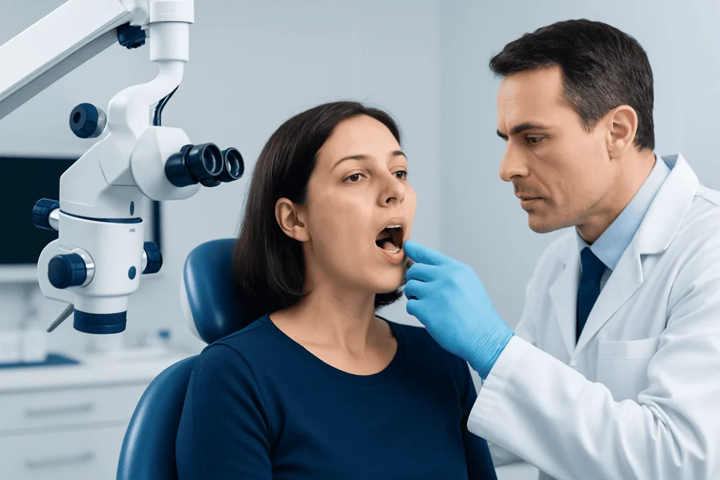
Diagnosis starts with a detailed oral examination. A dentist, oral medicine specialist, ENT doctor, or dermatologist may look at the spot under good lighting and ask when it first appeared, whether there was a bite or burn, what dental work has been done nearby, and whether there are any risk factors such as smoking, alcohol use, or a history of cancer.
The clinician may gently feel the lesion to assess whether it is flat, firm, soft, mobile, or attached to deeper tissue. They also inspect the rest of the mouth, gums, tongue, palate, and lips to see whether there are other pigmented areas or signs of irritation. A review of medications and medical conditions can provide further clues.
When the cause is not obvious, imaging or a biopsy may be recommended. A biopsy means removing a small sample of tissue so a pathologist can examine it under a microscope. This is the most reliable way to tell whether a pigmented lesion is benign or needs treatment. If a suspicious area is near restored teeth, dental imaging may help identify an amalgam tattoo or another local explanation.
In selected cases, patients may be referred for specialist evaluation and tissue testing, especially if the lesion persists or has concerning features. Depending on the findings, management can overlap with services such as biopsy or specialist assessment in ear, nose, and throat care.
Treatment options depend on the cause
Treatment is guided by the diagnosis rather than the color alone. A bruise or blood blister from trauma often needs no specific treatment beyond avoiding further irritation and allowing time to heal. If a rough tooth edge, dental appliance, or habit such as cheek biting is causing repeated injury, addressing that trigger is an important part of care.
Benign pigmentation, such as a stable melanotic macule or amalgam tattoo, may simply be observed after professional confirmation. In some cases, a clinician may photograph or measure the lesion and check it again later to confirm that it remains unchanged.
If the lesion is suspicious, enlarging, symptomatic, or cosmetically troubling, removal or biopsy may be advised. When a systemic disease or medication contributes to pigmentation, treatment focuses on the underlying cause in coordination with the appropriate specialist. If a malignant process is diagnosed, care may involve a multidisciplinary team and options such as oral cancer treatment.
Near the end of the care pathway, some patients may seek coordinated evaluation at a center that can combine dental, ENT, pathology, and oncology expertise. Acibadem International’s multidisciplinary specialists and JCI-accredited hospitals evaluate and treat oral lesions for international patients when further investigation is needed.
Self-care, monitoring, and prevention
Good self-observation can be useful, but it should be simple and safe. If a dark spot appears after clear minor trauma, it may be reasonable to watch it briefly while avoiding further irritation. Taking a dated photo can help track whether it is shrinking, staying the same, or changing.
Daily oral care supports overall mouth health. Gentle toothbrushing, flossing, staying hydrated, and having regular dental checkups make it easier to notice new changes early. If dentures, braces, or broken teeth are rubbing the cheek, arranging a dental review can help prevent repeated injury.
Prevention also includes avoiding tobacco and limiting other irritants to the mouth. Smoking is associated with pigment changes and raises the risk of significant oral disease. People who frequently bite their cheeks during stress or sleep may benefit from discussing behavioral strategies or dental protection with a clinician.
- Do not try to scrape, puncture, or burn a dark spot at home.
- Avoid very hot, sharp, or spicy foods if the area feels irritated.
- Seek professional advice if the cause is uncertain or the lesion does not clearly improve.
When to seek medical care
A black spot inside the cheek should be checked promptly if it has been present for more than two weeks, keeps returning, or is getting larger or darker. Medical or dental review is also important if the area bleeds, becomes painful, develops an ulcer, or feels firm or raised.
Urgent assessment is especially important for people with a history of tobacco use, heavy alcohol exposure, previous oral lesions, or immune system problems. Anyone who notices trouble swallowing, a persistent mouth sore, unexplained weight loss, or a lump in the neck should seek care without delay.
When in doubt, it is reasonable to arrange an examination rather than wait. Most causes are not dangerous, but mouth lesions can look similar to one another, and early evaluation is the safest way to clarify what the spot represents.
Frequently asked questions
Is a black spot on the inside of the cheek always serious?
No. Many dark spots are harmless and may come from cheek biting, minor bleeding under the tissue, natural pigmentation, or dental materials. A professional exam is still wise if the spot is new, changing, or does not go away.
How long should someone wait before getting a dark mouth spot checked?
If the spot does not clearly improve within about two weeks, it should be examined by a dentist or doctor. Earlier evaluation is appropriate if it is painful, bleeding, growing, or has no obvious cause.
Can a dental filling cause a black mark inside the cheek?
Yes. Tiny particles from certain dental materials can become embedded in nearby tissue and create a blue-gray or black area, sometimes called an amalgam tattoo. This is usually benign, but a clinician may confirm the diagnosis if the appearance is uncertain.
What does an oral biopsy involve?
An oral biopsy removes a small sample of tissue so it can be examined under a microscope. It is commonly done when a spot looks unusual, persists, or cannot be identified confidently by examination alone.
Can smoking cause dark spots inside the mouth?
Yes. Smoking can contribute to increased pigmentation in the mouth and also increases the risk of more serious oral disease. Any smoker with a new or persistent oral lesion should have it assessed.
Should someone try home remedies for a black spot inside the cheek?
Home care should be limited to gentle observation and avoiding further irritation if there is a clear minor injury. People should not scrape, puncture, or apply harsh substances to the spot, because this can worsen the tissue and delay diagnosis.
This article is for general information only and is not a substitute for professional medical advice. Please consult a qualified doctor about your individual situation.
Explore treatments in Turkey — costs, top hospitals & a free quote
JCI-accredited · board-certified surgeons · reply within 24h
Add us as a Preferred Source to see more of our trusted health content across Google Search, AI Overviews and Discover.

More from the Health Library

Fluticasone Propionate: What Patients Need to Know

High Functioning Autism: An Evidence-Based Guide for Patients

Golden Rice: A Complete Medical Overview

Trypsin Role in Digestion: What Patients Need to Know

Inferiority Complex: A Complete Medical Overview