Calcific Tendonitis: Early Signs, Risk Factors, and How It Is Treated

Calcific tendonitis happens when calcium deposits form within a tendon, usually in the rotator cuff of the shoulder. Pain may come on gradually or suddenly, especially when the deposit enters an active inflammatory phase.
Key Takeaways
- Calcific tendonitis happens when calcium deposits form within a tendon, usually in the rotator cuff of the shoulder.
- Pain may come on gradually or suddenly, especially when the deposit enters an active inflammatory phase.
- Diagnosis is usually confirmed with an exam and imaging such as X-ray or ultrasound.
- Most people improve with non-surgical care, including activity changes, pain relief, and physical therapy.
- Procedures such as ultrasound-guided needling or, less commonly, surgery may be considered if symptoms do not improve.
Calcific tendonitis is a condition in which calcium deposits form inside a tendon, most often in the shoulder, leading to pain, limited movement, and irritation of nearby tissues. Many cases improve with rest, physical therapy, and other non-surgical treatments, while persistent symptoms may need targeted procedures.
Overview
Calcific tendonitis is a painful tendon condition caused by calcium deposits forming within a tendon. It most often affects the rotator cuff tendons in the shoulder, especially the supraspinatus tendon, but it can also occur in other areas of the body. The condition may cause aching, sharp pain with movement, stiffness, and difficulty raising the arm.
For many people, the question is not only what calcific tendonitis is, but how it is treated. In most cases, treatment begins with non-surgical approaches such as rest, anti-inflammatory measures, guided exercise, and physical therapy. If pain is severe or long-lasting, a doctor may recommend procedures to break up or remove the deposit, and surgery is usually reserved for cases that do not respond to conservative care.
Calcific tendonitis is different from simple wear-and-tear tendon pain. The symptoms often fluctuate because the calcium deposit can pass through different stages, including a phase where the body starts to reabsorb it. During that resorptive phase, inflammation may increase and pain can become suddenly more intense even though the body is actively trying to clear the deposit.
Early Signs and Symptoms
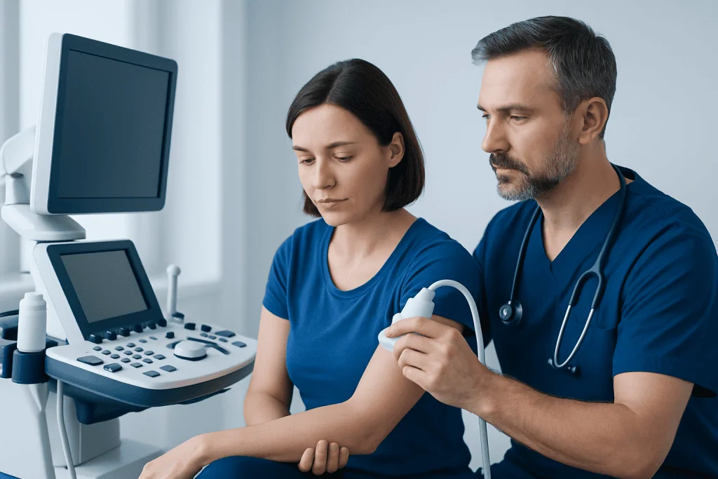
Early calcific tendonitis may cause mild discomfort that is easy to confuse with muscle strain or overuse. A person may notice pain when reaching overhead, putting on clothing, lifting objects, or sleeping on the affected shoulder. Some people feel only a vague ache at first, while others develop symptoms more abruptly.
As the condition progresses, the tendon and nearby bursa can become irritated. This may lead to restricted range of motion, tenderness, and weakness caused by pain rather than true loss of muscle strength. Night pain is common, especially when the shoulder is involved.
Symptoms can vary depending on the stage of the deposit. During quieter phases, the calcium may be present but cause little discomfort. During the inflammatory or resorptive stage, pain may become sharp and severe, sometimes making even small movements difficult.
- Shoulder pain that worsens with lifting or reaching
- Stiffness or reduced range of motion
- Pain when lying on the affected side
- Sudden flare-ups of intense pain
- Tenderness around the shoulder
Why It Happens and Who Is at Risk
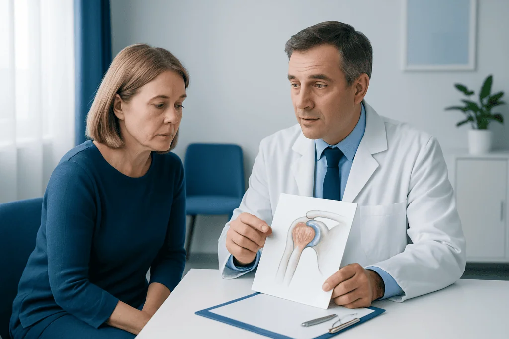
The exact cause of calcific tendonitis is not fully understood. It is not simply a matter of “too much calcium” in the diet or bloodstream. Instead, experts believe it involves changes within the tendon itself, including reduced oxygen supply, cell changes, and a process in which calcium crystals form and are later reabsorbed by the body.
Calcific tendonitis is most common in adults, especially in midlife, and it tends to occur more often in the shoulder than in other joints. Some people have no clear trigger. Others may have contributing factors such as repetitive overhead activity, tendon irritation, or biomechanical strain around the shoulder.
Certain conditions may overlap with or increase the likelihood of shoulder pain from tendons and surrounding structures. For example, doctors may also consider problems such as rotator cuff tear or shoulder impingement syndrome when symptoms are persistent or movement is limited. Having one shoulder disorder does not automatically mean a person has another, but similar symptoms can occur.
- Age-related tendon changes, often in adults between about 30 and 60
- Repetitive overhead use of the shoulder
- Previous tendon irritation or degeneration
- Possible association with metabolic or endocrine factors in some patients
- Female sex appears more commonly affected in some clinical series
How Doctors Diagnose Calcific Tendonitis
Diagnosis starts with a medical history and physical examination. A doctor will ask when the pain began, what movements make it worse, whether sleep is disturbed, and if there was any recent injury. During the exam, shoulder motion, strength, tenderness, and signs of impingement or bursitis are assessed.
Imaging is usually important because calcific tendonitis can often be seen directly. A plain X-ray is one of the most useful first tests because calcium deposits are often visible. Ultrasound can show the exact location of the deposit and whether there is associated inflammation, bursitis, or tendon thickening. In some cases, MRI may be used if the doctor needs to evaluate other causes of shoulder pain.
Diagnosis is not only about identifying calcium in the tendon. Small deposits may be present without causing symptoms, so the doctor also considers whether the imaging findings match the person’s pain and exam results. This careful approach helps guide treatment and avoid unnecessary procedures.
Treatment Options
Treatment depends on how severe the symptoms are, how long they have lasted, and whether the person is in a painful inflammatory phase. Many patients improve with conservative care. This may include a short period of activity modification, pain-relieving medicines advised by a doctor, ice during painful flare-ups, and a structured exercise program to maintain shoulder motion.
Physical therapy often plays a central role. Gentle stretching and strengthening may help reduce stiffness, improve shoulder mechanics, and prevent secondary problems caused by guarding the joint. In some cases, doctors may recommend image-guided procedures such as injection into an inflamed bursa or ultrasound-guided barbotage, a technique used to break up and aspirate calcium deposits. When appropriate, care may also involve physical therapy and rehabilitation or pain management as part of a broader non-surgical plan.
If symptoms continue despite adequate non-surgical treatment, other options may be considered. These may include extracorporeal shock wave therapy in selected cases or surgery to remove the deposit, often using a minimally invasive approach. When surgery is needed, doctors may also address related tendon or bursa problems at the same time, such as through arthroscopic surgery.
The best treatment is individualized. Factors such as deposit size, phase of the disease, level of pain, work demands, and response to earlier treatment all influence the plan. A qualified orthopedic specialist can help balance symptom relief with preserving tendon health and shoulder function.
Recovery, Prevention, and Self-care
Recovery time varies. Some episodes settle within weeks, while others improve more gradually over months, especially if the deposit is large or the shoulder has become stiff. Even when symptoms improve, continuing the prescribed exercises may help restore motion and lower the chance of ongoing discomfort.
There is no guaranteed way to prevent calcific tendonitis because the exact cause is complex. Still, shoulder-friendly habits may reduce strain and help overall tendon health. These include using good posture, avoiding repetitive painful overhead movements when possible, taking breaks during repetitive work, and strengthening the muscles that support the shoulder blade and rotator cuff.
Self-care should focus on symptom control and function, not on trying unproven remedies to dissolve calcium. It is also important not to stay completely inactive for too long, because prolonged immobility can lead to stiffness. A doctor or therapist can suggest when to rest and when to resume movement more actively.
- Use cold packs during painful flare-ups if advised
- Follow a guided home exercise program
- Return to sports or overhead work gradually
- Seek review if pain persists or range of motion worsens
When to Seek Medical Care
Medical assessment is a good idea if shoulder pain lasts more than a few weeks, interferes with sleep, or limits daily activities such as dressing, reaching, or lifting. An evaluation is also important when pain keeps returning after rest or simple home care.
More urgent care may be needed if there is significant weakness after an injury, marked swelling, fever, redness, or inability to move the shoulder. These symptoms can suggest a different or additional problem and should not be assumed to be calcific tendonitis.
Specialist review can be helpful when symptoms are severe or diagnosis is uncertain. In appropriate cases, assessment by orthopedics and traumatology may help confirm the cause and discuss targeted treatment options. Acibadem International’s multidisciplinary specialists in JCI-accredited hospitals also diagnose and treat calcific tendonitis for international patients.
Frequently asked questions
Is calcific tendonitis the same as regular tendonitis?
No. Calcific tendonitis involves calcium deposits forming inside a tendon, while other forms of tendonitis may involve irritation, overload, or degeneration without calcium buildup. The symptoms can overlap, which is why imaging is often helpful.
Where does calcific tendonitis usually occur?
It most commonly affects the shoulder, especially the rotator cuff tendons. Less often, it can occur in other tendons in the body. Shoulder involvement is the classic pattern seen in practice.
Can calcific tendonitis go away on its own?
Yes, in some people the body gradually reabsorbs the calcium deposit and symptoms improve over time. However, the process can take weeks or months, and painful flare-ups may happen along the way. Medical treatment can help control pain and maintain motion during recovery.
Does calcific tendonitis always require surgery?
No. Most cases are treated without surgery using rest, activity changes, medication guidance, and physical therapy. Surgery is usually considered only when symptoms remain significant despite appropriate non-surgical treatment.
What tests are used to confirm calcific tendonitis?
Doctors often use a physical exam together with imaging, especially X-rays and ultrasound. These tests can show the calcium deposit and help assess surrounding inflammation. MRI may be used if another shoulder problem is also suspected.
Is exercise safe with calcific tendonitis?
Usually yes, but the type and timing of exercise matter. Gentle, guided exercises are often recommended to prevent stiffness and support recovery. Painful or repetitive overhead activity may need to be reduced until the tendon settles.
References
- American Academy of Orthopaedic Surgeons
- National Institute of Arthritis and Musculoskeletal and Skin Diseases
- American College of Radiology
- Mayo Clinic
- Cleveland Clinic
This article is for general information only and is not a substitute for professional medical advice. Please consult a qualified doctor about your individual situation.
Joint and spine care in Turkey — expert assessment & treatment
JCI-accredited · board-certified surgeons · reply within 24h
Add us as a Preferred Source to see more of our trusted health content across Google Search, AI Overviews and Discover.