Realistic Heart: What Patients Need to Know

A realistic heart is most often an educational, imaging, or surgical-planning tool rather than a diagnosis. Doctors may use realistic heart images or 3D models to explain heart anatomy and individual heart conditions more clearly.
Key Takeaways
- A realistic heart is most often an educational, imaging, or surgical-planning tool rather than a diagnosis.
- Doctors may use realistic heart images or 3D models to explain heart anatomy and individual heart conditions more clearly.
- These models can be created from tests such as echocardiography, CT, or MRI.
- Realistic heart tools may help with planning procedures for congenital, structural, or complex heart disease.
- Chest pain, shortness of breath, fainting, or sudden palpitations need prompt medical assessment.
A realistic heart usually refers to a lifelike image, digital reconstruction, or 3D model of the human heart used to explain anatomy, support diagnosis, or help plan treatment. For patients, these tools can make heart conditions easier to understand and can improve communication with the care team.
Overview: what “realistic heart” usually means
A realistic heart is usually a lifelike representation of the human heart rather than a medical disorder itself. The term may describe a detailed illustration, a digital 3D reconstruction, a printed heart model, or an imaging-based view that closely matches a person’s actual anatomy. These tools are commonly used in patient education, medical training, and treatment planning.
For patients, the main value of a realistic heart model is clarity. The heart is a complex organ with chambers, valves, vessels, and electrical pathways that can be difficult to picture from words alone. When clinicians use a realistic model or image, it often becomes easier to understand what is normal, what has changed, and why a certain test or treatment is recommended.
This topic is different from general heart disease information because it focuses on how realistic visual tools are used in care. Rather than listing heart disorders in a broad way, it helps patients understand how modern imaging and modeling can personalize explanations and support decisions, especially in structural heart disease, congenital heart problems, or procedural planning.
How realistic heart models are used in patient care
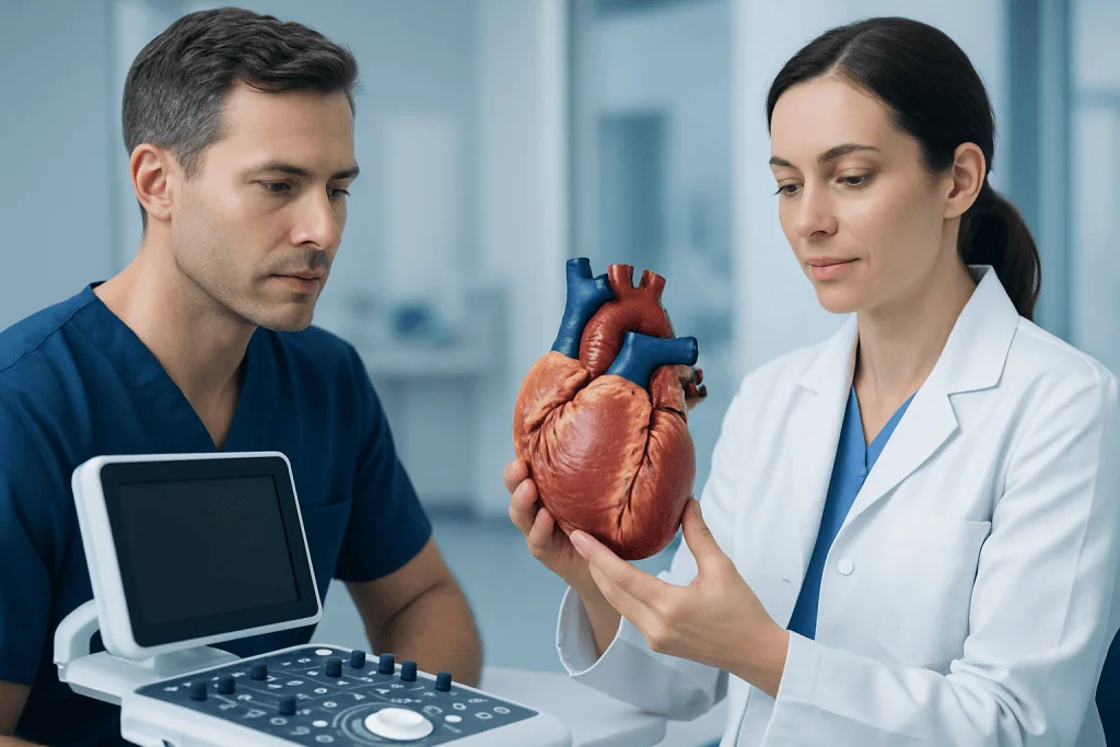
Realistic heart tools can be used at several stages of care. In the clinic, a doctor may show a patient a model to explain how blood moves through the heart, how a valve opens and closes, or how a defect changes normal circulation. This can make complex terms more understandable and help patients ask more informed questions.
In more advanced settings, a realistic heart may be built from a person’s own scan results. These patient-specific models can show the exact size, shape, and relationships of structures inside the chest. This is especially useful when anatomy is unusual or when a team is considering a catheter-based procedure or surgery.
Common uses include:
- Explaining normal heart anatomy and function
- Showing the location of a valve problem, blockage, or structural defect
- Planning procedures for congenital or complex heart disease
- Helping surgeons and cardiologists review the safest approach
- Supporting communication between patients, families, and specialists
In some cases, realistic models also support follow-up care. Patients who have had repairs for congenital heart disease or valve disease may benefit from seeing how the heart has changed over time and why ongoing monitoring matters.
What can a realistic heart show?

A realistic heart can show both normal structures and medical problems. It may display the four chambers, the valves, the major blood vessels, and the thickness or movement of the heart muscle. Depending on the imaging method, it can also highlight blood flow patterns, scarring, enlargement, or abnormal connections.
These details can be particularly helpful when discussing conditions such as heart valve disease or congenital structural problems. Instead of hearing only technical names, patients can often see where a valve narrows, where blood leaks backward, or how a wall between chambers differs from usual anatomy.
A realistic heart may also be useful in discussing rhythm-related concerns, though electrical activity itself is often shown through separate tests rather than a static model. For example, a patient with palpitations may still need an ECG or heart monitor even if the anatomy looks normal on imaging. The model helps with the structural picture, while other tests help explain function and rhythm.
It is important to remember that even a highly detailed model has limits. It is a tool that supports clinical judgment, not a replacement for examination, history-taking, or standard diagnostic testing.
How doctors create realistic heart images and models
Realistic heart representations are often created from medical imaging. Echocardiography uses ultrasound to show heart motion and valve function in real time. CT scans can provide detailed pictures of the heart and blood vessels, while cardiac MRI can offer precise information about structure, tissue characteristics, and function.
Once the images are collected, specialized software may reconstruct them into a 3D digital model. In selected cases, that digital file can be turned into a physical model using 3D printing. This approach is most often used when anatomy is complex and when a team wants to study a case carefully before an intervention.
Patients may hear about these tools when being evaluated with cardiac MRI or advanced scan-based planning. A model can help the team decide which procedure is technically feasible, what risks need special attention, and how to explain the plan in a patient-friendly way.
Not every heart condition requires this level of modeling. Many common concerns can be diagnosed and managed using a standard history, physical examination, ECG, and echocardiogram. The decision depends on the question the doctor is trying to answer and whether extra anatomical detail will change care.
Who may benefit most from a realistic heart approach?
Realistic heart tools are often most useful for people with complex or highly individual anatomy. This includes children and adults with congenital heart disease, patients with unusual valve or vessel anatomy, and those preparing for structural heart procedures. In these situations, standard diagrams may not fully reflect the person’s anatomy, while a personalized model can.
They may also benefit patients who are trying to understand an important treatment choice. For example, someone considering valve repair, valve replacement, or another structural procedure may feel more confident after seeing a realistic explanation of the problem and the planned approach. Good visualization does not make the decision for the patient, but it can improve informed consent and shared decision-making.
For some families, realistic heart models are especially valuable in pediatric care. Parents often want a clear explanation of what is happening and what the operation or catheter procedure aims to correct. A visual model can support these conversations in a calm and practical way.
When coronary artery disease is being assessed, physicians may rely on a combination of symptoms, risk factors, and imaging, sometimes including coronary CT angiography. In that setting, realistic imaging can help show where arteries narrow and how that may relate to symptoms.
What realistic heart tools cannot do
Although realistic heart models are helpful, they do not replace a full medical evaluation. A detailed picture of the heart does not automatically explain chest pain, dizziness, fatigue, or palpitations. Symptoms can come from blood pressure changes, arrhythmias, lung conditions, anxiety, anemia, thyroid problems, or many other causes that require broader assessment.
These tools also do not predict outcomes on their own. A model may show anatomy very clearly, but treatment decisions still depend on age, overall health, test results, symptom severity, and the experience of the care team. In other words, the model adds information; it is not the only information that matters.
Patients should also know that a realistic heart image is not always necessary. In straightforward situations, standard testing already provides enough information. Using more technology is only helpful when it answers an important clinical question or improves planning in a meaningful way.
When a procedure is being considered, the team may combine anatomical modeling with other forms of assessment, including stress testing, catheter-based measurements, or echocardiography. This balanced approach helps ensure that both structure and function are considered together.
When to seek medical care
People should seek medical care if they have symptoms that could point to a heart problem, whether or not they have seen realistic heart images online or during a consultation. Warning signs include chest pain or pressure, shortness of breath, fainting, unexplained swelling in the legs, a racing or irregular heartbeat, or marked fatigue with routine activity.
Urgent medical attention is especially important if chest discomfort is sudden, severe, or associated with sweating, nausea, breathlessness, or pain spreading to the arm, back, neck, or jaw. These symptoms do not always mean a heart emergency, but they should never be ignored.
Patients with known structural heart disease, repaired congenital heart disease, or arrhythmia should attend regular follow-up visits even when they feel well. Ongoing review helps identify changes early and guides timing for additional tests or treatment.
Near the end of a diagnostic journey, some people benefit from care in centers with coordinated cardiology, imaging, and surgical expertise. Acibadem International’s multidisciplinary specialists and JCI-accredited hospitals diagnose and treat heart conditions for international patients when advanced evaluation is needed.
Frequently asked questions
Is a realistic heart a medical condition?
Usually no. In most health contexts, realistic heart refers to a lifelike model, image, or digital reconstruction of the heart used for education, diagnosis, or treatment planning.
Why would a doctor show a patient a realistic heart model?
A realistic model can make heart anatomy and disease easier to understand. It may help explain where a problem is located, how it affects blood flow, and why a certain procedure or test is being recommended.
How is a patient-specific realistic heart created?
It is commonly created from imaging tests such as echocardiography, CT, or MRI. Specialized software can convert scan data into a 3D digital view, and in selected cases this can also be produced as a physical printed model.
Can a realistic heart model diagnose heart disease by itself?
No. It can support diagnosis by showing anatomy in detail, but doctors still need symptoms, examination findings, and other tests to reach an accurate conclusion.
Who benefits most from realistic heart imaging or modeling?
It is often most useful for people with congenital heart disease, valve disorders, complex anatomy, or those preparing for a structural heart procedure. It can also be helpful for patients who want a clearer explanation before making treatment decisions.
Should someone worry if they need advanced heart imaging?
Not necessarily. Advanced imaging is often used simply to get more precise information and to plan care safely. The need for extra imaging does not by itself mean that a condition is severe.
This article is for general information only and is not a substitute for professional medical advice. Please consult a qualified doctor about your individual situation.
Explore treatments in Turkey — costs, top hospitals & a free quote
JCI-accredited · board-certified surgeons · reply within 24h
Add us as a Preferred Source to see more of our trusted health content across Google Search, AI Overviews and Discover.

More from the Health Library

How to Prepare for a Routine Health Assessment

What’s the Difference Between Hemorrhoid and Polyp — Explained by Medical Evidence, Not Myths

Monocytes High: An Evidence-Based Guide for Patients

Scrofula: An Evidence-Based Guide for Patients

Wound Dehiscence: What Patients Need to Know