Fundoscopy: Benefits, Risks, and Recovery — A Complete Guide

Fundoscopy allows a doctor to examine the retina, optic nerve, macula, and retinal blood vessels. It may be performed with or without dilating eye drops, depending on the reason for the exam.
Key Takeaways
- Fundoscopy allows a doctor to examine the retina, optic nerve, macula, and retinal blood vessels.
- It may be performed with or without dilating eye drops, depending on the reason for the exam.
- The test can help detect eye conditions such as retinal disease, glaucoma-related optic nerve changes, and diabetic eye damage.
- Most people can return to normal activities soon after fundoscopy, although dilation can temporarily blur vision and increase light sensitivity.
- Complications are uncommon, but patients should seek medical care if severe eye pain, sudden vision loss, or persistent symptoms occur after the exam.
Fundoscopy is a common eye examination used to look at the back of the eye, including the retina, optic nerve, and blood vessels. It helps doctors detect eye disease and signs of wider health conditions, and it is usually quick, low risk, and easy to recover from.
Overview: What fundoscopy is and why it is done
Fundoscopy is an examination of the inside of the eye, especially the retina, optic nerve, macula, and retinal blood vessels. It is also called a fundus exam or ophthalmoscopy. Doctors use it to look for changes that may affect vision and to identify clues to general health conditions such as diabetes, high blood pressure, and some neurologic disorders.
Unlike many routine vision checks that focus on visual acuity or glasses prescriptions, fundoscopy looks directly at eye structures that cannot be seen from the outside. This makes it an important part of a comprehensive eye assessment when a person has visual symptoms, ongoing medical conditions, or risk factors for eye disease.
Fundoscopy may be done during a regular eye checkup, in an emergency setting, or as part of follow-up for known eye disease. Depending on the situation, the doctor may use a handheld ophthalmoscope, a special head-mounted light, a slit lamp with lenses, or retinal imaging tools. In many cases, the exam is brief and causes little discomfort.
How fundoscopy works and what doctors can see
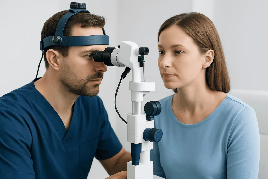
The goal of fundoscopy is to illuminate the back of the eye and magnify internal structures. Light passes through the pupil, allowing the clinician to inspect the fundus, which includes the retina, optic disc, macula, and retinal vessels. These parts of the eye can reveal both local eye disease and signs of disease elsewhere in the body.
There are several ways to perform the exam. Direct ophthalmoscopy uses a handheld device and gives a close, upright view of the fundus. Indirect ophthalmoscopy uses a light source and lens to provide a wider view, which can be especially helpful for examining the peripheral retina. Slit-lamp fundoscopy combines a microscope-like instrument with special lenses to produce a detailed view.
Dilating eye drops are often used to widen the pupils and improve visibility. This allows the doctor to examine more of the retina and may increase the ability to detect subtle abnormalities. In some situations, a non-dilated exam may be enough, but a dilated exam often provides more complete information.
Doctors may recommend fundoscopy when evaluating symptoms such as blurred vision, flashes, floaters, headache related to possible eye findings, vision loss, or eye pain. It is also commonly used to monitor conditions linked to retinal damage, including diabetes and glaucoma.
Who may need fundoscopy and when it is especially useful

Fundoscopy can be useful for many different people, not only those with known eye disease. It may be part of routine preventive eye care, especially in adults as they age, or in people with a family history of eye conditions. It is also frequently advised for anyone with symptoms that suggest a problem in the retina or optic nerve.
People with certain medical conditions often need fundoscopy more regularly because systemic diseases can affect the back of the eye. Examples include diabetes, hypertension, autoimmune conditions, and some neurological disorders. In these settings, retinal changes may appear before a person notices major visual symptoms.
Fundoscopy is especially valuable in urgent assessment when a patient reports sudden new floaters, light flashes, a curtain-like shadow in vision, sudden reduced vision, or severe headache with visual changes. These symptoms may point to retinal tears, retinal detachment, optic nerve swelling, or bleeding inside the eye, all of which need prompt medical review.
For patients already being evaluated by an ophthalmologist, fundoscopy may be combined with other tests such as retinal photography, optical coherence tomography, visual field testing, or a comprehensive eye examination. The exact combination depends on the clinical question and the structures the doctor needs to assess.
Step by step: what happens during the procedure
Before the exam, the doctor or eye care professional asks about symptoms, medications, allergies, and previous eye problems. Vision may be checked first, and the front of the eye may be examined. If dilation is planned, drops are placed in the eyes and usually take effect within about 15 to 30 minutes.
During direct ophthalmoscopy, the room is dimmed and the doctor shines a light through the pupil while looking through the handheld instrument. For slit-lamp fundoscopy, the patient rests the chin and forehead on a support while the doctor uses bright light and magnifying lenses to inspect the fundus in detail. Indirect ophthalmoscopy may involve a head-mounted light and a lens held near the eye for a broader view.
The exam itself is usually short, though the overall visit may be longer if dilation is needed or if additional imaging is performed. Bright lights can feel uncomfortable for a few moments, but the procedure should not be painful. Some people notice glare, watering, or mild temporary blurring during the test.
If the doctor finds an area that needs closer evaluation, the next step may include retinal imaging or referral for treatment. In patients with signs of retinal disease, this can lead naturally to further assessment such as retinal examination or specialist planning. The findings guide whether observation, medical treatment, laser treatment, or another intervention is most appropriate.
Benefits, findings, and possible risks
The main benefit of fundoscopy is that it allows direct inspection of structures essential to vision. This can help diagnose conditions early, sometimes before they cause major symptoms. Early recognition is important because many retinal and optic nerve disorders are more manageable when detected promptly.
Fundoscopy can reveal a wide range of findings, including diabetic retinopathy, hypertensive retinopathy, optic disc swelling, optic nerve cupping, retinal tears, retinal detachment, macular changes, hemorrhages, and inflammation. It may also support the evaluation of symptoms that are not caused by the eye alone, such as neurologic or vascular problems.
Risks from the examination itself are low. The most common effects are related to dilating drops and include temporary blurred vision, light sensitivity, and difficulty focusing on near objects for several hours. Rarely, dilation may trigger a sudden rise in eye pressure in susceptible people with narrow angles, which is why clinicians consider eye history and symptoms before using drops.
Allergic reactions to eye drops are uncommon but possible. Patients should tell the clinician about past reactions, glaucoma history, or eye surgery. If fundoscopy identifies urgent findings such as retinal tears or retinal detachment, treatment may be arranged quickly to help protect sight.
Recovery timeline and self-care after fundoscopy
Recovery after fundoscopy is usually simple. If no dilating drops are used, most people can resume their normal activities immediately. If the pupils are dilated, temporary effects may last a few hours and sometimes longer, depending on the drops used and the individual response.
Common short-term effects after dilation include blurred near vision, sensitivity to bright light, and larger-looking pupils. Wearing sunglasses outdoors can make the eyes more comfortable. Reading, detailed screen work, and driving may be more difficult until vision clears, so some patients prefer to arrange transportation home.
There is usually no need for special medication or downtime after a routine exam. Patients can return to work and daily tasks once their vision feels normal and safe for those activities. If the examination led to additional procedures or treatments, the doctor will provide more specific recovery instructions based on that care.
When fundoscopy identifies a condition that needs treatment, follow-up may include monitoring, medical therapy, laser procedures, or surgery. For some retinal problems, an ophthalmologist may discuss options such as retina surgery depending on the location and severity of the problem.
When to seek medical care
Medical advice should be sought promptly if a person develops sudden vision loss, a curtain or shadow over vision, a sudden shower of floaters, repeated flashes of light, or severe new eye pain. These symptoms can signal a retinal tear, retinal detachment, acute pressure rise, or another urgent eye problem that should not wait.
After a routine fundoscopy, mild blurring and light sensitivity can be normal if dilation was used. However, symptoms that are severe, unexpected, or do not improve should be assessed. Examples include intense headache, nausea with eye pain, markedly worsening vision, or redness that feels significant rather than mild irritation.
People with diabetes, high blood pressure, prior retinal disease, high myopia, or a family history of serious eye disease should keep regular eye appointments even if they feel well. Many retinal and optic nerve conditions begin quietly, and timely examinations can help detect changes before vision is affected.
If a specialist review is needed, multidisciplinary eye teams can coordinate diagnosis and treatment planning. Acibadem International’s ophthalmology specialists in JCI-accredited hospitals evaluate and treat international patients, including those who need advanced retinal assessment and follow-up.
Frequently asked questions
Is fundoscopy the same as a regular eye exam?
Fundoscopy is often one part of a regular eye exam, but it is not the whole exam. It specifically focuses on the back of the eye, including the retina and optic nerve, while a full eye exam may also check vision, eye pressure, and the front of the eye.
Does fundoscopy hurt?
Fundoscopy is usually not painful. The bright light can feel uncomfortable for a short time, and dilating drops may sting briefly, but most people tolerate the procedure well.
How long does blurred vision last after fundoscopy?
If dilating drops are used, blurred near vision and light sensitivity often last for a few hours. In some people, especially those sensitive to the drops, effects may last longer into the day.
Can I drive after fundoscopy?
Some people can drive after a non-dilated exam, but driving may be difficult after dilation because of blurred vision and glare. It is safest to follow the clinic’s advice and consider arranging transportation if dilation is planned.
Why would a doctor order fundoscopy for diabetes or high blood pressure?
Diabetes and high blood pressure can damage the small blood vessels in the retina. Fundoscopy helps doctors look for these changes, monitor progression, and decide whether further treatment or closer follow-up is needed.
What conditions can fundoscopy help detect?
Fundoscopy can help detect retinal tears, retinal detachment, diabetic retinopathy, hypertensive changes, macular problems, optic nerve swelling, and glaucoma-related optic nerve damage. It may also provide clues to some neurological and vascular conditions.
References
- American Academy of Ophthalmology
- National Eye Institute
- American Optometric Association
- Mayo Clinic
- MedlinePlus
This article is for general information only and is not a substitute for professional medical advice. Please consult a qualified doctor about your individual situation.
Explore treatments in Turkey — costs, top hospitals & a free quote
JCI-accredited · board-certified surgeons · reply within 24h
Check your numbers in seconds
BMI, calories, due date, blood pressure and 30+ more clinical calculators — free, instant, doctor-reviewed ranges.