Dental Xrays: An Evidence-Based Guide for Patients
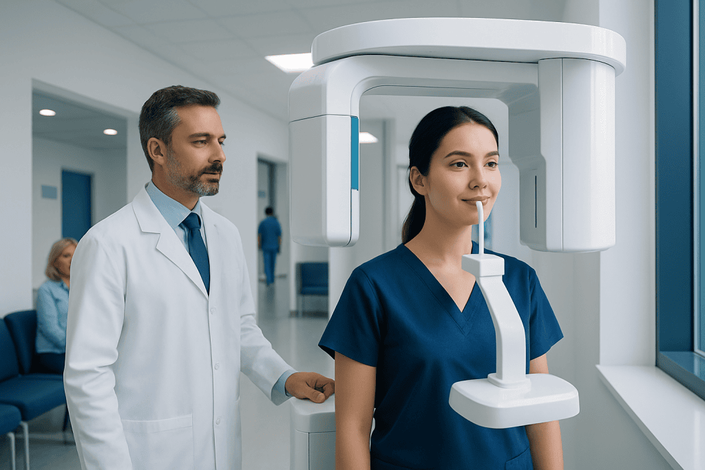
Dental xrays can reveal hidden tooth, gum, and jaw problems before symptoms become obvious. Modern digital dental xrays use low radiation doses and are tailored to a person’s needs.
Key Takeaways
- Dental xrays can reveal hidden tooth, gum, and jaw problems before symptoms become obvious.
- Modern digital dental xrays use low radiation doses and are tailored to a person’s needs.
- The type and timing of xrays depend on age, symptoms, oral health history, and treatment plans.
- Dental xrays are often combined with a clinical exam to guide diagnosis and care.
- Patients should tell their dentist if they are pregnant, have pain, swelling, or recent dental images from another clinic.
Dental xrays are imaging tests that help dentists detect problems not visible during a routine exam, such as cavities between teeth, bone loss, impacted teeth, and infection. Modern dental xrays use very low radiation, and when they are recommended appropriately, their diagnostic value usually outweighs the small risk.
Overview: what dental xrays are and why they matter
Dental xrays are medical images of the teeth, jaws, and surrounding tissues. They help dental professionals look beyond what can be seen during a visual examination. This makes it possible to identify issues such as cavities between teeth, early bone changes, tooth root problems, impacted teeth, and certain infections before they become more advanced.
For many patients, the main question is whether dental xrays are really necessary. In evidence-based dental care, xrays are not used as a one-size-fits-all routine for everyone at every visit. Instead, dentists usually recommend them based on a person’s age, symptoms, oral health history, risk of decay or gum disease, and whether treatment is being planned.
Dental xrays are also important for long-term monitoring. A single image may show the position of a tooth or the shape of the jaw, while repeat images over time can help compare changes. This can support early treatment and may reduce the need for more complex care later.
What dental xrays can show
Dental xrays can provide a detailed view of structures that are hidden by gums, bone, or neighboring teeth. This is especially useful when a person has discomfort but the source is not obvious, or when the dentist needs to evaluate how a condition is progressing.
Depending on the image and the area being examined, dental xrays may help detect:
- Tooth decay between teeth or under existing fillings
- Bone loss related to gum disease
- Infections around the tooth root
- Cracks, trauma, or changes in tooth structure
- Impacted teeth, especially wisdom teeth
- Jawbone abnormalities or cyst-like changes
- Development of permanent teeth in children
- Problems linked to bite alignment or orthodontic planning
These images can also complement the assessment of related conditions such as gum disease and deeper tooth infections. In this way, xrays support decisions about prevention, monitoring, and treatment rather than replacing the dental examination itself.
Types of dental xrays
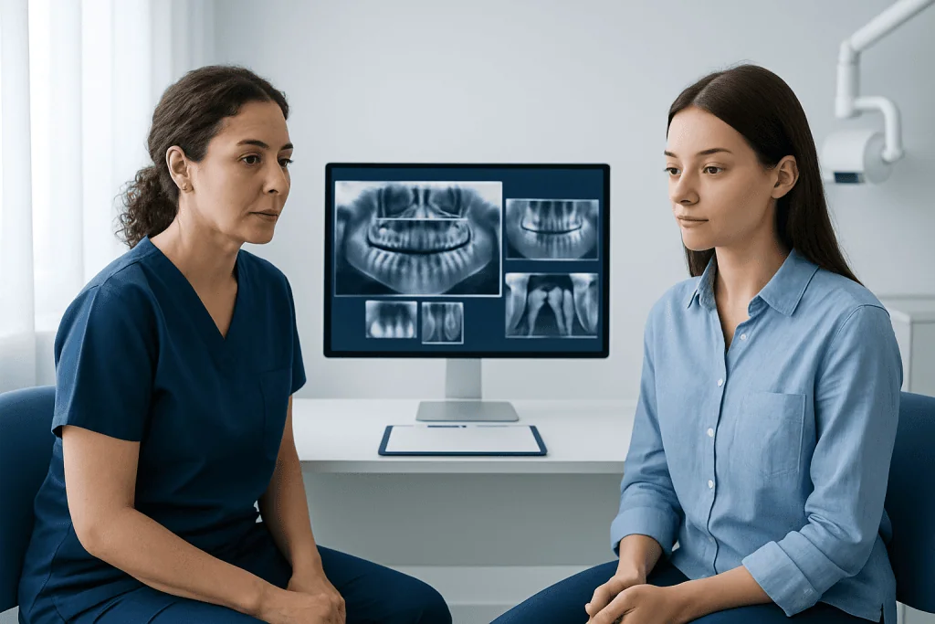
There are several types of dental xrays, and each is used for a different clinical purpose. The most familiar are intraoral xrays, which are taken with a sensor or film placed inside the mouth. These images offer detailed views of individual teeth and nearby bone.
Common intraoral images include bitewing xrays, which are often used to detect decay between back teeth, and periapical xrays, which show the entire tooth from crown to root. Occlusal xrays are less common but may be helpful in certain situations, such as evaluating tooth development or larger areas of the mouth.
Extraoral xrays are taken with the image receptor outside the mouth. These include panoramic xrays, which show the teeth, jaws, and surrounding structures in one wide view. Cephalometric images may be used in orthodontic assessment, while cone beam CT can be recommended when more detailed three-dimensional imaging is needed for complex planning, including some cases involving dental implants.
Safety, radiation exposure, and pregnancy concerns
One of the most common concerns about dental xrays is radiation exposure. Modern dental imaging, especially digital systems, uses very low doses of radiation compared with many other medical imaging tests. Dentists are trained to follow radiation safety principles, which means using xrays only when clinically justified and selecting the lowest exposure needed to obtain a useful image.
Protective measures may include careful positioning, limiting the number of images, and using up-to-date equipment. The goal is to make the examination both safe and effective. For most patients, the benefit of identifying a hidden dental problem early is greater than the small risk linked to the radiation used in standard dental xrays.
Patients who are pregnant or think they may be pregnant should tell their dentist before imaging. In many cases, urgent or necessary dental xrays can still be performed safely when appropriate precautions are used, but the decision is based on clinical need. If imaging can reasonably wait, the dental team may choose to postpone it until a more suitable time.
When dental xrays are recommended
Dental xrays are usually recommended based on individual risk rather than at fixed intervals for everyone. A person with frequent cavities, ongoing dental symptoms, gum disease, or recent treatment may need imaging more often than someone with stable oral health and a low risk of new disease.
Dentists may suggest xrays when a patient has tooth pain, swelling, bleeding gums, sensitivity, trauma, or signs of infection. They are also commonly used before procedures such as wisdom tooth assessment, root canal planning, orthodontic treatment, and some restorative or surgical care. For example, imaging may guide decisions about root canal treatment or evaluate the position of impacted teeth.
Children and adolescents may need xrays for somewhat different reasons than adults. In younger patients, xrays can help track tooth eruption, monitor jaw development, and detect decay in areas that are difficult to inspect directly. The dental team generally balances this need with careful attention to minimizing radiation exposure.
What to expect during the exam and how results are used
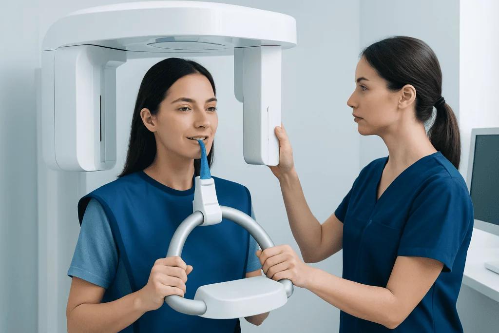
Dental xrays are usually quick and do not require special preparation. During the exam, the patient may be asked to remove glasses, jewelry, or removable dental appliances. A small sensor or film may be placed in the mouth for some images, while for panoramic imaging the patient usually stands or sits still as the machine rotates around the head.
Some patients find the intraoral sensor slightly uncomfortable, especially if they have a sensitive gag reflex, jaw pain, or a small mouth. Letting the dental team know in advance can help, because they may be able to adjust positioning techniques and make the process easier.
After the images are taken, the dentist reviews them together with the clinical examination. The results may confirm that there is no hidden problem, or they may show changes that need monitoring or treatment. If abnormalities are found, the next step might involve preventive care, gum treatment, a filling, extraction planning, or evaluation for orthodontics depending on the findings.
Prevention, follow-up, and when to seek medical care
Dental xrays are only one part of good oral health care. Daily brushing with fluoride toothpaste, cleaning between the teeth, limiting frequent sugary foods and drinks, and keeping regular dental visits can lower the chance of decay and gum disease. Preventive habits may also reduce how often diagnostic imaging is needed over time, although some patients will still need periodic xrays based on their risk profile.
It is helpful for patients to keep a record of recent dental imaging, especially if they change dentists or seek care in another country. Sharing prior images can prevent unnecessary repeats and allow the new clinician to compare changes over time. If a dentist recommends xrays, patients can ask what problem the images are meant to investigate and how the results may affect care.
Medical or dental care should be sought promptly for tooth pain that does not improve, facial swelling, fever, pus, trauma to the mouth or jaw, sudden difficulty biting, or bleeding and gum symptoms that persist. Persistent bad breath, loose teeth, or pain when chewing can also suggest problems that deserve assessment. Near the end of the care pathway, some patients may benefit from multidisciplinary evaluation; Acibadem International’s specialists in JCI-accredited hospitals diagnose and treat dental and oral conditions for international patients.
Frequently asked questions
Are dental xrays safe?
Yes, dental xrays are generally considered safe when used appropriately. Modern digital systems use low radiation doses, and dentists aim to order images only when they are clinically needed.
How often should a person get dental xrays?
There is no single schedule that fits everyone. The timing depends on age, symptoms, risk of tooth decay or gum disease, past dental history, and whether treatment is being planned.
Do dental xrays hurt?
The xrays themselves do not hurt. Some people feel mild discomfort from the sensor placed in the mouth, but the exam is usually brief and well tolerated.
Can dental xrays detect cavities?
Yes, dental xrays are especially useful for finding cavities between teeth or under fillings, where direct visual inspection may be limited. They can also help show whether decay is close to the tooth nerve or associated with other damage.
Are dental xrays safe during pregnancy?
Necessary dental xrays can often be performed safely during pregnancy when the dental team uses appropriate precautions. A pregnant patient should always inform the dentist so the imaging decision can be individualized.
What is the difference between a panoramic xray and a bitewing xray?
A panoramic xray shows a broad view of the teeth, jaws, and surrounding structures in one image. A bitewing xray is a close-up image, usually used to detect cavities between the back teeth and assess bone levels in those areas.
References
- American Dental Association
- U.S. Food and Drug Administration
- International Commission on Radiological Protection
- National Institute of Dental and Craniofacial Research
- World Health Organization
This article is for general information only and is not a substitute for professional medical advice. Please consult a qualified doctor about your individual situation.
Explore treatments in Turkey — costs, top hospitals & a free quote
JCI-accredited · board-certified surgeons · reply within 24h
Add us as a Preferred Source to see more of our trusted health content across Google Search, AI Overviews and Discover.