Arteriography: A Complete Medical Overview

Arteriography creates detailed images of arteries after contrast dye is injected through a thin catheter. It may be used to investigate narrowed, blocked, damaged or abnormally shaped arteries in many parts of the body.
Key Takeaways
- Arteriography creates detailed images of arteries after contrast dye is injected through a thin catheter.
- It may be used to investigate narrowed, blocked, damaged or abnormally shaped arteries in many parts of the body.
- The procedure is commonly performed with local anaesthetic and sedation rather than general anaesthesia.
- Some conditions, including certain artery narrowings or bleeding sites, can be treated during the same catheter-based procedure.
- Most people return home the same day or after a short observation period, depending on the reason for the test and treatment provided.
Arteriography, also called an arteriogram, is a minimally invasive imaging procedure that shows the inside of arteries using contrast dye and X-ray imaging. It can help identify blood vessel problems and, in some cases, allow treatment during the same procedure.
What Is Arteriography?
Arteriography is a medical imaging procedure used to examine arteries, the blood vessels that carry oxygen-rich blood from the heart to the body. During the test, a specialist inserts a thin, flexible tube called a catheter into an artery, injects a contrast dye and takes a series of X-ray images. The dye makes the blood vessels visible so the care team can assess their shape and blood flow.
The terms arteriography, arteriogram and angiography are often used closely together. An arteriogram is the image produced by the examination, while arteriography refers to the imaging procedure itself. Angiography is a broader term that may describe imaging of arteries, veins or other blood vessels. Modern procedures frequently use digital subtraction angiography, a technique that helps make blood vessels easier to see by reducing background detail from bones and soft tissues.
Arteriography is usually performed by an interventional radiologist, vascular specialist or cardiology team in a dedicated imaging suite. It may be recommended when non-invasive tests, such as ultrasound, CT angiography or MR angiography, do not provide enough information, or when a doctor believes that catheter-based treatment may be needed at the same time.
Why Arteriography May Be Recommended
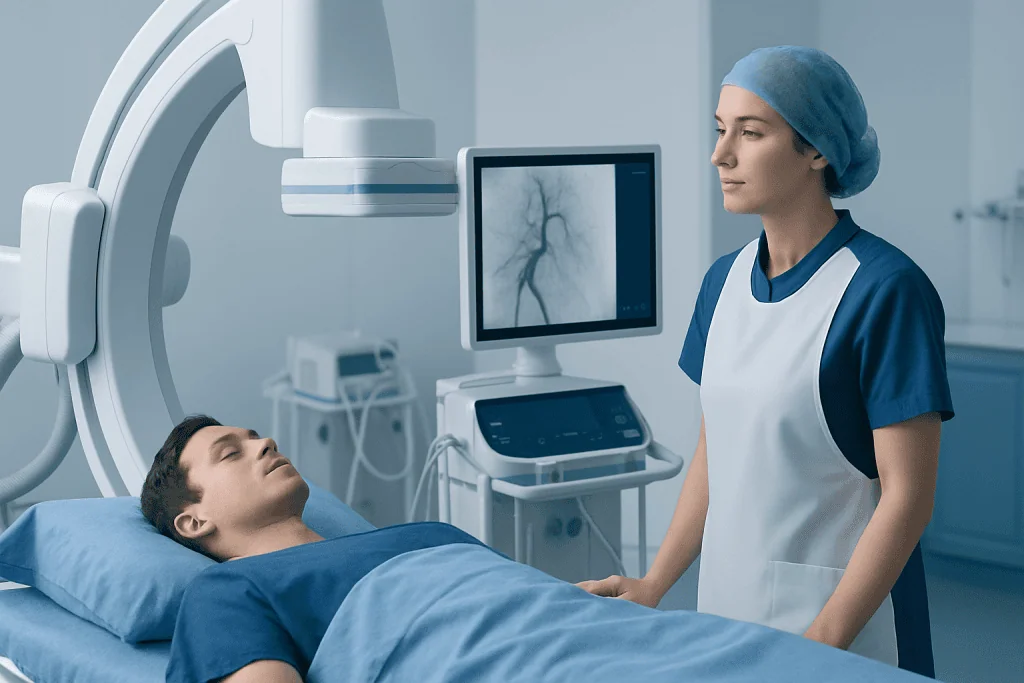
Doctors use arteriography to investigate symptoms and conditions related to reduced blood flow, abnormal blood vessels or internal bleeding. The area examined depends on the clinical question. For example, coronary arteriography focuses on the arteries supplying the heart, while peripheral arteriography evaluates arteries in the legs, arms, pelvis or abdomen. Cerebral arteriography can provide detailed imaging of arteries in the brain and neck.
The test may help identify narrowing caused by atherosclerosis, a buildup of fatty material within artery walls. It can also show complete blockages, blood clots, aneurysms, artery tears, congenital vessel abnormalities or vessels feeding a tumor. In urgent settings, it may assist in locating active internal bleeding after an injury or medical condition.
- Chest discomfort or suspected reduced blood flow to the heart
- Leg pain during walking, non-healing foot wounds or changes in skin color
- Possible aneurysm or abnormal widening of an artery
- Symptoms that could be related to narrowed neck or brain arteries
- Unexplained bleeding that may need catheter-based control
- Planning for vascular surgery or evaluating the results of a previous procedure
Arteriography is not always the first imaging test. The clinician will consider a person’s symptoms, medical history, kidney function, allergy history and results from prior scans before deciding whether it is appropriate.
How to Prepare for an Arteriogram

Preparation instructions vary according to the body area being examined, the urgency of the test and whether treatment may be performed. People are commonly asked not to eat or drink for a specified period before the procedure. They should follow the instructions given by their hospital, as fasting requirements may differ for each individual.
Before arteriography, the team will review medicines, including blood thinners, diabetes medicines, insulin, antiplatelet medicines and supplements. It is important not to stop prescribed medication independently. A clinician will explain whether any medicine should be adjusted before the procedure and how it should be restarted afterward.
Patients should tell their care team about previous reactions to contrast dye, asthma, kidney disease, diabetes, pregnancy or a possible pregnancy, bleeding disorders and any implanted medical devices. Blood tests may be arranged to check kidney function and blood clotting. In most cases, arranging for a responsible adult to provide transport home is recommended, especially if sedation is planned.
What Happens During Arteriography?
On arrival, the patient may change into a gown and have their blood pressure, pulse and oxygen level checked. An intravenous line may be placed for fluids or medication. The specialist will explain the procedure and obtain consent, including discussion of expected benefits, alternatives and possible risks.
The catheter is most often introduced through an artery in the groin or wrist. The skin is cleaned and covered with sterile drapes, then local anaesthetic is used to numb the area. Some people receive medicine to help them feel calm and comfortable, but they are usually awake enough to communicate with the team. Using X-ray guidance, the specialist moves the catheter through the artery to the area being studied.
When contrast dye is injected, a person may briefly notice warmth, flushing, a metallic taste or the sensation of needing to pass urine. These effects usually pass quickly. Multiple images are taken as the dye flows through the arteries. The imaging part may be relatively short, but total procedure time varies depending on the complexity of the vessels and whether treatment is needed.
If a significant problem is found, treatment may sometimes be performed immediately. This can include balloon angioplasty to widen a narrowed artery, placement of a stent to support the vessel, clot-removing techniques or embolization to block a bleeding or abnormal vessel. The doctor will discuss this possibility before the procedure when relevant.
Recovery, Results and Possible Risks
After the catheter is removed, pressure or a closure device is used to reduce bleeding at the access site. The patient is observed while nurses check the puncture area, pulse, blood pressure and comfort level. Depending on the access site and the procedure performed, they may need to lie flat for a period or keep the affected limb relatively still.
Many diagnostic arteriograms are completed as day procedures, although some people need an overnight stay. This is more likely after a complex intervention, if there are other health conditions requiring observation, or if the test was performed during an emergency admission. Drinking fluids when permitted can help the body clear contrast material, unless a clinician has advised fluid restriction.
Minor bruising, tenderness or a small lump at the catheter entry site can occur. More serious complications are uncommon but can include bleeding, infection, artery injury, blood clots, stroke or heart complications depending on the vessels examined, and kidney problems related to contrast dye. Allergic-type reactions to contrast are also possible. The team takes precautions to reduce these risks and will monitor people closely.
Images may be reviewed during the procedure, particularly if an immediate treatment decision is needed. A full explanation of the results is usually provided by the referring doctor or specialist, who will discuss what was found and whether follow-up, medication, lifestyle changes or further treatment is appropriate.
Caring for Yourself After the Procedure
After discharge, patients should follow the specific instructions provided by their care team. Resting for the remainder of the day is often advised after a diagnostic procedure. Strenuous exercise, heavy lifting and vigorous bending may need to be avoided for a short time, particularly when the groin has been used for catheter access.
The access site should be kept clean and dry according to the discharge guidance. Mild soreness can usually be managed with the pain relief recommended by the clinician. It is generally sensible to avoid driving, alcohol and important decisions for at least the rest of the day if sedating medication was given.
Long-term care depends on the result of the arteriography. When artery narrowing is related to atherosclerosis, measures such as stopping smoking, eating a heart-healthy diet, regular physical activity when medically appropriate, managing blood pressure and diabetes, and taking prescribed medicines can support vascular health. These steps do not replace individualized advice from the treating clinician.
When to Seek Medical Care
After arteriography, patients should contact their hospital team or seek prompt medical advice if the puncture site becomes increasingly painful, swollen, red or warm, or if there is ongoing bleeding. Firm pressure should be applied to active bleeding while urgent medical assistance is arranged. A rapidly enlarging lump, a cold or pale limb, numbness, weakness or severe pain below the access site also needs urgent assessment.
Emergency care is needed for symptoms such as chest pain, sudden shortness of breath, fainting, facial drooping, difficulty speaking, sudden weakness on one side of the body or severe new headache. These symptoms may have causes unrelated to the procedure, but should always be evaluated without delay.
People should also seek medical advice before the procedure if they develop a new illness, fever, possible pregnancy or a significant change in kidney-related symptoms. Acibadem International’s multidisciplinary specialists and JCI-accredited hospitals can assess vascular concerns and provide diagnostic and treatment planning for international patients.
Frequently asked questions
Is arteriography painful?
Arteriography is usually performed with local anaesthetic, so the catheter insertion area is numbed. A person may feel brief pressure, mild discomfort or warmth when contrast dye is injected. Sedation may be offered in some cases to improve comfort.
How long does an arteriography procedure take?
A diagnostic arteriography procedure may take from less than an hour to several hours, depending on the arteries being examined and the complexity of the case. If angioplasty, stent placement or another treatment is performed at the same time, it may take longer. The care team can provide a more individualized estimate beforehand.
Is arteriography the same as a CT angiogram?
Both tests create images of blood vessels using contrast material, but they are performed differently. CT angiography uses a CT scanner and contrast given through a vein, while conventional arteriography uses a catheter placed directly into an artery. Conventional arteriography may offer the option of immediate catheter-based treatment.
Can someone go home after an arteriogram?
Many people go home on the same day after a diagnostic arteriogram, once the access site is stable and recovery checks are satisfactory. Some may need longer monitoring, especially after treatment during the procedure or when other medical conditions are present. Driving home is usually not advised after sedation.
What are the risks of contrast dye in arteriography?
Most people tolerate contrast dye well, though some experience temporary warmth or nausea. Allergic-type reactions are possible, and contrast can affect kidney function in people who already have kidney disease or other risk factors. The medical team reviews relevant health history and may use preventive measures when needed.
Why might treatment be done during arteriography?
Arteriography provides real-time images that can show a narrowed artery, clot, bleeding vessel or other treatable problem. In selected cases, the specialist can use the catheter to perform procedures such as balloon angioplasty, stenting or embolization. This may avoid the need for a separate procedure, but it is only done when clinically appropriate.
References
- Radiological Society of North America
- American Heart Association
- National Heart, Lung, and Blood Institute
- Society of Interventional Radiology
- National Institute of Diabetes and Digestive and Kidney Diseases
This article is for general information only and is not a substitute for professional medical advice. Please consult a qualified doctor about your individual situation.
Explore treatments in Turkey — costs, top hospitals & a free quote
JCI-accredited · board-certified surgeons · reply within 24h
Add us as a Preferred Source to see more of our trusted health content across Google Search, AI Overviews and Discover.
Check your numbers in seconds
BMI, calories, due date, blood pressure and 30+ more clinical calculators — free, instant, doctor-reviewed ranges.