MRI Scans: What Patients Need to Know
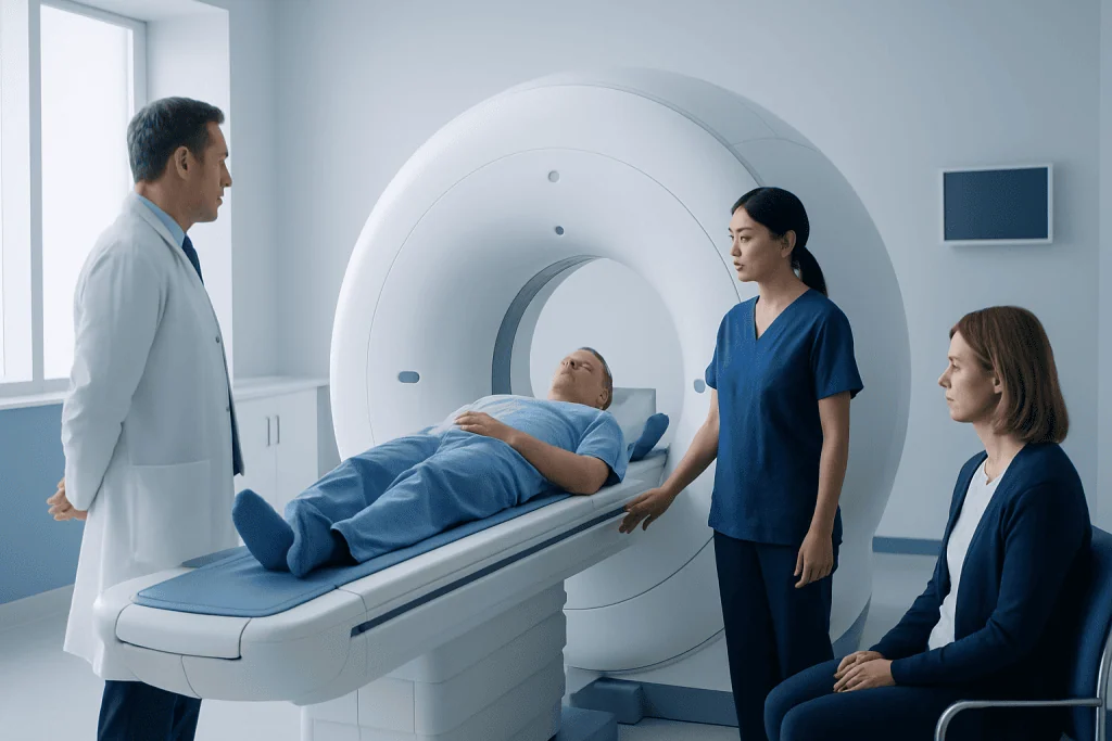
MRI scans provide highly detailed images without using X-rays or ionizing radiation. Patients should tell the imaging team about all implants, metal fragments, medical devices, pregnancy and kidney problems before an MRI.
Key Takeaways
- MRI scans provide highly detailed images without using X-rays or ionizing radiation.
- Patients should tell the imaging team about all implants, metal fragments, medical devices, pregnancy and kidney problems before an MRI.
- The scan is usually painless, although it can be noisy and may feel challenging for people with claustrophobia.
- Some MRI examinations use gadolinium-based contrast to make certain tissues or blood vessels easier to assess.
- Most people can return to their normal routine immediately after an MRI scan.
MRI scans are painless imaging tests that use a strong magnetic field and radio waves to produce detailed pictures of organs, soft tissues, joints and blood vessels. They do not use ionizing radiation, but careful safety screening is essential because the scanner’s magnet is very powerful.
MRI scans: the essentials
MRI scans, short for magnetic resonance imaging scans, are diagnostic tests that create detailed pictures of structures inside the body. They use a strong magnetic field, radiofrequency energy and computer processing rather than X-rays. This means an MRI does not expose the patient to ionizing radiation.
Doctors may request an MRI when they need a clear view of soft tissues, including the brain, spinal cord, nerves, muscles, ligaments, joints, heart, blood vessels and many abdominal or pelvic organs. MRI can help investigate symptoms, clarify an earlier test result, guide treatment planning or monitor a known condition over time.
An MRI examination is generally painless. The main preparation is completing a careful safety questionnaire and removing items that may be attracted to the magnet or affected by it. The imaging team will explain the planned scan, check whether contrast is needed and help the patient remain comfortable throughout the appointment.
Why a doctor may recommend an MRI
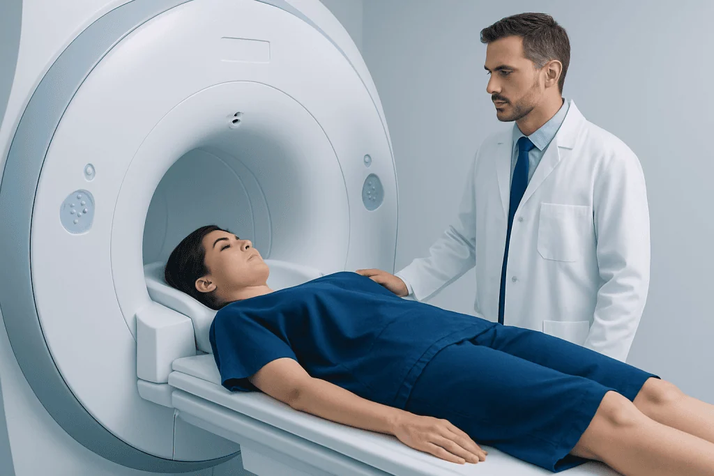
MRI is especially useful when different types of soft tissue need to be distinguished clearly. For example, it can show inflammation, swelling, tears, tumors, infection, bleeding or changes related to reduced blood flow. The best imaging test depends on the clinical question, so MRI is not always the first or only examination a doctor may consider.
In the brain and spine, MRI may be used to assess persistent neurological symptoms, headache patterns with concerning features, seizures, spinal pain with nerve symptoms, or possible stroke and inflammatory conditions. In the musculoskeletal system, it can help evaluate cartilage, tendons, ligaments, bone marrow and soft-tissue injuries that may not be visible on a standard X-ray.
MRI also has important roles in evaluating the heart, liver, kidneys, uterus, ovaries, prostate and other organs. Certain specialized MRI techniques can assess blood flow, heart muscle function or the bile ducts. A radiologist interprets the images and provides a report to the clinician who requested the examination, who then discusses what the findings mean in the context of the patient’s symptoms and medical history.
What happens during an MRI scan

Before the scan, the patient changes into suitable clothing if needed and removes metal-containing belongings. These may include jewelry, watches, hearing aids, removable dental appliances, hair accessories, coins, keys, bank cards and electronic devices. The team may ask additional questions about previous surgery, workplace injuries or any chance that metal entered the eye or body.
During the scan, the patient lies on a padded table that moves into the MRI machine. A body part may be placed inside a special device called a coil, which helps obtain clear images. The scanner makes repetitive loud tapping, knocking or humming sounds while images are being acquired. Earplugs or headphones are usually provided, and staff can communicate with the patient through an intercom.
Keeping still is important because movement can blur the images. The examination commonly takes between 20 and 60 minutes, depending on the body area and type of study, and may take longer for more detailed scans. Some imaging centers offer music, a call button, mirrors or relaxation support to help patients who feel anxious in the scanner.
In some cases, a contrast agent is injected through a small intravenous line partway through the examination. The patient may notice a brief cool sensation at the injection site, but contrast MRI does not usually cause the warm flushing sensation that can occur with some CT contrast studies. The imaging team monitors the patient and can pause if there is discomfort.
MRI safety, implants and contrast agents
The MRI magnet is always powerful, even when a scan is not taking place. Loose metal objects can become dangerous in the scan room, and certain implants or devices may move, heat up, malfunction or produce distorted images. For this reason, patients should answer screening questions carefully and should never enter the MRI area with unapproved metal or electronic items.
Many modern implants, including some joint replacements, dental fillings, surgical clips, vascular stents and certain pacemakers, can be scanned under specific conditions. However, this must be confirmed by the MRI team before the appointment. Patients should bring implant cards, device details or operative information when available. A history of shrapnel, bullets, metalworking injuries or surgery to the brain, eyes, ears, heart or blood vessels should always be mentioned.
Gadolinium-based contrast agents may be used when additional detail is needed, such as when evaluating inflammation, some tumors, blood vessels or active disease. Serious allergic-like reactions are uncommon, but patients should report any previous reaction to MRI contrast. Kidney function may need to be reviewed before contrast is given, particularly for people with significant kidney disease.
Pregnancy should also be discussed with the referring clinician and MRI team. MRI without contrast may be considered when medically necessary during pregnancy, while gadolinium contrast is generally avoided unless there is a strong clinical reason. People who are breastfeeding can ask their clinician or radiology team for individualized advice if contrast has been recommended.
How to prepare and feel more comfortable
Most MRI scans do not require extensive preparation. Patients should follow the instructions provided by the imaging center, as fasting may be requested for certain abdominal, pelvic or contrast-enhanced studies. Regular medicines can often be continued, but it is sensible to confirm this in advance, especially for people who use medicines for diabetes, anxiety or pain.
Comfortable clothing without metal fasteners may be appropriate, although the imaging center may provide a gown. Makeup, hair products and temporary tattoos can occasionally contain metallic particles, so staff may advise removing them. Patients should also inform the team about patches, insulin pumps, glucose monitors, medication pumps or other wearable devices because some must be removed or managed differently before scanning.
Claustrophobia is common and should be mentioned when the MRI is booked, not only on the day of the scan. Depending on the clinical need and local availability, options may include extra support from staff, a wider-bore scanner, relaxation techniques, a support person where permitted, or prescribed medicine for anxiety. Anyone taking a sedating medicine will usually need someone else to drive them home.
- Bring relevant implant or device information.
- Tell staff about allergies, kidney disease, pregnancy or possible pregnancy.
- Arrive early enough to complete safety screening without rushing.
- Ask questions if any instruction is unclear before the scan begins.
Results, aftercare and when to seek medical care
After an MRI scan, most patients can eat, drink, work and resume usual activities straight away. If contrast was used, the team may advise normal hydration unless the patient has been given fluid restrictions for another medical reason. If sedation was used, the patient should follow the discharge instructions and avoid driving, alcohol, important decisions and potentially hazardous activities for the recommended period.
A radiologist reviews the images and sends a report to the referring doctor. Timing varies according to the urgency and complexity of the examination. Patients should arrange follow-up with the clinician who ordered the MRI, as an imaging result is only one part of a full medical assessment and may need to be considered alongside examination findings, blood tests or other investigations.
Patients should seek prompt medical care for severe or rapidly worsening symptoms, such as sudden weakness or numbness on one side of the body, difficulty speaking, sudden loss of vision, a severe new headache, chest pain, major breathing difficulty, loss of bladder or bowel control with back pain, or symptoms after significant injury. These symptoms should not wait for a routine MRI appointment.
After contrast, urgent assessment is appropriate for symptoms of a serious allergic reaction, including trouble breathing, swelling of the lips or throat, widespread hives, faintness or severe dizziness. Mild symptoms such as temporary nausea or localized discomfort should still be reported to the imaging team or a clinician if they persist or cause concern.
Making sense of MRI findings
An MRI report may use technical terms that can sound concerning when read without context. Words such as “degenerative,” “incidental,” “signal change” or “lesion” describe imaging appearances and do not, by themselves, establish a diagnosis or determine whether treatment is needed. Many MRI findings are common with aging or may not be the source of a person’s symptoms.
The most useful next step is a discussion with the referring doctor, who can explain whether the result matches the patient’s symptoms and whether observation, further tests, specialist review or treatment is appropriate. Patients may wish to ask what the main finding means, whether it explains their symptoms, what alternatives are possible and when follow-up is needed.
For patients travelling for evaluation, Acibadem International’s multidisciplinary specialists and JCI-accredited hospitals can support the diagnosis and treatment of conditions that may require MRI assessment. Bringing prior scan images, reports, medication lists and implant information can help clinicians review the situation efficiently and safely.
Frequently asked questions
Do MRI scans use radiation?
No. MRI scans use magnetic fields and radiofrequency energy rather than ionizing radiation such as X-rays. This makes MRI different from CT scans and standard X-ray examinations, although MRI still requires important safety precautions.
How long does an MRI scan take?
Many MRI examinations take about 20 to 60 minutes. The exact time depends on the body part being examined, whether contrast is required and how many image sequences are needed. The imaging team can usually provide an estimate before the appointment.
Can a person with a pacemaker have an MRI?
Some people with modern MRI-conditional pacemakers and other cardiac devices can have an MRI under carefully controlled conditions. The device model, leads and clinical circumstances must be checked in advance. Patients should never assume a device is safe for MRI without confirmation from their healthcare team.
Is MRI contrast safe?
Gadolinium-based contrast is used when it can provide clinically useful information, and it is generally well tolerated. The imaging team reviews factors such as past contrast reactions and kidney function before giving it. Patients should report any new symptoms during or after the injection.
What if a patient is claustrophobic?
Patients should tell the doctor or imaging center before the scan is scheduled. Staff may offer practical support, communication throughout the scan, relaxation measures or, when clinically appropriate, medication prescribed by a doctor. Planning ahead often makes the experience more manageable.
Can a patient eat before an MRI scan?
For many MRI scans, normal eating and drinking are allowed. However, some abdominal, pelvic or contrast studies may require fasting for a period beforehand. Patients should follow the specific instructions from the imaging center and ask if they are unsure.
References
- Radiological Society of North America
- American College of Radiology
- U.S. Food and Drug Administration
- National Health Service
- World Health Organization
This article is for general information only and is not a substitute for professional medical advice. Please consult a qualified doctor about your individual situation.
Joint and spine care in Turkey — expert assessment & treatment
JCI-accredited · board-certified surgeons · reply within 24h
Add us as a Preferred Source to see more of our trusted health content across Google Search, AI Overviews and Discover.