Sagittal Craniosynostosis: Symptoms, Causes, and Treatment Options
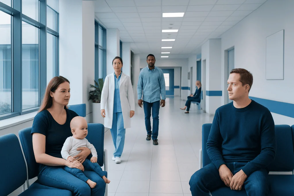
Sagittal craniosynostosis is the most common single-suture type of craniosynostosis. Its typical head shape is long and narrow, often with a prominent forehead or back of the head.
Key Takeaways
- Sagittal craniosynostosis is the most common single-suture type of craniosynostosis.
- Its typical head shape is long and narrow, often with a prominent forehead or back of the head.
- The condition is usually present at birth and is not caused by routine parental care during pregnancy or infancy.
- Diagnosis is based on a physical examination and, when needed, imaging to confirm which skull suture is fused.
- Surgery is the main treatment for most affected babies and is planned by a specialist craniofacial team.
- Early assessment is important, especially when an unusual head shape is noticed in the first months of life.
Sagittal craniosynostosis is a birth condition in which the sagittal suture, the seam running along the top of a baby’s skull, closes too early. It commonly causes a long, narrow head shape and should be assessed early by a craniofacial specialist, since treatment may help create space for normal brain growth and improve skull shape.
Overview: what is sagittal craniosynostosis?
Sagittal craniosynostosis, also called sagittal synostosis, occurs when the sagittal suture closes earlier than expected. Sutures are flexible bands of tissue between a baby’s skull bones. They normally remain open during early childhood, allowing the skull to expand as the brain grows.
The sagittal suture extends from the front to the back along the top center of the head. When it fuses too soon, skull growth is restricted from side to side. The skull may instead grow more from front to back, producing a long, narrow shape called scaphocephaly. The appearance can vary from subtle to more noticeable.
This condition is different from a head shape that develops because a baby regularly rests in one position. Positional flattening does not involve an early-fused skull suture and is managed differently. A pediatrician or craniofacial team can distinguish between these conditions and advise families on the appropriate next steps.
Head shape and other symptoms

The most recognizable feature of sagittal craniosynostosis is an unusually long, narrow head. The forehead may appear broad, prominent, or slightly pointed, and the back of the head may also project. Some babies have a raised ridge that can be felt along the top middle of the skull where the suture has fused.
Many infants are otherwise well, feeding normally and meeting early developmental milestones. Sagittal craniosynostosis does not automatically mean that a child will have developmental difficulties. However, the skull’s restricted growth pattern can affect its shape, and a small number of children may develop increased pressure inside the skull over time.
Possible concerns that merit medical assessment include persistent vomiting without another clear explanation, unusual sleepiness, marked irritability, feeding changes, headaches in an older child, or changes in vision. These symptoms have many possible causes and do not by themselves diagnose craniosynostosis, but they should not be ignored.
- Long, narrow skull shape
- Prominent forehead or back of the head
- Palpable ridge along the top of the skull
- Head shape that does not improve with repositioning
Why it happens and who may be affected

In most cases, the exact cause of sagittal craniosynostosis is not known. It is generally considered a developmental condition that begins before birth, when one skull suture fuses prematurely. It is not caused by how an infant sleeps, by handling a baby incorrectly, or by something a parent did or did not do during everyday pregnancy care.
Most cases occur on their own, meaning there is no known family history and no associated genetic syndrome. Less commonly, craniosynostosis can be part of a genetic condition involving more than one suture or other physical findings. A clinician may suggest genetic evaluation when there is a relevant family history, multiple fused sutures, unusual physical features, or other developmental concerns.
Sagittal synostosis is diagnosed more often in boys than girls. Having one child with the condition may slightly change the likelihood for a future pregnancy, but the individual family risk depends on whether an identifiable genetic cause is present. Genetic counseling can help families understand this in a clear, individualized way.
How specialists confirm the diagnosis
Assessment usually starts with a detailed history and physical examination. The clinician examines the baby’s head shape, feels the skull sutures and soft spots, measures head growth over time, and asks about pregnancy, birth, development, and family history. In some infants, an experienced craniofacial specialist can strongly suspect the diagnosis from examination alone.
Imaging may be used to confirm whether the sagittal suture is fused and to plan treatment. Depending on the child’s age and local practice, this may involve cranial ultrasound, low-dose computed tomography (CT), or other imaging. CT provides detailed views of the skull bones but is used thoughtfully because it involves radiation.
The care team also considers whether another condition could explain the head shape. Positional plagiocephaly, normal variation in head shape, and other forms of craniosynostosis can look similar at first. Accurate diagnosis matters because each condition has different monitoring and treatment needs.
Evaluation is often coordinated by a multidisciplinary craniofacial team that may include pediatric neurosurgeons, craniofacial or plastic surgeons, pediatricians, anesthesiologists, genetic specialists, ophthalmologists, and developmental professionals. This approach helps ensure that skull growth, vision, development, and family support are considered together.
Treatment options and timing
Surgery is the standard treatment for most babies with confirmed sagittal craniosynostosis. Its goals are to release the fused suture, create room for the growing brain, and improve the skull’s proportions. The exact approach depends on the child’s age, the severity and pattern of skull shape, imaging findings, and the experience of the specialist center.
For younger infants, some centers offer minimally invasive endoscopic suture surgery. Through small incisions, the fused suture is removed or opened. This approach is usually followed by a carefully fitted molding helmet that guides skull growth over subsequent months. It is generally only suitable within a limited early age window, so prompt referral is valuable.
Open cranial vault remodeling is another established option and may be recommended for older infants or when a broader reshaping procedure is needed. During this operation, surgeons reshape and reposition sections of skull bone to allow more balanced growth. The hospital team will explain expected recovery, pain management, blood management, follow-up imaging or examinations, and the specific risks relevant to the child.
Not every child requires the same procedure, and treatment decisions should be individualized rather than based on head shape alone. Acibadem International’s multidisciplinary specialists and JCI-accredited hospitals assess and treat craniosynostosis in international patients, with care planned around each child’s clinical needs.
Follow-up, development, and family support
After treatment, children need regular follow-up to monitor head growth, healing, skull shape, and overall development. Follow-up schedules differ by procedure and age. Children who use a molding helmet after endoscopic surgery need frequent adjustments as the head grows, as well as support for skin care and daily wear routines.
Most children recover well after appropriately planned treatment. Still, families should keep routine pediatric visits and raise any concerns about sleep, behavior, headaches, vision, learning, speech, or development. Some children benefit from additional assessment by ophthalmology, developmental pediatrics, speech and language therapy, or other services.
Parents and caregivers can support recovery by following wound-care instructions, attending scheduled appointments, giving only medicines recommended by the child’s clinical team, and allowing appropriate time for rest and gradual return to normal activities. It can also be helpful to ask the team which changes are expected during healing and which require a call.
Emotional support matters as well. Receiving a diagnosis involving a baby’s skull can feel overwhelming, even when treatment is available. Families may find it useful to bring questions to appointments, ask for written information, and seek support from reputable patient organizations or counseling services when needed.
When to seek medical care
A parent or caregiver should arrange a pediatric assessment if a baby’s head appears unusually long and narrow, if a ridge is felt along the top of the skull, or if the head shape seems increasingly different over time. Early evaluation is especially useful in the first months of life, when certain treatment options may be available.
Medical advice should also be sought if there are concerns about head growth, delayed milestones, feeding problems, unusual irritability, repeated vomiting, or changes in eye appearance or vision. These signs are not specific to sagittal craniosynostosis, but a clinician can determine whether they need further investigation.
Urgent medical care is appropriate if a child has severe or repeated vomiting, is difficult to wake, has a seizure, develops sudden weakness, has trouble breathing, or appears seriously unwell. These symptoms need prompt evaluation regardless of whether craniosynostosis has been diagnosed.
Frequently asked questions
Is sagittal craniosynostosis serious?
Sagittal craniosynostosis needs specialist assessment because the fused suture can affect skull growth and, in some children, may contribute to increased pressure inside the skull. Many children do very well with timely evaluation, appropriate treatment, and follow-up. The outlook depends on individual factors such as age at diagnosis and whether other conditions are present.
Can sagittal craniosynostosis correct itself without surgery?
A truly fused sagittal suture does not reopen on its own. Head shape changes caused by sleep position can improve with growth and repositioning, but this is a different condition. A craniofacial specialist can confirm the cause of the head shape and discuss whether surgery is recommended.
At what age is surgery performed for sagittal craniosynostosis?
The timing depends on the surgical technique and the child’s individual assessment. Minimally invasive approaches are generally considered in younger infants, while open cranial vault remodeling may be used later or when more extensive reshaping is appropriate. The treating team will recommend timing that balances skull growth, safety, and expected benefit.
Does sagittal craniosynostosis affect brain development?
Many children with isolated sagittal craniosynostosis develop normally, particularly with appropriate monitoring and treatment. Some children may need assessment for developmental, learning, or vision concerns during childhood. Regular follow-up helps identify and address any needs early.
Is sagittal craniosynostosis genetic?
Most cases are isolated and do not have a clearly identified genetic cause. In a smaller number of cases, craniosynostosis is associated with an inherited or new genetic change, especially when multiple sutures are involved or other findings are present. Genetic counseling may be offered when it is clinically appropriate.
How is sagittal craniosynostosis different from a flat spot on a baby’s head?
Sagittal craniosynostosis produces a long, narrow skull because a top skull suture has fused early. A flat spot from positioning usually causes asymmetry or flattening and does not involve a fused suture. Because the appearances can overlap, a clinician should assess persistent or unusual head shape changes.
References
- American Academy of Pediatrics
- Centers for Disease Control and Prevention
- National Institute of Neurological Disorders and Stroke
- American Association of Neurological Surgeons
- Craniofacial Foundation of America
This article is for general information only and is not a substitute for professional medical advice. Please consult a qualified doctor about your individual situation.
Explore treatments in Turkey — costs, top hospitals & a free quote
JCI-accredited · board-certified surgeons · reply within 24h
Add us as a Preferred Source to see more of our trusted health content across Google Search, AI Overviews and Discover.