Retrograde Urethrogram for Urethral Narrowing or Injury

A retrograde urethrogram uses contrast dye and X-ray imaging to outline the urethra. The test is often used to diagnose or map a urethral stricture, especially before treatment planning.
Key Takeaways
- A retrograde urethrogram uses contrast dye and X-ray imaging to outline the urethra.
- The test is often used to diagnose or map a urethral stricture, especially before treatment planning.
- It is usually an outpatient procedure and generally takes about 15 to 30 minutes.
- Mild temporary discomfort, a small amount of spotting, or brief burning during urination can occur afterward.
- Patients should tell their care team about pregnancy, contrast reactions, urinary infections, and medicines they take.
A retrograde urethrogram is a specialized X-ray examination of the urethra, the tube that carries urine from the bladder out of the body. It is commonly used to assess suspected urethral narrowing, injury, blockage, or an abnormal connection in the urinary tract.
What Is a Retrograde Urethrogram?
A retrograde urethrogram, often called an RUG test, is an imaging examination that helps a doctor see the urethra in detail. The urethra is the passage through which urine leaves the bladder. During the test, a radiopaque contrast liquid is gently introduced through the opening of the urethra while X-ray images are taken.
The contrast outlines the inside of the urethra on the images. This can show whether the passage is narrowed, blocked, torn, or leaking. The examination is called “retrograde” because the contrast moves backward from the urethral opening toward the bladder, rather than following the normal direction of urine flow.
A retrograde urethrogram is particularly useful for evaluating the male urethra, which is longer and can be affected by strictures or trauma at different points. It may also be used in selected women when a doctor needs detailed imaging of the urethra. The test does not usually require sedation and is commonly performed in a radiology department or urology setting.
Why a Doctor May Recommend This Test
The most common reason for a retrograde urethrogram is suspected urethral stricture. A stricture is a scar-related narrowing of the urethra that can slow or block urine flow. Imaging helps identify the stricture’s location, length, and severity, information that can guide further care for a urethral stricture.
A doctor may also request this examination after an injury to the pelvis, penis, or perineum, the area between the genitals and anus. In this situation, the test can help determine whether the urethra has been damaged before a urinary catheter is inserted. It may also be used to assess urine leakage from the urethra, previous surgical changes, certain congenital abnormalities, or persistent urinary symptoms when other tests have not provided enough information.
Symptoms that may lead to assessment include a weak or spraying urine stream, difficulty starting urination, straining to pass urine, a sense that the bladder does not empty fully, repeated urinary infections, or urinary retention. These symptoms can have several causes, so an RUG test is one part of a broader clinical evaluation rather than a diagnosis on its own.
How to Prepare for a Retrograde Urethrogram

Preparation is usually straightforward. Most people can eat and drink normally unless their doctor gives different instructions. The imaging team may ask about symptoms, previous urinary procedures, recent infections, allergies, and medicines, including blood-thinning medicines. A person should not stop prescribed medication unless their clinician specifically advises it.
It is important to tell the care team about a previous reaction to X-ray contrast material, iodine-containing products, latex, antiseptics, or local anesthetic medicines. A history of kidney disease should also be shared, although the amount of contrast used for a retrograde urethrogram is generally small and is placed into the urethra rather than injected into a vein.
Anyone who could be pregnant should inform the radiology team before the examination. X-rays involve radiation, and the team can decide whether the test should be postponed, modified, or replaced with another approach. If there are signs of an active urinary tract infection, such as fever, burning urination, or cloudy urine, the clinician may first evaluate and treat the infection or decide whether imaging should proceed.
What Happens During the Procedure
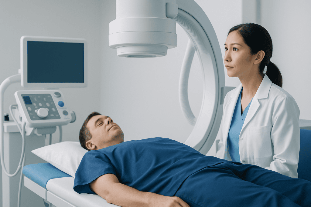
Before the examination, the patient changes into a gown and removes clothing or objects that could interfere with the X-ray images. They lie on an X-ray table, often on their back or slightly turned to one side. The genital area is cleaned using an antiseptic solution to reduce the risk of infection.
A clinician places a small soft tube, syringe tip, or catheter-like device just inside the urethral opening. This is not usually inserted deeply. Contrast liquid is then introduced slowly while a radiologist or radiology professional takes X-ray images. The patient may be asked to remain still or change position briefly to obtain clear views of different parts of the urethra.
People may notice pressure, fullness, coolness from the contrast, or short-lived discomfort during the test. Severe pain is not expected, and the patient should tell the team immediately if it occurs. The imaging portion commonly takes 15 to 30 minutes, though timing can vary depending on the reason for the examination and the findings.
Afterward, the equipment is removed and the patient can usually go home shortly afterward. Sedation is generally not needed, so most people can return to ordinary activities the same day unless their doctor gives individualized instructions.
Understanding the Results
A radiologist reviews the X-ray images and sends a report to the referring doctor, often a urologist. The report describes how contrast moved through the urethra and whether there is narrowing, an irregular area, a complete blockage, contrast leakage, or another structural finding. Results may be available promptly in urgent trauma situations, while routine reports may take longer.
If a stricture is present, the report may describe its site and approximate length. This information matters because treatment choices can differ for a short, simple narrowing and for a longer or recurrent narrowing. Doctors also consider symptoms, urine-flow testing, bladder function, past procedures, and overall health when discussing next steps.
Not every abnormal image requires immediate treatment. For example, a mild narrowing may be monitored if symptoms are limited and bladder emptying is adequate. When treatment is appropriate, options can include endoscopic procedures, dilation in selected cases, or reconstructive surgery. A urologist can explain whether urethroplasty surgery or another approach is suitable for the individual situation.
Risks, Side Effects and Aftercare
Retrograde urethrogram is generally considered a safe procedure. Some people have mild burning when they urinate for a short time afterward, minor spotting of blood at the urethral opening, or temporary soreness. Drinking fluids as normally advised by a clinician may help maintain regular urine flow, unless fluid intake is restricted for another health reason.
Less common risks include urinary tract infection, irritation of the urethra, bleeding, or an allergic-type reaction to contrast or other materials used during the procedure. The risk of radiation exposure from a single diagnostic X-ray examination is low, and the imaging team uses the lowest practical amount needed to answer the clinical question.
Patients should follow any specific discharge instructions provided by the care team. It is sensible to contact the doctor if discomfort does not improve, if there is persistent or increasing bleeding, or if new urinary symptoms develop. In people with complex urinary conditions, the RUG findings may be combined with cystoscopy, ultrasound, urine testing, or other imaging to create a complete care plan.
When to Seek Medical Care
Prompt medical assessment is important for an inability to pass urine, severe lower abdominal pain with a full bladder, blood at the urethral opening after a pelvic or genital injury, or difficulty urinating after major trauma. In these circumstances, emergency clinicians may use a retrograde urethrogram to check the urethra before attempting catheter placement.
A non-urgent appointment with a doctor or urologist is appropriate for a gradually weaker urine stream, straining, repeated urinary infections, recurrent urinary retention, or bothersome changes in urination. These concerns are common and can result from several conditions, so early evaluation can help clarify the cause and avoid unnecessary discomfort.
After an RUG test, urgent medical advice is warranted for fever or chills, worsening pain, inability to urinate, heavy bleeding, swelling, or symptoms of a significant allergic reaction such as trouble breathing or facial swelling. Acibadem International’s multidisciplinary specialists and JCI-accredited hospitals can assess urinary tract concerns and coordinate diagnostic imaging and urology care for international patients when needed.
Frequently asked questions
Is a retrograde urethrogram painful?
Most people experience pressure or brief discomfort rather than severe pain. Some may feel temporary burning or fullness as contrast is introduced. Patients should tell the imaging team if they feel significant pain, as the procedure can be adjusted or stopped if necessary.
How long does a retrograde urethrogram take?
The imaging procedure commonly takes around 15 to 30 minutes. The total appointment may be longer because of registration, preparation, positioning, and discussion of aftercare. In urgent injury assessment, the timing may depend on the person’s overall condition.
Can a retrograde urethrogram diagnose a urethral stricture?
Yes. It is one of the main tests used to identify a urethral stricture and assess where it is located. It can also help estimate the length and degree of narrowing, which supports treatment planning.
Do patients need to fast before a retrograde urethrogram?
Fasting is not usually required for this test. However, preparation instructions can vary if the examination is combined with another procedure or if a person has particular health needs. The imaging center or referring doctor should provide personalized instructions before the appointment.
Can a person drive home after a retrograde urethrogram?
Most people can drive themselves home because sedation is not routinely used. If a person receives medication that affects alertness or feels unwell afterward, they should arrange support and follow the advice of the care team. People recovering from trauma may have separate restrictions.
What is the difference between a retrograde urethrogram and a voiding cystourethrogram?
A retrograde urethrogram shows the urethra as contrast is introduced through the urethral opening. A voiding cystourethrogram usually fills the bladder with contrast and takes images while the person urinates. The two tests may be used separately or together depending on the clinical question.
References
- American Urological Association
- European Association of Urology
- Radiological Society of North America
- National Institute of Diabetes and Digestive and Kidney Diseases
This article is for general information only and is not a substitute for professional medical advice. Please consult a qualified doctor about your individual situation.
Explore treatments in Turkey — costs, top hospitals & a free quote
JCI-accredited · board-certified surgeons · reply within 24h
Add us as a Preferred Source to see more of our trusted health content across Google Search, AI Overviews and Discover.