Private mri scan in Turkey: Hospitals, Safety & What to Expect

MRI uses strong magnetic fields and radio waves, not X-rays, to create detailed images. Safety screening is essential, particularly for people with implants, metal fragments, kidney disease or pregnancy.
Key Takeaways
- MRI uses strong magnetic fields and radio waves, not X-rays, to create detailed images.
- Safety screening is essential, particularly for people with implants, metal fragments, kidney disease or pregnancy.
- Most MRI examinations are outpatient procedures, and normal daily activities can usually resume immediately.
- A contrast agent may be recommended for some scans; clinicians assess whether it is appropriate before use.
- MRI findings should be interpreted alongside symptoms, examination findings and previous test results.
A private MRI scan in Turkey can provide detailed images of internal structures without using ionising radiation. Safe care depends on appropriate medical screening, qualified imaging teams, clear reporting and follow-up with the clinician who requested the scan.
Overview: private MRI scans in Turkey
A private MRI scan in Turkey is an outpatient diagnostic imaging examination that uses a powerful magnetic field and radio waves to create detailed images of organs, soft tissues, joints, blood vessels and the nervous system. It does not use ionising radiation, unlike X-rays and CT scans. MRI may be requested to investigate symptoms, clarify another test result, monitor a known condition or support treatment planning.
For international patients, a well-organised pathway usually includes sharing the referral question and relevant medical records before travel, completing a safety questionnaire on arrival, undergoing the scan and receiving a radiology report. The most useful scan is not simply the fastest available appointment: it is one that is appropriately selected, safely performed and reviewed by the clinician responsible for ongoing care.
People considering imaging as part of a broader health assessment can learn more about the purpose and organisation of private MRI scanning through the relevant service page. The scan should always be matched to an individual clinical question, rather than used as a substitute for medical consultation.
How MRI works and what it can show
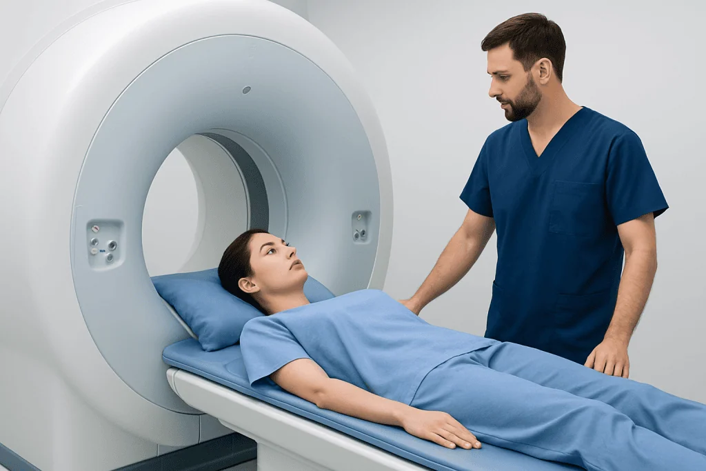
An MRI scanner contains a strong magnet. When a person lies inside the scanner, radiofrequency pulses briefly alter the alignment of hydrogen atoms in body tissues. As the atoms return to their usual state, they emit signals that a computer converts into highly detailed images. Different imaging sequences can highlight structures such as fluid, fat, muscles, ligaments, nerves, the brain and spinal cord.
MRI is particularly valuable for many brain, spine, joint, pelvic and abdominal questions. For example, it may help assess a suspected soft-tissue injury, inflammation, a neurological concern or certain organ abnormalities. The body area, symptoms and clinical question determine whether MRI, ultrasound, CT, X-ray or another test is most suitable.
Some examinations use intravenous contrast, usually a gadolinium-based agent, to make blood vessels, inflammation or certain tissue changes easier to see. Contrast is not needed for every MRI. The radiology and clinical teams weigh its potential value against factors such as kidney function, allergy history and the type of scan requested.
Who may benefit and important safety checks

MRI may be appropriate when a doctor needs more detail than is available from a physical examination or other imaging tests. A referral should state the symptoms, duration of the problem, relevant previous diagnoses and the specific area to be examined. Bringing prior imaging reports and, where possible, image files can help radiologists compare findings over time.
Before the scan, the imaging team asks about all implanted or attached devices and any possible metal in the body. Many modern devices are MRI-conditional, meaning that MRI can be performed only under particular conditions. However, some cardiac devices, neurostimulators, cochlear implants, vascular clips, infusion pumps and older implants may require specialist assessment or may make MRI unsuitable.
Patients should also report previous surgery, metalwork, tattoos or permanent cosmetics, bullet or shrapnel injuries, metal exposure at work, pregnancy, breastfeeding, kidney disease and previous reactions to contrast agents. People who may have metal fragments in their eyes may need additional assessment before entering the MRI room. These checks are routine and are central to MRI safety.
- MRI is often suitable for many people with joint replacements, dental fillings or braces, but these must still be declared.
- Claustrophobia, anxiety, pain or difficulty lying flat can often be managed with advance planning.
- Children and adults unable to remain still may need an individual plan, sometimes involving sedation supervised by an appropriate team.
What happens during a private MRI appointment
Before travelling, patients may be asked to provide a referral, medical history, existing reports and details of implants or medicines. They should confirm the body region being scanned and whether contrast is anticipated. Travel plans should allow enough time for registration, safety screening, the examination itself and collection or secure delivery of reports.
At the hospital, a radiology professional reviews the MRI safety questionnaire. Patients remove metal objects, including jewellery, watches, hearing aids, bank cards and some removable dental items, and may change into a gown. If contrast is required, a small cannula is placed in a vein. The team explains how to communicate during the scan and provides hearing protection because the machine makes loud tapping and knocking sounds.
The patient lies on a moveable table that enters the scanner. It is important to remain as still as possible, although the radiographer can speak with the patient throughout the examination. Different image sequences are taken in short blocks, and some scans involve breath-holding instructions. Scan duration varies according to the body part and complexity, but the imaging team can give a clearer estimate on the day.
After the scan, a radiologist interprets the images and prepares a report. Patients should ask how and when reports and image files will be provided, especially if their referring doctor is in another country. An MRI report describes imaging findings; it does not always establish a diagnosis on its own.
Benefits, limitations and possible risks
The main benefit of MRI is its ability to produce detailed views of many soft tissues without ionising radiation. This can support clinical decisions while avoiding unnecessary invasive procedures in some situations. Modern scanners and trained staff also offer options that may improve comfort, such as communication systems, positioning aids and planning for anxiety.
MRI has limitations. Incidental findings, which are unexpected changes that may be harmless, can lead to further tests or worry. A normal MRI does not rule out every cause of symptoms, and an abnormal finding may not be responsible for the person’s symptoms. Results are most meaningful when interpreted in the context of a full medical assessment.
The magnetic field can pose a serious hazard if unsafe metal objects or incompatible implants enter the scan area, which is why screening must never be rushed. The scan can also be noisy and may feel enclosed. Contrast reactions are uncommon but possible, and gadolinium-based contrast is used cautiously in people with severe kidney impairment. Staff should be told immediately about itching, rash, breathing difficulty, dizziness, pain or unusual symptoms during or after contrast administration.
Recovery, results and planning a stay in Turkey
Most people can return to normal activities immediately after a standard MRI. There is no recovery period from the magnetic field itself. If contrast has been given, the team may provide individual aftercare advice. People who receive sedative medication should not drive, operate machinery or make important decisions until the effects have fully worn off, and they should arrange a responsible adult to accompany them.
For medical travel, planning should include the timing of the scan, report availability, accommodation needs and any follow-up appointment. Patients with limited mobility, ongoing pain, complex medical conditions or a need for sedation may benefit from allowing additional time rather than arranging same-day onward travel. It is sensible to keep copies of the referral, questionnaire, report and image files for future care.
JCI accreditation is one indicator that a hospital has undergone assessment against international quality and patient-safety standards, but patients may still wish to ask practical questions about radiologist reporting, language support, emergency procedures and coordination with their home clinician. Acibadem International’s multidisciplinary specialists and JCI-accredited hospitals diagnose and treat international patients, with care planning that can include imaging, accommodation and follow-up arrangements.
When to seek medical care
An MRI appointment should not delay urgent assessment. People should seek emergency medical care for sudden weakness or numbness on one side of the body, new trouble speaking, a sudden severe headache, loss of consciousness, seizures, chest pain, severe breathing difficulty, or new loss of bladder or bowel control with significant back pain or leg weakness.
A timely medical appointment is also appropriate for persistent or worsening pain, unexplained neurological symptoms, a new lump, ongoing joint limitation, or symptoms that interfere with sleep, work or daily activities. A clinician can determine whether MRI is needed and which body area and imaging protocol are most appropriate.
After an MRI, patients should arrange follow-up with the referring clinician to discuss the report and next steps. Further tests, observation, specialist review or treatment may be recommended depending on the findings and the person’s symptoms; no single imaging result should be interpreted in isolation.
Frequently asked questions
Is a private MRI scan in Turkey safe?
MRI is generally considered safe when the required screening is completed and the examination is performed by trained imaging professionals. The strongest safety concern is the magnetic field, which is why implants, metal fragments and devices must be disclosed before the scan.
Does an MRI scan use radiation?
No. MRI uses magnetic fields and radio waves rather than ionising radiation. This differs from X-rays and CT scans, which use ionising radiation to create images.
How should a patient prepare for an MRI?
Patients should provide accurate information about implants, previous surgeries, possible metal exposure, kidney problems, pregnancy and prior contrast reactions. They should also follow any instructions from the imaging centre about food, drink, medicines and arrival time, particularly if contrast or sedation is planned.
Can someone with claustrophobia have an MRI?
Often, yes. Patients should mention claustrophobia before booking so the imaging team can discuss practical comfort measures, scan planning and whether further support may be appropriate. Remaining still is important for clear images, so advance planning can be helpful.
How long does it take to receive MRI results?
Timing varies according to the type of examination, whether comparison with earlier imaging is needed and the reporting process. Patients should confirm the expected reporting timeframe and how their images and report will be shared with their referring clinician.
Is MRI contrast always necessary?
No. Many MRI scans are performed without contrast. When contrast may improve the diagnostic value of the scan, the medical team reviews the individual situation, including kidney health and any history of contrast reactions.
References
- World Health Organization
- U.S. Food and Drug Administration
- Radiological Society of North America
- American College of Radiology
- National Health Service
This article is for general information only and is not a substitute for professional medical advice. Please consult a qualified doctor about your individual situation.
Joint and spine care in Turkey — expert assessment & treatment
JCI-accredited · board-certified surgeons · reply within 24h
Add us as a Preferred Source to see more of our trusted health content across Google Search, AI Overviews and Discover.