

Can an Ultrasound Detect Cancer: Preparation, Procedure and Results

Ultrasound uses sound waves rather than radiation to create real-time images of organs and tissues. It can identify suspicious changes, but ultrasound findings alone cannot confirm or rule out every cancer.
Key Takeaways
- Ultrasound uses sound waves rather than radiation to create real-time images of organs and tissues.
- It can identify suspicious changes, but ultrasound findings alone cannot confirm or rule out every cancer.
- The test is especially useful for evaluating many abdominal organs, the thyroid, breasts, testicles, pelvis, and lymph nodes.
- CT, MRI, mammography, endoscopy, blood tests, or biopsy may be recommended depending on the body area and symptoms.
- Ultrasound-guided biopsy helps clinicians collect tissue from an abnormal area accurately and with minimal invasiveness.
Can an ultrasound detect cancer? Ultrasound can reveal abnormal masses, enlarged lymph nodes, fluid collections, or changes in organs that may suggest cancer, but it cannot diagnose cancer with certainty. A biopsy and, in some cases, additional imaging or laboratory tests are usually needed to confirm the cause of an abnormal finding.
Overview: Can an Ultrasound Detect Cancer?
Ultrasound can detect changes that may be associated with cancer, including a lump, irregular mass, enlarged lymph node, or an abnormal area within an organ. It is often an important first imaging test because it is painless, widely used, and does not expose the patient to ionising radiation.
However, an ultrasound does not usually provide a definite cancer diagnosis. Some non-cancerous conditions, such as cysts, inflammation, scar tissue, fibroids, or benign growths, can also appear abnormal. When an ultrasound finding is concerning, a clinician may recommend further imaging and, most importantly, a biopsy to examine cells or tissue under a microscope.
Ultrasound is also valuable after a cancer diagnosis. It may help assess nearby lymph nodes, monitor certain known lesions, evaluate symptoms, or guide a needle safely during a tissue-sampling procedure.
How Ultrasound Works and Who May Need It
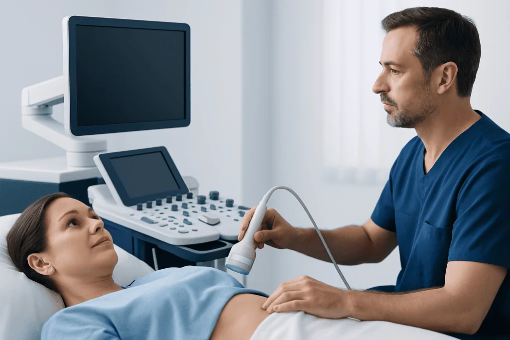
Ultrasound imaging uses a handheld device called a transducer. The transducer sends high-frequency sound waves into the body and receives returning echoes. A computer converts these echoes into live images, allowing the clinician to see movement, blood flow, and the appearance of soft tissues in real time.
A doctor may request an ultrasound when a person has a new lump, pain, swelling, unexplained bleeding, jaundice, altered bowel or urinary symptoms, or abnormal results from another test. It may also be used to investigate an organ seen incompletely on another scan or to follow a previously identified lesion.
The examination is particularly helpful for structures close to the body surface and for many organs in the abdomen and pelvis. Its usefulness can be reduced when bowel gas, body size, overlying bone, or the depth and location of an abnormality limit the sound waves reaching the target area.
- Abdominal ultrasound may assess the liver, gallbladder, kidneys, pancreas, spleen, and pelvic organs.
- Breast ultrasound may help assess a breast lump or clarify a finding on mammography.
- Thyroid and neck ultrasound can evaluate thyroid nodules and lymph nodes.
- Testicular ultrasound is commonly used for a new testicular lump or swelling.
Will Cancer Show Up on Ultrasound?
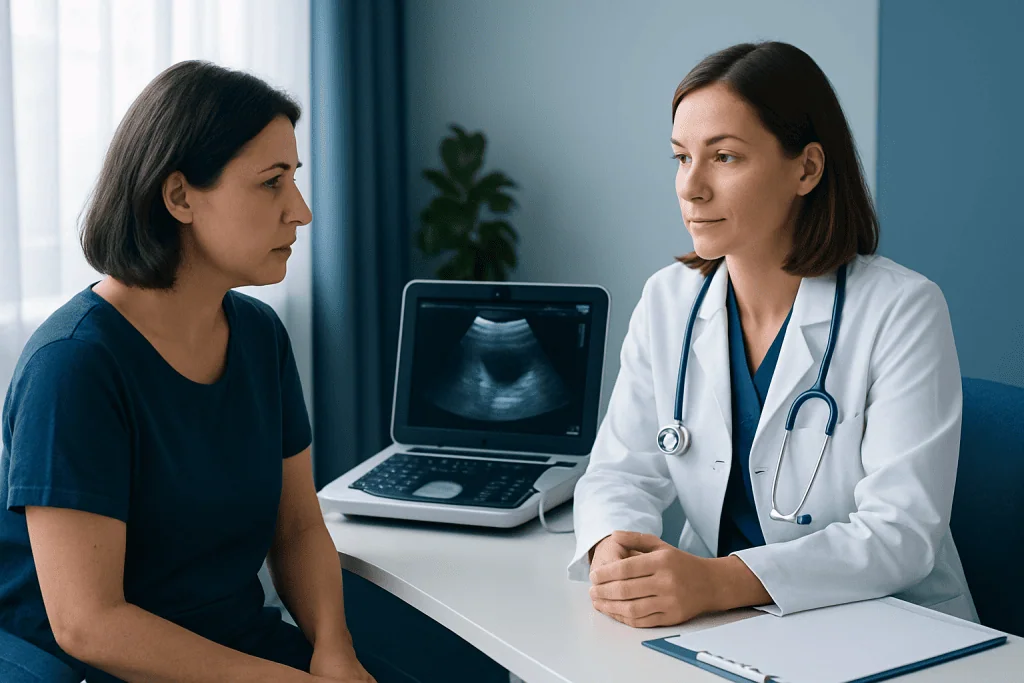
Cancer may show up on ultrasound as an area that looks different from surrounding tissue. Depending on the location, a suspicious finding may be solid rather than fluid-filled, have irregular edges, distort normal anatomy, show unusual blood flow, or be associated with enlarged lymph nodes. These features help the radiologist decide whether further assessment is needed.
Still, ultrasound images cannot reliably distinguish every benign lesion from cancer. For example, some harmless lumps can look solid or irregular, while some cancers can appear subtle or may not be visible at all. The report may describe a finding as benign-appearing, indeterminate, or suspicious rather than naming it as cancer.
If a lesion needs clarification, the next step may be repeat imaging, mammography, CT, MRI, endoscopy, or tissue sampling. Ultrasound-guided biopsy is often used when a visible abnormality can be reached safely with a needle. This approach enables the care team to obtain tissue while watching the needle position in real time.
What Cancers Cannot Be Detected in Ultrasound?
Ultrasound cannot detect every cancer, and a normal examination does not rule out cancer in all situations. Its limitations depend on the organ being examined, the size and location of a tumour, the quality of the scan, and whether gas, bone, air-filled lungs, or deep tissue blocks the sound waves.
Cancers within the brain and spinal cord are not evaluated with standard ultrasound because the skull and spine limit the technique; MRI or CT is generally more suitable. Ultrasound is also not a primary test for most lung cancers because air in the lungs interferes with sound transmission. Small bowel cancers and many cancers inside the stomach or colon are usually assessed with endoscopy and/or CT rather than routine ultrasound.
Very small tumours may be missed, particularly if they do not alter the shape of an organ. In addition, ultrasound may not fully show some deep abdominal or pelvic areas. A clinician interprets ultrasound findings alongside symptoms, physical examination, personal history, family history, and other test results rather than using the scan in isolation.
Can Ultrasound Pick Up Pancreatic Cancer?
An abdominal ultrasound can sometimes identify pancreatic cancer, particularly when a tumour is large enough to be seen or when it causes indirect changes such as widening of the bile ducts or pancreatic duct. It may be requested early when someone has upper abdominal symptoms, jaundice, unexplained weight loss, or abnormal liver blood tests.
However, the pancreas sits deep in the abdomen, and bowel gas can obscure parts of it. This means a normal abdominal ultrasound cannot reliably exclude pancreatic cancer. If clinical concern remains, a doctor may recommend a contrast-enhanced CT scan, MRI with MRCP, or endoscopic ultrasound.
Endoscopic ultrasound places an ultrasound probe close to the pancreas through a flexible scope passed into the digestive tract. It can provide detailed images and may be used to guide a biopsy when appropriate. Evaluation and treatment planning for pancreatic cancer typically involves gastroenterology, radiology, pathology, surgery, and oncology specialists.
What Is the Best Test to Check for Cancer?
There is no single best test to check for all cancers. The most appropriate test depends on the body area, symptoms, age, individual risk factors, and whether the goal is screening, investigating a symptom, or confirming an imaging finding. A doctor selects tests carefully to answer the specific clinical question.
For population screening, examples include mammography for breast cancer, cervical screening tests, stool-based tests or colonoscopy for colorectal cancer, and low-dose CT for certain people at high risk of lung cancer. These tests are designed for particular cancers and are not interchangeable. Routine whole-body scanning is not a proven replacement for recommended screening programmes.
When an abnormality is found, diagnostic imaging may include ultrasound, CT, MRI, mammography, PET imaging, or endoscopy. Although imaging can indicate how likely cancer may be and whether it has spread, biopsy is generally the definitive test for many solid tumours because it identifies the cell type and guides treatment decisions. Assessment for breast cancer or other suspected malignancies should be tailored to the individual findings.
Preparation, Procedure, Recovery, Risks and Benefits
Preparation depends on the type of ultrasound. For an abdominal scan, a person may be asked not to eat for several hours, as fasting can improve views of the gallbladder and upper abdominal organs. For pelvic or bladder imaging, they may be asked to drink water and arrive with a comfortably full bladder. The imaging centre provides specific instructions in advance.
During the procedure, the person usually lies on an examination couch while gel is applied to the skin. The sonographer moves the transducer over the area being examined and may ask the person to change position, hold their breath briefly, or point to the site of symptoms. Most examinations take around 15 to 45 minutes, depending on the area assessed and the complexity of the study.
There is no recovery time after an external ultrasound, and normal daily activities can usually resume immediately. The test is considered very safe because it does not use radiation. Mild pressure or temporary discomfort can occur if the transducer is pressed over a tender area. If a biopsy is performed, the team will explain separate aftercare instructions and possible risks, such as bruising, bleeding, infection, or pain.
The main benefit of ultrasound is that it provides real-time, radiation-free imaging and can guide procedures precisely. Its main limitation is that results may be inconclusive, making additional testing necessary. Acibadem International’s multidisciplinary specialists and JCI-accredited hospitals evaluate suspicious findings and coordinate diagnostic and treatment planning for international patients.
When to Seek Medical Care
A person should arrange medical assessment for a new or persistent lump, unexplained swelling, ongoing pain, unusual bleeding, persistent changes in bowel or bladder habits, unexplained weight loss, jaundice, difficulty swallowing, or symptoms that do not improve as expected. These symptoms have many possible causes and do not necessarily mean cancer, but timely assessment helps identify the reason.
Urgent medical advice is appropriate for severe abdominal pain, new jaundice, coughing up blood, significant unexplained bleeding, severe shortness of breath, or rapidly worsening symptoms. The appropriate urgency depends on the symptom, its severity, and the person’s overall health.
People who have received an abnormal ultrasound result should discuss it with the doctor who requested the scan. The report should be interpreted in context, and the clinician can explain whether follow-up imaging, specialist referral, or biopsy is appropriate.
Frequently asked questions
Can an ultrasound detect cancer?
Ultrasound can detect abnormalities that may be cancer, such as a mass, enlarged lymph node, or change in an organ. However, it cannot usually confirm cancer by itself. A biopsy or additional testing may be needed to establish the diagnosis.
Can ultrasound tell if a lump is cancerous?
Ultrasound can show whether a lump is fluid-filled, solid, or mixed, and can identify features that may warrant further investigation. Many benign lumps can resemble suspicious lesions on imaging. Tissue testing is often needed when cancer cannot be confidently excluded.
Does a normal ultrasound rule out cancer?
No. A normal ultrasound can be reassuring, but it does not exclude every type of cancer or every small, deep, or poorly visible lesion. If symptoms persist or risk is high, a clinician may recommend further evaluation despite a normal scan.
How soon are ultrasound results available?
Images are obtained during the examination, but the formal report is usually prepared by a radiologist and sent to the requesting clinician. Timing varies by healthcare setting and by whether additional review is needed. The requesting doctor can explain the findings and next steps.
Is ultrasound safer than CT for cancer assessment?
Ultrasound does not use ionising radiation, while CT uses X-rays. However, the best test is not always the one with the least radiation; it is the test that provides the information needed for the clinical situation. Doctors balance the expected benefit, limitations, and any potential risks when choosing imaging.
Can an ultrasound replace a biopsy?
In most cases, no. Ultrasound can help identify a suspicious area and guide a needle to it, but it does not examine cells directly. A biopsy is often necessary to confirm cancer type and support treatment planning.
References
- World Health Organization
- American Cancer Society
- National Cancer Institute
- Radiological Society of North America
- American College of Radiology
This article is for general information only and is not a substitute for professional medical advice. Please consult a qualified doctor about your individual situation.
Ultrasound in Turkey — costs, top hospitals & a free quote
JCI-accredited · board-certified surgeons · reply within 24h

Check-up & Preventive Medicine
Comprehensive health screenings designed for early detection and prevention.
3 specialists in this unitMore from the Health Library



Related Specialists

Assoc. Prof. Dr. Caner Baran
Urology
Assoc. Prof. Dr. Gökşen Gökşenoğlu
Physical Medicine and Rehabilitation
Dr. Abdullah Derin
Internal Medicine
Prof. Dr. Bülent Güçyetmez
Anesthesiology