Pregnant Xray: Preparation, Procedure and Results

Tell the referring clinician and radiology team about a known, possible, or uncertain pregnancy before any X-ray examination. A medically necessary X-ray should not usually be delayed solely because of pregnancy, especially after injury or with concerning symptoms.
Key Takeaways
- Tell the referring clinician and radiology team about a known, possible, or uncertain pregnancy before any X-ray examination.
- A medically necessary X-ray should not usually be delayed solely because of pregnancy, especially after injury or with concerning symptoms.
- The first trimester is the most sensitive period for fetal development, but the actual risk depends mainly on the body area imaged and radiation dose.
- Most routine chest, dental, and limb X-rays involve little or no direct fetal exposure.
- No recovery time is needed after a standard X-ray, and results are interpreted by a radiologist and shared with the referring clinician.
A pregnant xray may be recommended when the information it provides is important for the pregnant person’s health and cannot be obtained adequately another way. Diagnostic X-rays outside the abdomen and pelvis, such as many chest or limb X-rays, generally expose the fetus to very low scattered radiation; the imaging team will use the lowest appropriate dose and consider alternatives when suitable.
Pregnant Xray: an answer-first guide
A pregnant xray is usually postponed or replaced with ultrasound or magnetic resonance imaging (MRI) when those tests can answer the clinical question. However, when an X-ray is medically necessary, it can often be performed safely during pregnancy using careful planning, appropriate shielding practices where indicated, and the lowest radiation exposure that produces a useful image.
The decision is not simply whether radiation is present. Clinicians weigh the likely benefit of finding or ruling out an important condition against the small potential risk from the examination. For many studies that image parts of the body away from the uterus, including chest and extremity X-rays, fetal exposure is extremely low because the fetus is outside the direct X-ray beam.
It is important to tell the clinician, radiographer, and radiologist about pregnancy before imaging begins, including if pregnancy is possible but unconfirmed. This allows the team to select the most appropriate examination and adjust the technique if needed.
How X-rays work during pregnancy
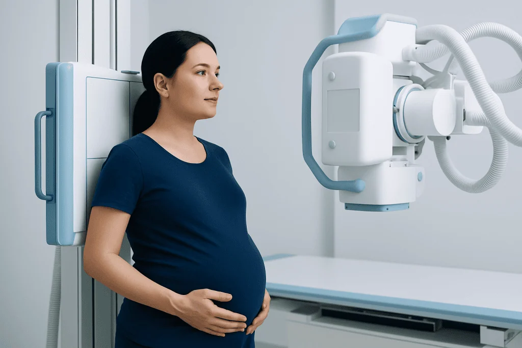
An X-ray uses a controlled beam of ionizing radiation to create images of internal structures. Dense materials, including bone, absorb more radiation and appear lighter on the image, while air-filled areas such as the lungs appear darker. X-rays can help assess suspected fractures, chest infections, bowel problems, dental concerns, and other conditions when a prompt diagnosis matters.
Radiation exposure is measured in terms of dose, and risk depends on the total dose received, the part of the body examined, and the stage of pregnancy. Diagnostic imaging departments follow the principle of keeping radiation exposure as low as reasonably achievable while still obtaining an image that can guide care.
Pregnancy xrays are not all the same. A hand, ankle, or chest X-ray generally produces far less fetal exposure than an examination in which the abdomen or pelvis is in or near the beam. If abdominal or pelvic imaging is needed, the clinical team may first consider ultrasound or MRI, depending on the symptoms and urgency.
A normal X-ray result can be reassuring, but it does not always exclude every possible cause of symptoms. The radiologist interprets the images in the context of the reason for the test, and the referring clinician explains what the findings mean and whether further assessment is needed.
When may a pregnant person need an X-ray?

A clinician may recommend an X-ray during pregnancy if delaying diagnosis could cause more harm than the imaging exposure. Examples include a possible broken bone after a fall, persistent chest symptoms, a suspected serious lung condition, severe dental infection, or evaluation after significant trauma. The clinical question, not pregnancy alone, determines whether imaging is appropriate.
Before ordering the test, the clinician considers symptoms, examination findings, previous imaging, and safer alternatives. Ultrasound does not use ionizing radiation and is often the first choice for pelvic, abdominal, or pregnancy-related questions. MRI also does not use ionizing radiation and can be useful for many soft-tissue, brain, spine, and abdominal concerns, although it may not be the best or fastest test in every situation.
Chest imaging is a common concern. A pregnancy test before chest x ray is not routinely necessary for every person of reproductive age, but departments may ask about the possibility of pregnancy as part of standard safety screening. If pregnancy is known or possible, the team can confirm that a chest X-ray is justified and use an optimized technique.
In an emergency, imaging should not be withheld if it is needed to diagnose a potentially serious problem. Prompt assessment may protect both the pregnant person and the pregnancy by identifying conditions that require urgent treatment.
Which trimester is most sensitive to radiation?
The first trimester, especially the earliest weeks after conception when organs are forming, is generally considered the most sensitive period for possible effects from substantial radiation exposure. The developing fetus changes rapidly during this time, which is why clinicians take particular care to avoid unnecessary ionizing radiation early in pregnancy.
That said, sensitivity does not mean that a needed diagnostic X-ray is automatically unsafe. Most single, properly performed diagnostic X-rays deliver doses well below levels associated with fetal developmental effects. The body part being examined is especially important: imaging of the chest or limbs typically gives the fetus only minimal scattered exposure, while direct imaging of the abdomen or pelvis deserves more individualized consideration.
Later in pregnancy, fetal development remains important and unnecessary radiation is still avoided. Clinicians continue to use the lowest suitable dose in every trimester and may consult a medical physicist when a person has had multiple examinations or a higher-dose study is being considered.
Anyone concerned about an imaging test already performed should speak with the referring clinician or radiology department. They can review the type of examination, the area imaged, and whether a dose estimate or additional counseling is appropriate.
What is the 10-day rule for radiology during pregnancy?
The 10-day rule was a historical radiation-protection approach that aimed to schedule certain non-urgent abdominal or pelvic X-ray examinations during the first 10 days after menstruation began, when pregnancy was considered least likely. Its purpose was to reduce the chance of exposing an unrecognized early pregnancy to radiation.
Modern practice is more individualized. Many professional radiology guidance systems do not apply the 10-day rule rigidly because menstrual cycles vary, pregnancy testing is available when appropriate, radiation doses from many diagnostic examinations are low, and delaying important care can be harmful. Instead, teams assess the possibility of pregnancy, the urgency of the examination, the body region being imaged, and whether another modality can answer the question.
For tests with possible direct uterine exposure, the radiology team may ask about the date of the last menstrual period and may discuss a pregnancy test prior to x ray if the result would change the imaging plan. Testing is not a substitute for clinical judgment: when urgent imaging is necessary, care should proceed with appropriate dose optimization.
Patients should not cancel a recommended examination based only on the 10-day rule. Discussing the reason for the test and pregnancy status with the medical team supports a safe, informed decision.
Preparation for X-rays and the step-by-step procedure
Preparation for x rays is usually simple. The patient should tell the healthcare team about a confirmed pregnancy, a missed period, a possible pregnancy, prior recent imaging, allergies to contrast material if contrast has been discussed, and any relevant medical conditions. For a standard X-ray, fasting and special medication changes are usually not required.
On arrival, the radiographer confirms identity, the body area to be examined, and the reason for the test. The patient may be asked to remove jewelry, clothing with metal fasteners, or other objects that could obscure the image. A gown may be provided. The team explains positioning and helps the patient stay as comfortable and stable as possible.
During the examination, the radiographer positions the body part and briefly steps behind a protective barrier to operate the equipment. The patient is asked to remain still and may be asked to hold their breath for a few seconds during a chest image. The exposure itself lasts only a fraction of a second, and several views may be needed.
Protective measures are selected according to the examination and current imaging protocols. The most effective ways to reduce exposure are ensuring the X-ray is clinically justified, limiting the beam to the needed area, and using optimized equipment settings. The procedure is painless, although positioning an injured area may be uncomfortable.
Results, benefits, risks and recovery timeline
There is no recovery period after a routine X-ray. Patients can normally eat, drink, travel, work, and continue usual activities immediately unless their underlying injury or illness requires different advice. Standard X-rays do not leave radiation in the body and do not make a person radioactive.
A radiologist reviews the images and prepares a report for the clinician who requested the examination. Timing varies by setting: urgent findings may be communicated promptly, while non-urgent reports are often available within a short period. The clinician discusses the findings, explains whether they account for the symptoms, and recommends next steps if necessary.
The main benefit of a pregnant xray is that it can provide information needed to make timely decisions about care. The potential risk is exposure to ionizing radiation, but for most routine diagnostic studies this exposure is low, particularly when the abdomen and pelvis are not directly imaged. Unnecessary examinations are avoided, while needed examinations are optimized rather than automatically deferred.
If a study involving the abdomen, pelvis, fluoroscopy, or computed tomography is being considered, the patient may wish to ask why it is needed, whether ultrasound or MRI could be suitable, and how the team will minimize exposure. These questions can support shared decision-making without delaying urgent care.
When to seek medical care
Pregnant people should seek urgent medical assessment after a significant fall, vehicle collision, or other injury, even if they are worried about needing imaging. They should also seek prompt care for severe or worsening shortness of breath, chest pain, coughing up blood, fainting, high fever with concerning symptoms, severe abdominal pain, heavy vaginal bleeding, or a sudden inability to use an injured limb.
For non-urgent concerns, a clinician can help decide whether imaging is necessary and which test is best. Patients should mention pregnancy early when booking an appointment and again on arrival. This includes people who are trying to conceive or are unsure whether they may be pregnant.
Acibadem International’s multidisciplinary specialists and JCI-accredited hospitals can assess symptoms and coordinate appropriate diagnostic imaging and treatment for international patients. The right investigation is chosen according to the individual clinical situation, pregnancy stage, and need for timely care.
Frequently asked questions
Can an X-ray harm a fetus?
The possibility of harm depends on the radiation dose, the body area being examined, and the stage of pregnancy. Most routine diagnostic X-rays, particularly of the chest or limbs, involve very low fetal exposure and are not expected to cause developmental effects. A clinically necessary examination should be discussed with the medical team rather than avoided without advice.
Do I need a pregnancy test before an X-ray?
A pregnancy test is not needed before every X-ray. It may be considered when pregnancy is possible and the planned examination could expose the abdomen or pelvis to radiation, especially if the result would alter the imaging plan. The radiology team will ask pregnancy-screening questions and explain any recommended testing.
Is a chest X-ray safe during pregnancy?
When clinically needed, a chest X-ray can usually be performed during pregnancy with an optimized technique. Because the uterus is not in the direct X-ray beam, fetal exposure is generally very low. The clinician will consider whether the test is necessary and whether an alternative study could provide the needed information.
Should I tell the radiographer if I might be pregnant?
Yes. A person should tell the radiographer and referring clinician about a known pregnancy, a possible pregnancy, or uncertainty about pregnancy before the examination. This information helps the team select the most suitable test and reduce exposure where possible.
Can I have an X-ray after a fall while pregnant?
Yes, if the clinician believes it is needed to identify or exclude an injury. Untreated fractures, chest injury, or other trauma can pose significant health risks, so necessary imaging should not be delayed solely because of pregnancy. The team will use the most appropriate low-exposure approach.
What happens if I had an X-ray before knowing I was pregnant?
A single routine diagnostic X-ray before pregnancy was recognized is often not a cause for alarm, particularly if the abdomen and pelvis were not directly imaged. Contact the clinician or imaging department with the date and type of examination so they can review the exposure and provide individualized reassurance or advice. Do not make decisions about the pregnancy based on imaging exposure without qualified medical counseling.
References
- American College of Radiology
- American College of Obstetricians and Gynecologists
- International Atomic Energy Agency
- Radiological Society of North America
- World Health Organization
This article is for general information only and is not a substitute for professional medical advice. Please consult a qualified doctor about your individual situation.
Explore treatments in Turkey — costs, top hospitals & a free quote
JCI-accredited · board-certified surgeons · reply within 24h
Add us as a Preferred Source to see more of our trusted health content across Google Search, AI Overviews and Discover.