Hepatoma Ultrasound: Preparation, Procedure and Results
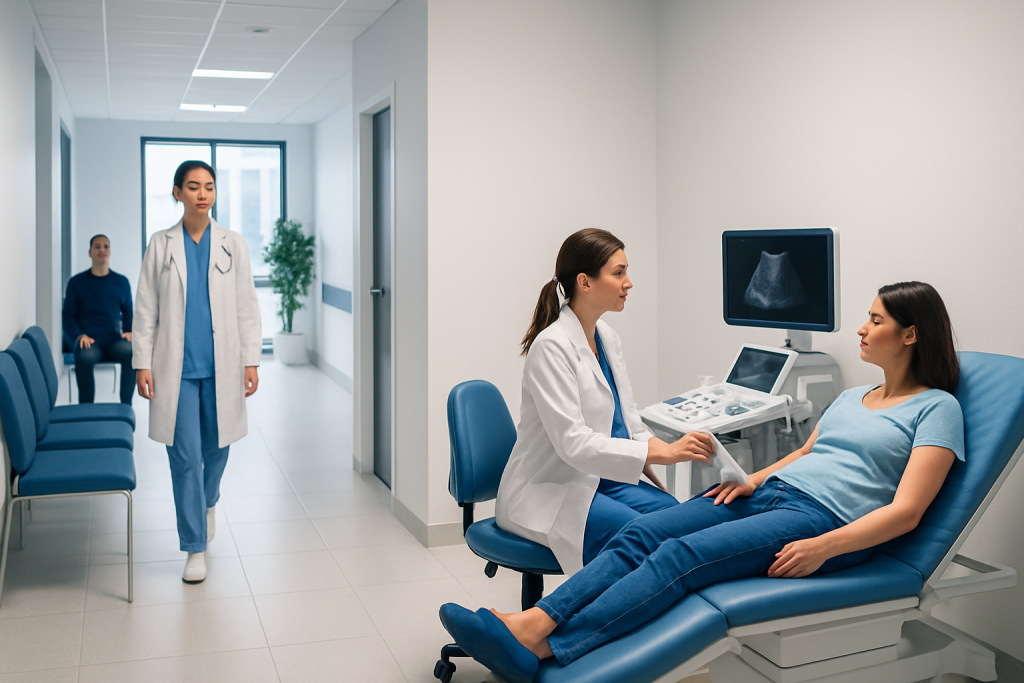
Ultrasound is painless, does not use radiation, and usually takes about 15 to 30 minutes. Fasting is commonly requested because food, drinks, coffee, and sometimes water can reduce image quality.
Key Takeaways
- Ultrasound is painless, does not use radiation, and usually takes about 15 to 30 minutes.
- Fasting is commonly requested because food, drinks, coffee, and sometimes water can reduce image quality.
- Ultrasound can identify a liver lesion, but it cannot always confirm whether a lesion is hepatocellular carcinoma.
- The radiologist usually reviews images after the scan, so final results may not be available immediately.
- People with cirrhosis, chronic hepatitis B, or other significant liver disease may need regular liver cancer surveillance.
A hepatoma ultrasound is a noninvasive imaging test that examines the liver for masses or changes that may be associated with hepatocellular carcinoma, also called hepatoma. It is commonly used for liver cancer surveillance and initial assessment, while CT, MRI, blood tests, and sometimes biopsy may be needed to clarify an abnormal result.
Overview: What Is a Hepatoma Ultrasound?
A hepatoma ultrasound is an abdominal ultrasound focused on the liver. It uses high-frequency sound waves to produce real-time images of liver tissue, blood vessels, and nearby structures. The test may be used when a person has symptoms, abnormal liver blood tests, known chronic liver disease, or a need for regular screening for hepatocellular carcinoma (HCC), the most common primary liver cancer.
The term hepatoma is often used to refer to hepatocellular carcinoma. Ultrasound is particularly valuable as a first-line test because it is widely available, does not expose the patient to ionizing radiation, and does not usually require injections or contrast material. However, an ultrasound finding alone does not always establish a cancer diagnosis.
For people at increased risk of liver cancer, especially those with cirrhosis or chronic hepatitis B, clinicians may recommend liver ultrasound surveillance at regular intervals. The exact plan depends on the person’s liver condition, medical history, and specialist guidance. More information about liver malignancy may be found in liver cancer care.
How It Works and Who May Need It
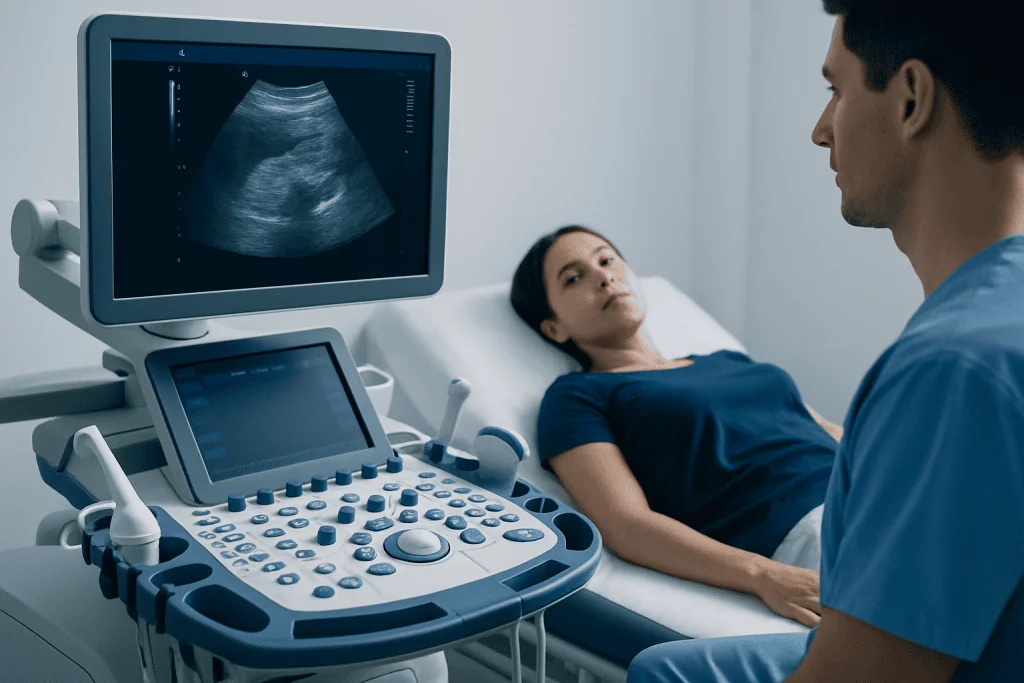
During an ultrasound, a transducer placed on the skin sends sound waves into the body and receives the echoes that return from internal tissues. A computer converts these echoes into moving images. Different liver tissues reflect sound differently, helping the sonographer and radiologist assess the liver’s size, surface, texture, blood flow, and any focal lesion.
A hepatoma ultrasound may be appropriate for people with cirrhosis, chronic hepatitis B or C, fatty liver disease with advanced scarring, a previous liver lesion, or a history of liver cancer treatment. It may also be requested to investigate upper abdominal discomfort, unexplained weight loss, jaundice, abdominal swelling, or abnormal liver function tests. These symptoms are not specific to cancer and often have other explanations.
Ultrasound may be less detailed in some circumstances, including marked obesity, extensive bowel gas, or a deeply positioned lesion. In these cases, the clinical team may recommend contrast-enhanced CT or MRI for a clearer evaluation. Depending on the findings, a specialist may also discuss liver cancer treatment options.
How to Prepare for Ultrasound of the Liver

When learning how to prepare for ultrasound of the liver, the most important step is to follow the instructions provided by the imaging center. Many liver ultrasounds require fasting for several hours before the appointment. Fasting helps reduce gas in the stomach and intestines and may improve visualization of the liver, gallbladder, pancreas, and surrounding structures.
Why can’t you drink water before a liver ultrasound?
Some imaging departments ask patients not to drink water shortly before a liver ultrasound because fluid and swallowed air can affect the appearance of the upper abdomen or contribute to bowel activity. Other centers allow small sips of plain water, particularly for essential medicines. Instructions vary by the type of scan and whether the gallbladder or other abdominal organs are being examined, so the patient should confirm the local guidance rather than making assumptions.
Can I drink coffee while fasting for a liver ultrasound?
Coffee is generally not recommended during the fasting period, including black coffee. It can stimulate digestion and gallbladder contraction, which may make parts of an abdominal examination more difficult to interpret. Tea, juice, milk, chewing gum, smoking, and mints may also be restricted. Prescribed medicines should not be stopped without medical advice; the imaging team or prescribing clinician can explain how to take them safely before the test.
- Wear comfortable, loose-fitting clothing that allows access to the upper abdomen.
- Bring prior scans, reports, and a list of medicines if requested.
- Tell the team about pregnancy, recent procedures, and relevant liver conditions.
- Arrive early enough to complete registration and review preparation details.
What Happens During the Procedure?
A liver ultrasound is usually performed in an outpatient imaging department. After checking in, the patient may be asked to lie on an examination table and expose the upper abdomen. A clear water-based gel is applied to the skin; this helps transmit sound waves and prevents air from interfering with the scan.
The sonographer moves the transducer across the abdomen and may ask the patient to turn slightly, raise an arm, or hold their breath briefly. These position changes can improve the view between the ribs and help capture images of different parts of the liver. Mild pressure from the probe is normal, but the examination should not be painful. The patient should mention any tenderness or discomfort.
A standard examination commonly takes about 15 to 30 minutes, although the length can vary. Doppler ultrasound may be added to evaluate blood flow in vessels such as the portal vein and hepatic veins. The sonographer obtains the images but typically does not provide a diagnosis during the procedure; a radiologist interprets the completed study.
Hepatoma Ultrasound Appearance and What the Images Can Show
Can you see hepatocellular carcinoma on ultrasound?
Ultrasound can sometimes show a lesion that raises concern for hepatocellular carcinoma, but it cannot reliably confirm HCC in every case. The hepatoma ultrasound appearance can vary according to the lesion’s size, blood supply, background liver disease, and technical image quality. A mass may appear darker, brighter, mixed in texture, or different from surrounding liver tissue, but these appearances are not unique to cancer.
Hepatoma ultrasound images may show a new focal nodule, irregular liver texture, changes associated with cirrhosis, or fluid in the abdomen. The scan can also detect many noncancerous findings, such as cysts, hemangiomas, fatty changes, or regenerative nodules. This is why an unexpected lesion should be assessed in the full clinical context rather than interpreted from one image alone.
When a suspicious nodule is found, clinicians often use multiphase CT or contrast-enhanced MRI to look for characteristic enhancement patterns. Blood tests, including alpha-fetoprotein in selected situations, may support assessment but cannot diagnose or exclude HCC by themselves. A biopsy is occasionally needed when imaging results remain uncertain or when another diagnosis is possible.
Results, Recovery, Benefits and Limitations
Do you get liver ultrasound results immediately?
Patients may be told that the scan has been completed, but final liver ultrasound results are commonly issued after a radiologist reviews the images and prepares a report. The timing varies among centers and can range from the same day to several days. The referring doctor then explains what the findings mean and whether follow-up imaging, laboratory tests, or specialist review is needed.
There is no recovery period after a routine hepatoma ultrasound. The gel is wiped off, and most people can return to work, driving, meals, and normal activities straight away. If fasting was required, eating and drinking can usually resume once the test is finished unless the clinician has given other instructions.
The main benefits are that ultrasound is noninvasive, painless, repeatable, and radiation-free. Its limitations are equally important: small lesions may be difficult to see, image quality may be reduced by body habitus or bowel gas, and the test cannot always distinguish cancer from benign liver changes. An abnormal result is a reason for timely follow-up, not a diagnosis by itself.
When to Seek Medical Care
A person should contact a doctor promptly if they have known cirrhosis, chronic hepatitis B, chronic hepatitis C, or another condition that places them at increased risk of liver cancer and they have missed recommended surveillance. Regular monitoring can help identify liver changes at an earlier stage, when more management options may be available.
Medical assessment is also appropriate for persistent upper abdominal pain or fullness, unexplained loss of appetite or weight, new jaundice, worsening fatigue, abdominal swelling, vomiting, or black stools. These symptoms can arise from many conditions and do not necessarily mean cancer, but they should not be ignored, especially in someone with existing liver disease.
Urgent care is needed for severe abdominal pain, vomiting blood, confusion, fainting, or rapidly worsening jaundice. Acibadem International’s multidisciplinary specialists and JCI-accredited hospitals support international patients with diagnostic assessment and coordinated care for liver conditions, including interventional radiology procedures when clinically appropriate.
Frequently asked questions
What is a hepatoma ultrasound used for?
A hepatoma ultrasound is used to examine the liver for nodules, masses, cirrhosis-related changes, and other abnormalities. It may be part of routine surveillance for people at higher risk of hepatocellular carcinoma or an investigation after symptoms or abnormal blood tests. It is usually a first assessment rather than a stand-alone cancer diagnosis.
How long does a liver ultrasound take?
Most liver ultrasounds take approximately 15 to 30 minutes. The duration can be longer if Doppler blood-flow imaging is needed or if additional images are required. Preparation and registration may add time to the appointment.
Do I need to fast before a hepatoma ultrasound?
Many centers ask patients to fast for several hours before a liver ultrasound, but the exact instructions differ. Fasting can reduce bowel gas and help the sonographer obtain clearer images. Patients should follow the instructions from their own imaging center, especially regarding water and medicines.
Can ultrasound tell if a liver mass is cancerous?
Ultrasound can identify a liver mass or suspicious area, but it often cannot determine with certainty whether it is cancerous. CT or MRI with contrast can provide more detailed information about the lesion’s blood-flow pattern and characteristics. Sometimes additional blood tests or a biopsy are considered.
What happens if a liver ultrasound shows a lesion?
A lesion does not automatically mean cancer, because benign cysts, hemangiomas, fatty changes, and other conditions can also appear on imaging. The doctor may compare prior scans, request CT or MRI, arrange blood tests, or refer the patient to a liver specialist. The next step depends on the lesion’s size, appearance, growth, and the person’s liver health.
Is liver ultrasound safe?
Yes. Ultrasound uses sound waves rather than radiation and is considered safe for most people, including during pregnancy when clinically needed. The scan is noninvasive and usually causes no side effects beyond brief pressure from the probe.
References
- World Health Organization
- American Cancer Society
- American College of Radiology
- European Association for the Study of the Liver
- National Cancer Institute
This article is for general information only and is not a substitute for professional medical advice. Please consult a qualified doctor about your individual situation.
Ultrasound in Turkey — costs, top hospitals & a free quote
JCI-accredited · board-certified surgeons · reply within 24h
Add us as a Preferred Source to see more of our trusted health content across Google Search, AI Overviews and Discover.

Check-up & Preventive Medicine
Comprehensive health screenings designed for early detection and prevention.
1 specialists in this unit