Gamma Radiation and X Rays: How It Works, Results and What to Expect

X rays and gamma rays are both ionizing radiation, meaning they can remove electrons from atoms and affect living cells. Diagnostic X rays use very low, brief exposures designed to provide useful images with minimal radiation.
Key Takeaways
- X rays and gamma rays are both ionizing radiation, meaning they can remove electrons from atoms and affect living cells.
- Diagnostic X rays use very low, brief exposures designed to provide useful images with minimal radiation.
- Gamma radiation can be focused or delivered in planned doses to damage cancer cells or treat selected conditions.
- Most people do not feel an X ray; unusual sensations afterward are more often related to anxiety, positioning, contrast material, or another health issue.
- X-ray image availability ranges from immediate to several days depending on the examination and reporting process.
- Radiation procedures are planned and supervised to balance expected benefit with the lowest practical exposure.
Gamma radiation and X rays are high-energy forms of ionizing radiation, but they are used differently in healthcare. X rays are most often used to create diagnostic images, while carefully planned gamma radiation may be used in certain cancer treatments and targeted brain procedures.
Overview: Gamma Radiation and X Rays
Gamma radiation and X rays are types of electromagnetic energy with enough strength to ionize atoms. In healthcare, this property can be useful: a short, low-dose X-ray exposure can create images of structures inside the body, while accurately targeted gamma radiation can help treat some tumors and other selected conditions.
The main difference is their origin. X rays are produced by medical equipment when electricity accelerates particles, whereas gamma rays are released from unstable atomic nuclei or radioactive materials. Both can pass through tissue, but their use, intensity, duration, and safety controls differ greatly between diagnostic imaging and radiation treatment.
Radiation is not automatically harmful or beneficial; its effects depend mainly on dose, the part of the body exposed, how long exposure lasts, and the clinical reason for using it. Healthcare teams use radiation only when the expected information or treatment benefit outweighs the potential risk.
How Gamma Radiation and X Rays Work
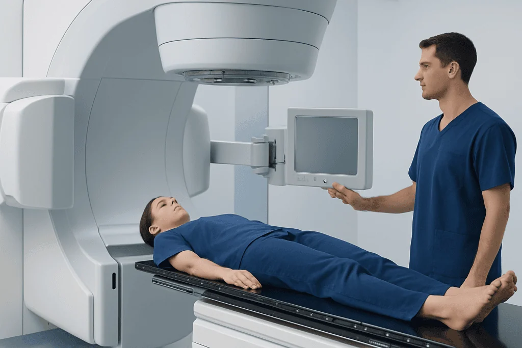
When ionizing radiation passes through the body, it transfers energy to cells and tissues. Different tissues absorb or allow radiation through at different rates. In an X-ray image, dense materials such as bone absorb more radiation and appear lighter, while air-filled areas appear darker. This contrast helps clinicians assess fractures, chest conditions, dental concerns, and many other problems.
Gamma rays are physically similar to X rays in that both are high-energy photons. Their source and medical delivery are different. In radiation oncology, gamma radiation may be directed from carefully planned external sources toward a defined target. The goal is to damage the DNA of abnormal cells so they can no longer divide effectively, while protecting nearby healthy tissue as much as possible.
Some highly focused approaches, such as stereotactic radiosurgery, use many precisely aimed radiation beams that converge at a target. Despite its name, radiosurgery does not involve an incision. It may be considered for selected brain tumors, blood vessel abnormalities, or certain nerve conditions after specialist assessment.
What Does Gamma Radiation Do to Your Body?

Gamma radiation deposits energy in tissue and can damage cellular DNA directly or indirectly through the formation of reactive molecules. Cells that divide frequently, including many cancer cells, may be more vulnerable to this damage. Radiation treatment is therefore often delivered in carefully designed sessions that allow normal tissues time to repair between exposures.
The body’s response depends on the radiation dose, the exposed area, and whether radiation is used for a diagnostic test or treatment. The tiny, brief exposure from a routine X ray does not cause the same effects as therapeutic radiation. In contrast, radiation therapy may cause temporary effects in the treated region, such as tiredness, skin changes, irritation, or organ-specific symptoms.
Modern planning uses imaging, computer calculations, positioning devices, and quality checks to reduce exposure to healthy tissue. A radiation oncologist explains expected benefits, possible side effects, and alternatives before treatment begins. People who are pregnant or may be pregnant should tell the imaging or treatment team before any radiation-based procedure.
Candidacy and the Step-by-Step Procedure
Diagnostic X rays may be recommended after an injury, for persistent symptoms, before certain procedures, or to monitor a known condition. A clinician chooses the type of imaging based on the question that needs answering. An X ray may be enough for a suspected bone injury, while ultrasound, CT, MRI, or laboratory testing may be more suitable in other situations.
Gamma radiation treatment is not a general-purpose procedure. Candidacy is determined by a multidisciplinary team that may include a radiation oncologist, medical oncologist, surgeon, radiologist, physicist, and other specialists. They consider the diagnosis, location and size of the target, prior treatment, overall health, and the likely value of radiation compared with other options.
For an X ray, the person is usually asked to remove metal objects or clothing that could obscure the image, wear a gown if needed, and hold still in one or more positions. The radiographer leaves the room briefly to operate the equipment, but communication remains possible. The exposure itself generally lasts a fraction of a second; positioning may take longer.
Before gamma radiation treatment, planning commonly includes detailed imaging and sometimes a fitted mask, frame, or body-positioning device. The team creates a treatment plan and verifies positioning at each visit. During treatment, the person lies still while the machine delivers radiation; the procedure is painless, and the team monitors from outside the room.
Benefits, Risks and Radiation Safety
The main benefit of X-ray imaging is rapid, noninvasive information that can guide diagnosis and treatment. It can identify many fractures, joint changes, lung findings, bowel obstruction patterns, and problems involving teeth or other structures. The images may also help avoid unnecessary procedures or support timely referral to the appropriate specialist.
For appropriately selected patients, gamma radiation therapy can control or shrink certain tumors, reduce symptoms, or treat a precisely defined target without conventional surgery. The expected outcome varies widely according to the underlying condition. Radiation may be used alone or alongside surgery, systemic therapy, and supportive care.
Ionizing radiation has potential risks, including a small long-term cancer risk that rises with greater cumulative dose. Therapeutic doses can also cause treatment-area side effects, and rarely long-term tissue injury. Teams follow the principle of using the lowest radiation exposure reasonably achievable for diagnostic tests and use detailed planning for treatment.
People should keep a record of significant imaging and radiation treatments when possible, although medically necessary studies should not be avoided solely because of past imaging. The ordering clinician and radiology team can explain why a test is recommended and whether another approach may be appropriate.
Why Do I Feel Weird After an X-ray?
A standard X ray is painless, very brief, and usually does not cause a sensation during or afterward. The radiation dose from a routine examination is not expected to make a person feel dizzy, weak, nauseated, or unwell immediately afterward. Feeling unusual may instead be related to pain from the original injury or illness, stress, fasting, an uncomfortable position, or another unrelated cause.
Some imaging examinations involve contrast material, especially certain CT, fluoroscopy, or nuclear medicine studies. Contrast can sometimes cause a temporary warm feeling, altered taste, nausea, itching, or other reactions. These examinations are different from a simple X ray, so it is important to tell staff exactly which test was performed and what symptoms occurred.
New or worsening symptoms should not be assumed to be caused by the X ray. Urgent medical assessment is appropriate for trouble breathing, facial or throat swelling, fainting, severe rash, chest pain, severe headache, or rapidly worsening symptoms. For nonurgent concerns, contacting the imaging department or the clinician who ordered the test is a sensible first step.
How Long Do X-Rays Take to Get Results?
The image itself is created immediately, and the radiographer may check that it is technically adequate before the person leaves. However, the final interpretation is usually provided by a radiologist, who reviews the images in clinical context and sends a report to the referring clinician.
Timing varies by setting and urgency. In emergency care, preliminary findings may be available quickly to guide immediate decisions. For outpatient imaging, the formal report may be ready the same day or within several days. Complex examinations, additional image review, and comparison with prior studies can extend this timeline.
The clinician who ordered the X ray is generally best placed to explain what the report means for the individual. A result may be normal, may show a clear cause of symptoms, or may require follow-up imaging or assessment. A report should be interpreted alongside symptoms, examination findings, and medical history rather than in isolation.
How Long Do Gamma Rays Last?
Gamma rays are not stored in the body after an external-beam radiation treatment or ordinary X-ray examination. Once the machine stops, the external radiation exposure stops. A person is not radioactive after an external X ray, CT scan, or external-beam radiation therapy and can usually be around family members normally unless their clinical team advises otherwise.
Gamma rays themselves travel at the speed of light, so the exposure occurs only while the radiation source is active and directed toward the body. In treatment, the biological effects in tissues can continue afterward because cells need time to respond to DNA damage. This is why treatment response and some side effects may develop gradually over days, weeks, or longer.
A different situation applies to some nuclear medicine tests and internal radiation treatments, in which a radioactive substance may temporarily remain in the body. The team provides specific safety instructions when this applies. These instructions may include hydration, hygiene measures, or temporary limits on close contact with others.
Recovery, Follow-Up and When to Seek Medical Care
No recovery period is usually needed after a routine X ray. Most people can eat, drink, drive, work, and resume usual activities right away unless their underlying condition or another procedure requires different advice. If imaging involved contrast material, sedation, or an invasive component, the care team will provide individual aftercare instructions.
Recovery after gamma radiation treatment depends on the target area, dose plan, and other treatments. Some people continue normal daily activities, while others experience fatigue or local symptoms that build gradually. Follow-up appointments, physical examinations, and repeat imaging help the team assess response and manage side effects safely.
Medical care should be sought promptly for severe or worsening symptoms after an imaging or radiation procedure, especially breathing difficulty, swelling of the face or throat, fainting, severe pain, new weakness, confusion, seizures, persistent vomiting, or a high fever. People receiving radiation therapy should also contact their oncology team about new skin breakdown, severe diarrhea, painful swallowing, urinary difficulty, or symptoms that interfere with eating, drinking, or daily function.
Acibadem International’s multidisciplinary specialists and JCI-accredited hospitals support international patients who need diagnostic imaging, radiation planning, and treatment for appropriate conditions. A qualified clinician can explain whether a radiation-based test or treatment is suitable for the person’s individual needs.
Frequently asked questions
Are gamma rays stronger than X rays?
Gamma rays and X rays are both ionizing radiation and can overlap in energy range. The main distinction is their source: gamma rays come from atomic nuclei, while X rays are generated by equipment. Medical safety depends on dose, exposure time, and the body area involved rather than the name alone.
Do X rays cause radiation to stay in the body?
No. A routine X ray uses external radiation, and no radiation remains in the body after the image is taken. The person does not become radioactive and can be near other people normally.
Can gamma radiation cure cancer?
Radiation using gamma rays or other high-energy beams can cure some cancers, control others, or relieve symptoms, depending on the cancer type and stage. It is often one part of a broader treatment plan. The oncology team can discuss the realistic goals of treatment for an individual diagnosis.
Why do I feel weird after an X-ray?
A routine X ray generally does not produce immediate symptoms. Feeling unwell may be related to the illness or injury being investigated, anxiety, positioning, fasting, or a different part of the examination such as contrast material. Severe or rapidly worsening symptoms should be medically assessed promptly.
How long do X-rays take to get results?
The images are made immediately, but the final report requires review by a radiologist. Emergency results may be communicated quickly, while outpatient reports often take from the same day to several days. The referring clinician will explain the result and any next steps.
How long do gamma rays last after treatment?
After external radiation treatment, gamma rays do not remain in the body once the machine has stopped. Tissue changes from treatment may continue over time as cells respond and heal. Internal radiation or nuclear medicine procedures are different and may require temporary safety instructions.
References
- International Atomic Energy Agency
- World Health Organization
- U.S. Food and Drug Administration
- Radiological Society of North America
- National Cancer Institute
This article is for general information only and is not a substitute for professional medical advice. Please consult a qualified doctor about your individual situation.
Oncology care in Turkey — second opinion and treatment plan
JCI-accredited · board-certified surgeons · reply within 24h
Add us as a Preferred Source to see more of our trusted health content across Google Search, AI Overviews and Discover.