Ureteral Obstruction Treatment: How It Works, Results and What to Expect

A blocked ureter can cause urine to back up into the kidney, sometimes leading to hydronephrosis, pain, infection or declining kidney function. Urgent drainage is often needed when obstruction occurs with infection, fever, severe pain, a single functioning kidney or reduced kidney function.
Key Takeaways
- A blocked ureter can cause urine to back up into the kidney, sometimes leading to hydronephrosis, pain, infection or declining kidney function.
- Urgent drainage is often needed when obstruction occurs with infection, fever, severe pain, a single functioning kidney or reduced kidney function.
- Temporary drainage with a stent or nephrostomy tube may be followed by treatment for stones, scar tissue, tumors or other causes.
- Many ureteral obstructions can be treated successfully, but the best approach depends on the location, cause, duration and effect on kidney function.
- Follow-up imaging and kidney function tests help confirm that urine is draining well after treatment.
Ureteral obstruction treatment aims to relieve a blockage that prevents urine from draining from the kidney to the bladder. Depending on the cause and urgency, care may involve a ureteral stent, nephrostomy tube, stone treatment or reconstructive surgery to protect kidney function and address the source of obstruction.
Overview: How ureteral obstruction treatment works
Ureteral obstruction treatment relieves a blockage in one of the ureters, the narrow tubes that carry urine from the kidneys to the bladder. Treatment first restores drainage when necessary, then addresses the reason for the blockage. This may help ease symptoms, reduce the risk of infection and preserve as much kidney function as possible.
A blockage can be partial or complete, sudden or gradual, and may affect one or both ureters. Kidney stones are a common cause, but scarring, a narrowed ureter, enlarged nearby structures, blood clots, congenital differences, inflammation and cancers can also interfere with urine flow. The approach is individualized after imaging and laboratory testing identify the likely cause and severity.
Some people need immediate decompression using a ureteral stent or a tube placed through the back into the kidney, called a percutaneous nephrostomy. Others can proceed directly to treatment of the cause, such as stone removal or ureteral reconstruction. The goal is not simply to remove a blockage, but to create durable drainage while safely managing any underlying illness.
What will happen if the ureter is blocked?
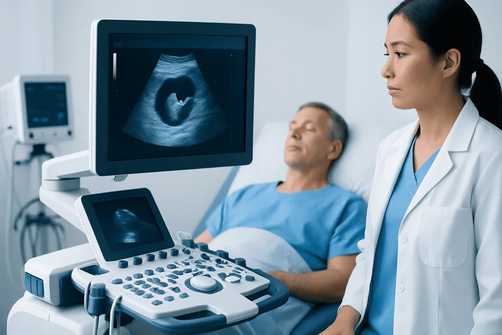
When a ureter is blocked, urine may build up above the obstruction and stretch the kidney’s drainage system. This enlargement is called hydronephrosis. It may cause flank or abdominal pain, nausea, urinary changes, or no noticeable symptoms at all, particularly when the blockage develops slowly.
Persistent pressure can impair kidney function. If bacteria are also present, an obstructed urinary system can develop a serious infection because urine cannot drain normally. For this reason, fever or chills with suspected obstruction needs prompt medical assessment.
The effects vary widely. A small stone may pass and allow normal drainage to return, while a complete or longstanding obstruction may require prompt intervention. Imaging, urine testing and blood tests help clinicians determine whether urgent drainage is needed and what treatment is most appropriate.
How long does it take for ureteral obstruction to cause kidney damage?

There is no single timeline because kidney injury depends on whether the blockage is complete or partial, whether one or both kidneys are affected, the person’s baseline kidney health and whether infection is present. A complete obstruction can begin affecting kidney function within days to weeks, while partial obstruction may cause gradual damage over a longer period.
When both kidneys are obstructed, or when a person has one functioning kidney, kidney function can decline more quickly and urgent care is important. Infection behind an obstruction is also an emergency because it can worsen rapidly without drainage and antibiotics.
Clinicians do not rely on symptoms alone to judge risk. They use ultrasound, CT or other imaging to look for swelling, along with blood tests such as creatinine and urine studies. Early assessment and relief of significant obstruction offer the best chance of protecting kidney function.
Who may need treatment and how the cause is diagnosed
People may be evaluated for ureteral obstruction because of colicky flank pain, blood in the urine, recurrent urinary infections, nausea, reduced urine output or an imaging finding of hydronephrosis. However, not everyone with these symptoms has an obstruction, and some obstructions cause little discomfort. A clinician will consider symptoms alongside medical history and test results.
Testing often includes urinalysis and urine culture, kidney function blood tests, and imaging. Ultrasound can identify kidney swelling, while CT may define the level and cause of a blockage, especially when stones are suspected. In selected situations, contrast imaging, MRI or a camera examination of the bladder and ureter may be used.
Candidacy for a particular procedure depends on the obstruction’s cause, size, location and severity. It also depends on infection status, pregnancy, kidney function, bleeding risk, previous operations and overall health. People with kidney stones may need a different pathway from those with a scar-related narrowing or a mass compressing the ureter.
A urologist commonly coordinates care. Depending on the cause, the care team may also include radiology, nephrology, oncology, gynecology, colorectal surgery or other specialists.
Treatment options and the procedure journey
Treatment commonly has two stages: urgent drainage when needed, followed by definitive treatment of the cause. A ureteral stent is a soft internal tube placed through the bladder into the ureter during cystoscopy. It holds the drainage pathway open and allows urine to pass from the kidney to the bladder. In some cases, ureteral stent placement is used as a temporary bridge before stone removal or reconstructive surgery.
A percutaneous nephrostomy is another drainage option. Under imaging guidance, an interventional radiologist places a small tube through the skin of the back into the kidney. Urine drains into an external collection bag. This may be especially helpful when a stent cannot be placed promptly or cannot pass through the blockage.
Once drainage is secured, the underlying problem is treated. Stones may be managed with observation, medication in appropriate cases, ureteroscopy, shock-wave treatment or other stone procedures. A narrowed or scarred ureter may be treated with endoscopic dilation or incision, or with reconstruction such as ureteral reimplantation or pyeloplasty. Obstruction from a tumor requires a coordinated plan focused on both drainage and treatment of the tumor.
Before a procedure, the team reviews imaging, medications and medical conditions, and treats infection if possible. Stent placement and ureteroscopy are often performed with anesthesia. A nephrostomy may be performed with local anesthetic, sedation or anesthesia depending on the circumstances. The exact plan should be discussed with the treating team, including whether a stent or tube will need later exchange or removal.
Can a blocked ureter be fixed?
Yes. Many blocked ureters can be relieved and, in many cases, the cause can be treated or repaired. The outlook is often favorable when obstruction is recognized early and urine drainage is restored, especially if the kidney has not sustained substantial long-term damage.
The term “fixed” can mean different things. A stone can be removed, a temporary stent can restore drainage while inflammation settles, or a narrowed section of ureter can be surgically reconstructed. In some situations, such as obstruction caused by advanced cancer or recurrent scarring, ongoing stent changes, nephrostomy drainage or additional treatment may be needed.
Follow-up is important even after symptoms improve. A clinician may arrange imaging, urine tests and kidney function blood tests to ensure the kidney is draining and to identify recurrence early. The care plan also addresses preventable causes, such as reducing the risk of future stones when relevant.
Benefits, risks and recovery after treatment
The main benefits of treatment are restoration of urine flow, relief of pressure on the kidney, treatment or prevention of infection-related complications, and an opportunity to correct the cause of obstruction. Pain and nausea often improve once effective drainage is established, though the timing varies with the condition and the procedure used.
After a ureteral stent, temporary urinary frequency, urgency, burning with urination, blood-tinged urine or discomfort in the bladder or flank can occur. These symptoms are often manageable but should be discussed with the treating team. After a nephrostomy, patients receive instructions on tube and skin care, drainage bag handling and signs that the tube may be blocked or displaced.
Potential procedure-related risks include bleeding, infection, pain, injury to the urinary tract, stent movement or blockage, and the need for further procedures. Stents and nephrostomy tubes are not usually permanent and must be monitored, exchanged or removed on schedule to reduce complications. The team can explain the expected recovery timeline based on the chosen procedure and individual health needs.
Recovery after simple stent placement may involve a short period of reduced activity, while recovery after reconstructive surgery may take longer. Patients should follow individualized instructions about fluids, activity, work, pain management and follow-up appointments rather than relying on a standard timeline.
Can you still urinate if you have a blockage?
Yes. Many people with a blockage in one ureter can still urinate normally because the other kidney continues to make urine and drain into the bladder. Even with one blocked ureter, urine output may therefore appear unchanged. Normal urination does not rule out ureteral obstruction.
If both ureters are blocked, or if a person has only one functioning kidney and that ureter is blocked, urine output may become markedly reduced or stop. This requires urgent medical assessment. Reduced urine output can also have other causes, so evaluation is needed to determine the reason.
Changes in urine color, blood in the urine, burning, frequency or urgency can occur with obstruction or with a stent, but these symptoms are not specific. New fever, worsening pain, vomiting or inability to pass urine should not be managed with self-care alone.
When to seek medical care
Prompt medical care is needed for severe or persistent flank pain, fever, chills, vomiting, visible blood in the urine, markedly reduced urine output or an inability to urinate. These symptoms can occur with ureteral obstruction, infection or other urinary conditions that need timely assessment.
Emergency assessment is particularly important when fever or chills occur with suspected kidney stone symptoms, because infection with an obstructed kidney can require urgent drainage. People who are pregnant, have a single functioning kidney, known kidney disease, a transplanted kidney or a weakened immune system should seek advice early if they develop possible obstruction symptoms.
For ongoing follow-up, patients should keep all appointments for stent or nephrostomy care and contact their team if they develop worsening pain, fever, leaking around a tube, a tube that stops draining, or a tube that is pulled out. Acibadem International’s multidisciplinary specialists and JCI-accredited hospitals assess and treat urinary tract obstruction for international patients, with care tailored to the underlying cause.
Frequently asked questions
Is ureteral obstruction treatment always surgery?
No. The first treatment may be drainage with a ureteral stent or nephrostomy tube rather than reconstructive surgery. Some small stones pass on their own, while others need endoscopic removal or another procedure. The need for surgery depends on the cause, location and persistence of the blockage.
How quickly is a ureteral stent placed for an obstruction?
Timing depends on symptoms, kidney function and whether infection is suspected. Obstruction with infection, severe kidney impairment, a single functioning kidney or uncontrolled symptoms may require urgent drainage. In less urgent cases, the clinician can plan treatment after completing appropriate imaging and testing.
Will a ureteral stent hurt?
A stent can cause temporary bladder pressure, frequent urination, urgency, flank discomfort or blood-tinged urine. Experiences vary, and symptoms often improve with time or supportive treatment recommended by the care team. Severe pain, fever or inability to urinate should be reported promptly.
Can hydronephrosis go away after the blockage is treated?
Hydronephrosis often improves after urine drainage is restored, but the degree and speed of improvement depend on how long and how severely the kidney was obstructed. Follow-up imaging helps show whether swelling is resolving. Some dilation can remain even when drainage is adequate, so results are interpreted alongside kidney function and symptoms.
What causes recurrent ureteral obstruction?
Recurrent obstruction can result from repeated kidney stones, scar tissue after inflammation or surgery, congenital narrowing, external compression or an untreated underlying disease. Identifying the cause is important because prevention and follow-up differ for each condition. People with recurrent stones may benefit from evaluation of stone type, diet and metabolic risk factors.
Can a ureteral obstruction return after repair?
It can return in some circumstances, particularly when scar tissue reforms, stones recur or the original cause continues. Follow-up appointments, imaging and kidney function tests are used to monitor recovery and detect recurrence. New flank pain, fever or urinary changes should be discussed with a clinician rather than assuming they are expected.
References
- National Institute of Diabetes and Digestive and Kidney Diseases
- American Urological Association
- European Association of Urology
- National Health Service
- Merck Manual Consumer Version
This article is for general information only and is not a substitute for professional medical advice. Please consult a qualified doctor about your individual situation.
Heart care in Turkey — expert evaluation and treatment
JCI-accredited · board-certified surgeons · reply within 24h
Add us as a Preferred Source to see more of our trusted health content across Google Search, AI Overviews and Discover.